
Case Report | DOI: https://doi.org/10.31579/2690-4861/509
1Department of Radiation Oncology, Memorial Sloan Kettering Cancer Center, New York, NY, United States.
2St. George’s University School of Medicine, Grenada, Caribbean.
3Department of Radiation Oncology, American University of Beirut Medical Center, Beirut, Lebanon.
4University of Balamand, Balamand, Lebanon.
*Corresponding Author: Neal Kim, Department of Radiation Oncology, Memorial Sloan Kettering Cancer Center, New York, NY, United States.
Citation: Neal Kim, Zachary McSween, Lara Hilal, Joseph Hajj, Marsha Reyngold, et al., (2024), Reirradiation for De novo Anorectal Cancers in Patients with a History of Prostate Radiation Therapy: Dosimetric Considerations and Practical Radiation Planning Techniques, International Journal of Clinical Case Reports and Reviews, 18(5); DOI:10.31579/2690-4861/509
Copyright: © 2024, Neal Kim. This is an open-access article distributed under the terms of the Creative Commons Attribution License, which permits unrestricted use, distribution, and reproduction in any medium, provided the original author and source are credited.
Received: 24 July 2024 | Accepted: 01 August 2024 | Published: 09 August 2024
Keywords: prostate radiation therapy; anal cancer; rectal cancer; reirradiation; second course of pelvic RT; toxicity
This case report examines the treatment planning approach for patients receiving anorectal radiotherapy with a history of prostate radiotherapy. Three cases are presented: two of these patients received prior 50.4 Gy in 28 fractions via external beam radiation therapy, where one proceeded to obtain a cone-down of 30.6 Gy in 17 fractions, while the other received 100 Gy of low-dose-rate brachytherapy; the third patient only received prior low-dose-rate brachytherapy to an unknown dose due to absent prior planning records. This paper explores the unique challenges this reirradiation setting poses, outlines treatment planning strategies to overcome these obstacles, and reviews retreatment dose constraints from the literature.
RT, radiation therapy; EBRT, external beam radiation therapy; OAR, organ-at-risk; LDR, lose-dose rate; PTR, prior treatment review; IDL, isodose line; EQD2, equivalent dose in 2 Gy fractions; IMRT, Intensity-modulated radiation therapy; 3D-CRT, 3-dimensional conformal radiation therapy; GTV, gross tumor volume; PTV, planning target volume.
There is an increasing incidence of patients presenting with rectal cancer with a history of prostate radiotherapy [1]. There is no consensus about whether there is a direct relation between prior prostate radiotherapy and second pelvic cancer incidence. Omer et al. described common limitations in methodology among published studies, such as inconsistencies in defining the latency period and the extent of patient follow-up [2]. As prostate radiotherapy continues to advance and experience associated gains in survival paired with screening initiatives leading to earlier cancer diagnoses in younger patients, the option of pelvic reirradiation overall is expected to become an increasingly more common topic of consideration [2]. These cases can be particularly challenging for treatment planning as organs-at-risk (OARs) in pelvic reirradiation are primarily arranged in serial functional subunits, where injury to any segment of a serial organ can result in serious complications including, but not limited to, fistula formation, perforation, and radionecrosis [3,4].
This case report outlines the treatment planning approaches undertaken in managing this unique population at our institution. We selected three patients who recently received anorectal radiotherapy at our institution having a history of prostate radiotherapy. Two of the three patients received prior 50.4 Gy in 28 fractions via external beam radiation therapy (EBRT), where one received a cone-down of 30.6 Gy in 17 fractions of EBRT, and the other received 100 Gy of low-dose-rate (LDR) brachytherapy. The third patient received only prior LDR brachytherapy (dose unknown due to absent planning records). The patients’ differences in prior prostate radiotherapy modality led to their inclusion in this case study report as it resulted in diverging treatment planning considerations (see Table 1-3 for treatment summarization). We also compile pelvic reirradiation constraints cited in the literature with their corresponding interval between radiation courses, follow-up interval, and Grade 3+ toxicity incidences to contextualize our cases better and provide insight on best practices to manage this unique patient population (see Tables 4-6).
| Case | Prostate RT | Pelvic RT | Initial Clinical Response | In-field POD | Out-of-field POD | Grade 3+ Toxicity | OS from RT end |
| 1 | 2012: -EBRT 50.4 Gy/28 fx + 30.6 Gy/17 fx | 2022: -Anorectal + LN 50/45 Gy/25 fx QD | Partial | No POD | 8 mo; skin (Merkel cell) | Grade 3 acute dermatitis | 11 mo |
| 2 | 2001: -EBRT 50.4 Gy/28 fx + 100 Gy LDR | 2022: -Rectum + LN 45 Gy/30 fx BID | Complete | No POD | No POD | N | 17 mo (since last f/u) |
| 3 | 2001: -LDR dose unavailable | 2017: -Rectum + LN 45 Gy/25 fx QD | Complete | No POD | No POD | N | 60 mo |
Table 1. Treatment characteristics
Table 1 Abbreviations: RT, radiation therapy; POD, progression of disease; OS, overall survival; EBRT, external beam radiation therapy; LN, lymph nodes; QD, once a day; LDR, low-dose-rate (brachytherapy); BID, = twice a day; f/u, follow-up.
| Case | Structure | Prior-RT Brachytherapy Dmax Gy3 EQD2 | Prior-RT EBRT Dmax Gy3 EQD2 | Re-RT Dmax Gy3 EQD2 | Total Gy3 EQD2 |
| 1 | Bladder | 77.8 | 46.2 | 123.9 | |
| Rectum | 77.8 | 55.2 | 133 | ||
| Urethra | 77.8 | 31.8 | 109.6 | ||
| 2 | Bladder | 47.8 (D2cc) | 60.7 | 40.5 | 149 |
| Rectum | 47.8 (D2cc) | 55.7 | 40.5 | 143.9 | |
| Urethra | 47.8 (D2cc) | 55.2 | 40.5 | 143.5 | |
| 3 | Bladder | N/A | 50.5 | N/A | |
| Rectum | N/A | 50.7 | N/A | ||
| Urethra | N/A | 13.3 | N/A |
Table 2. Dosimetric details
Table 2 Abbreviations: RT, radiation therapy; EQD2, equivalent dose in 2 Gy fractions.
| Case | Planning Scan | Fusion | Contrast | Orientation | Immobilization | Anal BB Marker | Foley Catheter | Full Bladder | Imaging |
| 1 | CT | PET | IV & Oral | Head-first supine (Frog-leg) | Alpha cradle | Y | N | Y | Every treatment, 2D kV Imaging |
| 2 | MRI T1 & T2 | IV | Head-first prone | Belly board | Y | ||||
| 3 | N | N | Head-first prone | Belly board | N |
Table 3. Simulation and setup details
| Structure | n | Metric | Median Gy3 EQD2 | Range | Interquartile Range |
| Bladder & Bladder wall | 10 | Dmax-D1cc < | 116.3 | 80-147.4 | 110-137.1 |
| Rectum & Rectal wall | 14 | Dmax-D1cc < | 124.5 | 88.4-169.2 | 111.5-137.4 |
| Urethra | 9 | Dmax-D1cc < | 144.8 | 134.1-165.1 | 144.8-158.8 |
Table 4. Summary of cumulative dose constraints
Table 4 Abbreviations: EQD2, equivalent dose in 2 Gy fractions.
| OAR | Re-RT Cited Constraint | Re-RT Rx (Median) | Re-RT Constraint in EQD2 | (Estimated) Prior-RT Rx (Median) | Cumulative Constraint in EQD2 |
| Bladder11 | Dmax < 36 Gy | 30 Gy/5 fx | Dmax < 73.4 Gy3 EQD2 | N/A | Dmax < 147.4 Gy3 EQD2 |
| Bladder wall12 | Dmax < 34 Gy | 34 Gy/5 fx | Dmax < 66.6 Gy3 EQD2 | 75.6 Gy/40 fx | Dmax < 140.6 Gy3 EQD2 |
| Bladder wall13 | Dmax < 34 Gy | 34 Gy/5 fx | Dmax < 66.6 Gy3 EQD2 | 73.8 Gy/40 fx | Dmax < 138.1 Gy3 EQD2 |
| Bladder10 | Dmax < 110 Gy3 EQD2 | Dmax < 110 Gy3 EQD2 | |||
| Bladder14 | Dmax < 25 Gy | 25 Gy/5 fx | Dmax < 40 Gy3 EQD2 | 50.4 Gy/28 fx | Dmax < 88.4 Gy3 EQD2 |
| Bladder15 | D0.1cc < 33 Gy | 36-38 Gy/6 fx | D0.1cc < 56.1 Gy3 EQD2 | 78 Gy/39 fx | D0.1cc < 134.1 Gy3 EQD2 |
| Bladder16 | D0.5cc < 80 Gy3 EQD2 | D0.5cc < 80 Gy3 EQD2 | |||
| Bladder15 | D0.5cc < 28 Gy | 36-38 Gy/6 fx | D0.5cc < 42.9 Gy3 EQD2 | 78 Gy/39 fx | D0.5cc < 120.9 Gy3 EQD2 |
| Bladder16 | D0.5cc < 110 Gy3 EQD2 | D0.5cc < 110 Gy3 EQD2 | |||
| Bladder15 | D1cc < 24 Gy | 36-38 Gy/6 fx | D1cc < 33.6 Gy3 EQD2 | 78 Gy/39 fx | D1cc < 111.6 Gy3 EQD2 |
| Rectum17 | Dmax < 40 Gy | 30 Gy/5 fx | Dmax < 88 Gy3 EQD2 | 70 Gy/25 fx | Dmax < 169.2 Gy3 EQD2 |
| Rectum18 | Dmax < 40.5 Gy | 36 Gy/6 fx | Dmax < 79 Gy3 EQD2 | 74 Gy/40 fx | Dmax < 150.8 Gy3 EQD2 |
| Rectal wall12 | Dmax < 34 Gy | 34 Gy/5 fx | Dmax < 66.6 Gy3 EQD2 | 75.6 Gy/40 fx | Dmax < 140.6 Gy3 EQD2 |
| Rectal wall13 | Dmax < 34 Gy | 34 Gy/5 fx | Dmax < 66.6 Gy3 EQD2 | 73.8 Gy/40 fx | Dmax < 138.1 Gy3 EQD2 |
| Rectal wall17 | Dmax < 30 Gy | 30 Gy/5 fx | Dmax < 54 Gy3 EQD2 | 70 Gy/25 fx | Dmax < 135.2 Gy3 EQD2 |
| Rectum11 | Dmax < 30 Gy | 30 Gy/5 fx | Dmax < 54 Gy3 EQD2 | N/A | Dmax < 128 Gy3 EQD2 |
| Rectum19 | Dmax < 22.5 Gy | 30 Gy/5 fx | Dmax < 33.8 Gy3 EQD2 | 80 Gy/40 fx | Dmax < 113.8 Gy3 EQD2 |
| Rectum10 | Dmax < 100 Gy3 EQD2 | Dmax < 100 Gy3 EQD2 | |||
| Rectum20 | Dmax < 22.5 Gy | 30 Gy/5 fx | Dmax < 33.8 Gy3 EQD2 | 66 Gy/33 fx | Dmax < 99.8 Gy3 EQD2 |
| Rectum14 | Dmax < 25 Gy | 25 Gy/5 fx | Dmax < 40 Gy3 EQD2 | 50.4 Gy/28 fx | Dmax < 88.4 Gy3 EQD2 |
| Rectum15 | D0.1cc < 33 Gy | 36-38 Gy/6 fx | D0.1cc < 56.1 Gy3 EQD2 | 78 Gy/39 fx | D0.1cc < 134.1 Gy3 EQD2 |
| Rectum15 | D0.5cc < 28 Gy | 36-38 Gy/6 fx | D0.5cc < 42.9 Gy3 EQD2 | 78 Gy/39 fx | D0.5cc < 120.9 Gy3 EQD2 |
| Rectum21 | V36 < 1cc | 35-36.25 Gy/5 fx | V73.4 Gy3 EQD2 < 1cc | 70 Gy/40 fx | V141.3 Gy3 EQD2 < 1cc |
| Rectum15 | D1cc < 24 Gy | 36-38 Gy/6 fx | D1cc < 33.6 Gy3 EQD2 | 78 Gy/39 fx | D1cc < 111.6 Gy3 EQD2 |
| Urethra12 | Dmax < 40.8 Gy | 34 Gy/5 fx | Dmax < 91.1 Gy3 EQD2 | 75.6 Gy/40 fx | Dmax < 165.1 Gy3 EQD2 |
| Urethra13 | Dmax < 40.8 Gy | 34 Gy/5 fx | Dmax < 91.1 Gy3 EQD2 | 73.8 Gy/40 fx | Dmax < 162.6 Gy3 EQD2 |
| Urethra19 | Dmax < 37.5 Gy | 30 Gy/5 fx | Dmax < 78.8 Gy3 EQD2 | 80 Gy/40 fx | Dmax < 158.8 Gy3 EQD2 |
| Urethra17 | Dmax < 36 Gy | 30 Gy/5 fx | Dmax < 73.4 Gy3 EQD2 | 70 Gy/25 fx | Dmax < 154.6 Gy3 EQD2 |
| Urethra11 | Dmax < 36 Gy | 30 Gy/5 fx | Dmax < 73.4 Gy3 EQD2 | N/A | Dmax < 147.4 Gy3 EQD2 |
| Urethra20 | Dmax < 37.5 Gy | 30 Gy/5 fx | Dmax < 78.8 Gy3 EQD2 | 66 Gy/33 fx | Dmax < 144.8 Gy3 EQD2 |
| Urethra15 | Dmax < 33 Gy | 36-38 Gy/6 fx | Dmax < 56.1 Gy3 EQD2 | 78 Gy/39 fx | Dmax < 134.1 Gy3 EQD2 |
| Urethra22 | V36 < 1cc | 30 Gy/5 fx | V73.4 Gy3 EQD2 < 1cc | N/A | V147.4 Gy3 EQD2 < 1cc |
| Urethra23 | V36 < 1cc | 36 Gy/6 fx | V64.8 Gy3 EQD2 < 1cc | 75.6 Gy/40 fx | V138.7 Gy3 EQD2 < 1cc |
Table 5. Individual cumulative dose constraints
Table 5 Abbreviations: RT, radiation therapy; Rx, prescription; EQD2, equivalent dose in 2 Gy fractions.
| Source | Median Prior and Re-RT Interval | Median Re-RT Follow-up Interval | Grade 3+ Toxicity | Comments |
| Abusaris (2011)10 | Between 1st & 2nd course, 15 mo (1.5-81.5); between 2nd & 3rd course, 7 mo (1.5-40) | After 2nd course, 16 mo (7.5-57); after 3rd course, 7 mo (3.5-49.5) | (n=23) After three radiation courses, 4% of the patients experienced acute Grade 3 pain and 7% of the patients acute Grade 3 dysuria. Grade 3 late skin toxicity was experienced in 4% of the patients. | Cumulative constraint directly cited in Gy3 EQD2. Note that these values are adjusted for time (i.e., dose reduction of 25/50% for a re-irradiation after 6-12 months/12 months+, respectively). |
| Loi (2018)11 | 76 mo (9-205) | 21.3 mo (6.1-49.2) | (n=50) One patient experienced both Grade 3 acute and chronic bladder toxicity, consisting of acute urinary retention and macro-hematuria, respectively. | Prior RT: Median Rx dose = 74 Gy4 EQD2 (60-80). Fractions unspecified, though likely close to 2 Gy/fx and calculating for using Gy3 EQD2 a/b = 3 should yield similar value. |
| Fuller (2020)12 | 98 mo (31-241) | 44 mo (3-110) | (n=50) Late toxicity was limited to the GU domain, with 5-year Grade 3+ GU rates of 8%. | Prior RT: Median Rx dose = 75.6 Gy (64.8-81). Fractions unspecified, though 92% received conventionally fractionated RT, thereby 40 fx estimated for calculation. |
| Fuller (2015)13 | 88 mo (32-200) | 24 mo (3-60) | (n=29) One patient with acute and late Grade 3 toxicity (urethral obstruction with suprapubic catheter, hemorrhagic cystitis), and one with late Grade 4 toxicity (hemorrhagic cystitis with subsequent cystoprostatectomy). | Prior RT: Median Rx dose = 73.8 Gy (64.8-81). Fractions unspecified, though expect mostly conventionally fractionated based on paper context, thereby 40 fx estimated for calculation. |
| Dagoglu (2015)14 | 22 mo (15-336) | 38 mo (6-86) | (n=18) One patient had small bowel perforation and required surgery (Grade 4), two patients had symptomatic neuropathy (one Grade 3) and one patient developed hydronephrosis from ureteric fibrosis requiring a stent (Grade 3). | Prior RT: Median Rx dose = 50.4 Gy (25-100.4). Fractions unspecified, though expect mostly conventionally fractionated based on paper context, thereby 28 fx estimated for calculation. |
| Bergamin (2020)15 | 99.6 mo (54-163.2) | 25 mo (13-46) | (n=25) There was one Grade 3 GI toxicity (4%), who developed tenesmus 60 days post reirradiation and was found to have a rectal ulcer overlying the hydrogel. | Prior RT: Median Rx dose expected = 78 Gy. Fractions unspecified, though expect mostly conventionally fractionated based on paper context, thereby 40 fx estimated for calculation. |
| Slevin (2021)16 | Consensus paper. Cumulative constraint cited directly in Gy3 EQD2. Note that for the constraint Bladder D0.5cc < 80 80 Gy3 EQD2 as “ideally and assuming no recovery,” 72% of experts agreed-strongly agreed, which was considered “not consensus.” | |||
| Cozzi (2023)17 | 73.8 mo (21-146) | 26.7 mo (7-50) | (n=20) No ≥ Grade 3 GU/GI acute/late toxicities reported. | Prior RT: Median Rx = 70 Gy (35-78.2)/25 fx (5-39). |
| Scher (2019)18 | 82.5 mo (29-207) | 21 mo (3-31) | (n=42) One patient experienced Grade 3 acute and late urinary toxicity (urinary incontinence). | Prior RT: Median Rx dose = 74 Gy (65-76). Fractions unspecified, though 83% were treated with 3D-CRT, thereby 40 fx estimated for calculation. |
| Vavassori (2010)19 | From biochemical failure to re-RT: 13.5 mo (2.7-38.4) | 11.2 mo (9.6-18.6) | (n=6) No ≥ Grade 3 GU/GI acute/late toxicities reported. | Prior RT: Median Rx dose = 80 Gy (70-80). Fractions unspecified, though expect mostly conventionally fractionated based on paper context, thereby 40 fx estimated for calculation. |
| Arcangeli (2015)20 | 36 mo | 6 mo | (n=1) No ≥ Grade 3 GU/GI acute/late toxicities reported. | Prior RT: Rx = 66 Gy/33 fx, though n=1. |
| Janoray (2016)21 | 11.7 mo (2.5-46.5) | 111 mo (38-398) | (n=21) No ≥ Grade 3 GU/GI acute/late toxicities reported. | Prior RT: Median Rx dose = 71.1 Gy (45-76.5). Fractions unspecified, though expect mostly conventionally fractionated based on paper context, thereby 40 fx estimated for calculation. |
| Michalet (2022)22 | 88 mo (21-240) | 12 mo | (n=33) One patient experienced a Grade 3 hematuria requiring an intervention. | Prior RT: Median Rx dose = 74 Gy EQD2. Fractions and a/b unspecified, though physical Rx dose ranged from 66-80, and calculating for using Gy3 EQD2 a/b = 3 should yield similar value. |
| Leroy (2017)23 | 65 mo (28-150) | 22.6 mo (6-40) | (n=23) Two patients presented with Grade 3 toxicities (two cystitis and one neuralgia) | Prior RT: Median Rx dose = 75.6 Gy (70-75.6). Fractions unspecified, though expect mostly conventionally fractionated based on paper context, thereby 40 fx estimated for calculation. |
Table 6. Reirradiation outcomes
Table 6 Abbreviations: RT, radiation therapy; Rx, prescription; EQD2, equivalent dose in 2 Gy fractions; fx, fractions; GU, genitourinary; GI, gastrointestinal; 3D-CRT, 3-dimensional conformal radiation therapy.
Our institution follows a general workflow when addressing reirradiation cases for planning, referred to as prior treatment review (PTR). When target volumes are finalized by the radiation oncologist, a medical physicist or dosimetrist planner initiates PTR by first assessing the overlap between the prior and current treatments. This step can be limited due to the need for prior planning records that relevant treatment parameters and dosimetry, such as dose-volume histograms (DVHs), digitally reconstructed radiographs (DRRs), and isodose distributions. This limitation is often associated with prior radiotherapy delivered at an outside institution. In contrast, patients previously treated in-house have their plans readily accessible to assess treatment overlap more reliably by reproducing treatment fields, isodoses, and structures on the reirradiation CT simulation scan. The degree of treatment overlap is communicated to the radiation oncologist. It may result in changes to the target volume/field aperture, isodose distribution, and prescription dose/fractionation to meet retreatment constraints. These are defined in equivalent dose in 2 Gy fractions (EQD2) and utilized for reirradiation cases cross-campus-wide for a given anatomical site.
Planners can convert between physical dose and EQD2 using the following equation [5]:
Equation 1
Where D is the total physical dose (Gy), d is the physical dose per fraction (Gy), and α/β ratio refers to the responsivity of a given tissue to cell killing. The cumulative dose received by an OAR can be calculated by summing the doses, often for the maximum dose (Dmax), in Gy EQD2 from every prior plan and the proposed current plan, at which this cumulative value should be less than the retreatment constraint in question. If the constraint is exceeded, consensus must be achieved with another radiation oncologist with site expertise in a peer review process before the patient can start treatment.
An 80-year-old male with a past medical history of prostate cancer (cT1cN0M0) underwent a colonoscopy in 2022. A rectal mass was discovered, and a biopsy revealed a moderately to poorly differentiated adenocarcinoma with mucinous features. Perineal and digital rectal exams revealed a fungating lesion starting at the anal verge that extended to the anal margin and involved the sphincter. CT showed a 3.4x2.9cm perianal mass at the sphincter complex without metastases, staged as locally advanced anorectal adenocarcinoma. The patient was shortly started on neoadjuvant FOLFOXx8. The patient declined abdominoperineal resection to preserve the sphincter and instead opted for definitive radiation with concurrent 5-fluorouracil.
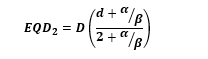
In the latter half of 2022, the patient underwent a CT/PET simulation: positioned supine in a frog-leg position, immobilized using an alpha cradle, with a full bladder, and without a Foley catheter. The patient was prescribed 45 Gy in 25 fractions to the pelvic lymph nodes (PTV45) and simultaneously boosted the anorectal region to 50 Gy (PTV50). PTV45 included the perirectal, presacral, internal iliac, external iliac, and inguinal nodes. PTV50 was generated from an expansion of the anal canal and the connecting partial segment of the rectum (see Figure 1A). EMR documentation revealed that the patient received prostate EBRT in 2012 at an outside institution, delivering a total of 81 Gy in 45 fractions via IMRT: 50.4 Gy in 28 fractions were delivered to the prostate and seminal vesicles, followed by a cone-down of 30.6 Gy in 17 fractions delivered to the prostate alone. This was the extent of prior treatment information, as planning records were unavailable.
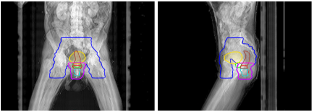
Figure 1A: Case report 1: yellow = bladder, brown = rectum, teal = anal canal, red = GTV, blue = PTV45, magenta = PTV50, and green = prostate.
The prior target was assessed to have largely overlapped with the current treatment, and for calculating cumulative dose, the prior contribution to OARs was assigned 81 Gy, i.e., to have received a Dmax of the prior prescription. Our intuition’s retreatment constraints for genitourinary (GU) are higher than that of gastrointestinal (GI), where for GU: bladder and rectum Dmax < 150 and 140 Gy3 EQD2, respectively; for GI: both bladder and rectum Dmax < 100 Gy3 EQD2. Reirradiation cases discussed in this study can be considered a blend of these two categories, and from a planning perspective, questions arise on which retreatment constraint values to use. Following peer review, delivering full prescription coverage was determined acceptable, where the bladder and rectum received a Dmax of 123.9 and 133 Gy3 EQD2, respectively. The urethra and prostate were also limited to Dmax < 110 and 120 Gy3 EQD2, respectively, in concern of urinary complications. This resulted in some compromise of target coverage: V100% of GTV and PTV50 were 98.1% and 92.6%, respectively; D95% of GTV and PTV50 were 101.7% and 97.0%, respectively.
About two-thirds into the treatment course, the patient notably experienced diarrhea (4-6 stools per day over baseline) and moist desquamation not confined to the skin folds (Grade 3 acute dermatitis), which was treated with Silvadene and resolved mostly by the time of completion of radiotherapy. After fraction 22 of 25, the patient was admitted to a hospital for severe acute respiratory syndrome, and chemoradiation was paused for one month before proceeding to deliver the remaining fractions. Four months after completing radiotherapy, the patient reported feeling significantly better without any significant rectal or urinary issues. A sigmoidoscopy and CT around this time showed partial clinical response without evidence of new disease. The patient was shortly started on capecitabine but could not tolerate it due to severe diarrhea, which required an 11-day admission at a local hospital. At the eight-month mark, the patient was diagnosed with Merkel cell carcinoma via biopsy. This presented as subcutaneous nodules across the lower abdomen to the bilateral groins and pubis, and the patient was experiencing unbearable pain. The patient was started on carboplatin etoposide in response, but treatment was stopped indefinitely within two months due to his rapidly deteriorating condition. The patient passed away shortly afterward, which was approximately 11 months after the completion of pelvic reirradiation, and has experienced Grade 3+ toxicity in acute dermatitis.
An 87-year-old man with a past medical history of prostate cancer underwent a colonoscopy in 2022. An ulcerated, non-obstructive rectal mass was discovered, and biopsy results revealed moderately differentiated adenocarcinoma of the rectum. MRI showed a tumor that was 4.1cm in craniocaudal length, 7.4cm distance between the inferior border to the anal verge, staged as cT4bN+M0. The patient was started on neoadjuvant FOLFOXx8. As a poor surgical candidate, the patient opted to pursue definitive radiation with concurrent capecitabine.
Later that year, the patient underwent a CT simulation: positioned prone on a belly board, with a full bladder and no Foley catheter. A same-day MRI was obtained, to which the T1 and T2 sequences were fused to the planning CT scan. The patient was prescribed 45 Gy in 30 fractions BID [6] to both the rectum and pelvic lymph nodes (PTV45), which included the perirectal, presacral, internal iliac, and external iliac nodes (see Figure 1B). EMR documentation revealed that the patient received prostate-only EBRT followed by brachytherapy in 2005 at an outside institution. IMRT was used to deliver 50.4 Gy in 25 fractions, followed by 100 Gy LDR brachytherapy via Pd-103 seeds.
Figure 1B: Case report 2: yellow = bladder, brown = rectum, teal = anal canal, red = GTV, blue = PTV45, magenta = PTV50, and green = 50% IDL of brachytherapy.
Unlike prior EBRT doses seen with Case Report 1, Equation 1 cannot calculate EQD2 for LDR brachytherapy as there are no fractions, lacking the variable “d” (dose per fraction). Hilal et al. circumvent this, building upon the methodology outlined in Stock et al., using Equations 2and 3 below to first calculate BED and then convert to EQD2 [1,7]:
Equation 2
Where λ is the radioactive decay constant, calculated from ln(2)/T1/2, where T1/2 is the radioisotope’s half-life, and μ is the repair rate constant, calculated from ln(2)/t1/2, where t1/2 is the tissue repair half-time. Stock et al. specify T1/2 and t1/2 for Pd-103 as 17 days and 1 hour, respectively, used for brachytherapy calculations in this paper [7]. The BED is then converted to EQD2 through the following equation (rearrangement of Equation 1):
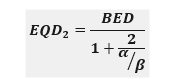
Equation 3
Utilizing the prior plan reports, Dmax to the bladder, rectum, and urethra were 148, 150, and 158 Gy3, which are 99.5, 101, and 107 Gy3 EQD2, respectively, after applying Equations 2 and 3. When summed with the prior combination EBRT doses, the bladder, rectum, and urethra total 160.2, 156.7, and 162.2 Gy3 EQD2, respectively. Therefore, the contribution of EQD2 from only the prior radiotherapy course exceeds the GU retreatment constraints used at our institution (bladder and rectum Dmax < 150 and 140 Gy3 EQD2, respectively) before even factoring in current doses. TG-137 recommends reporting brachytherapy doses that D2cc rather than Dmax act as the primary planning parameter when evaluating rectum dose [8]. Their rationale is that D2cc of irradiated volume is more clinically relevant over D0.1cc (a proxy for Dmax), which is instead listed as a secondary parameter [8]. Applying that understanding here, when D2cc is used for prior brachytherapy doses delivered (approx. 75 Gy), prior bladder, rectum, and urethra doses total 108.5, 103.4, and 103 Gy3 EQD2, respectively.
Between peer review and consensus with the brachytherapy team, the consensus was to reproduce the 50% (50 Gy) isodose line (IDL) of the brachytherapy treatment achieved in part by referencing the prior plan report’s dose distribution and considering the current anatomy and position of the prostate and seed implants. This region was limited to D1cc < 45 Gy and resulted in some compromise of target coverage: V100% of GTV and PTV45 were 60.2% and 83.2%, respectively; D95% of GTV and PTV45 were 90.9% and 90.6%, respectively; Dmin of GTV was 87.9% (39.6 Gy). Note that PTV45 comprises the rectum and pelvic lymph nodes, as the structure set did not contain a separate PTV for only the rectum. Optimizing the dose distribution in this manner also resulted in lesser doses to the urethra and prostate. As the prior 50% brachytherapy IDL was mainly limited to less than 45 Gy, the bladder, rectum, and urethra could be said to have received 40.5 Gy3 EQD2, which would total 149, 143.9, and 143.5 Gy3 EQD2, respectively.
By the end of the radiotherapy treatment course, the patient had notably experienced hemorrhoids, diarrhea (4-6 stools per day over baseline), and fatigue limiting instrumental ADL. One month after radiotherapy, the patient was admitted to a local hospital for four days, receiving treatment for colitis with IV Flagyl after initially presenting at their emergency department for fatigue, weakness, and persistent loose stools. At the six-month mark, the patient reported a significant improvement in overall well-being and denied any pain, diarrhea, or blood in the stool. Sigmoidoscopy and MRI findings at this time were consistent with a complete clinical response. The patient showed no evidence of disease or side effects during the latest follow-up visit, which was approximately 17 months after the completion of pelvic reirradiation and has not experienced radiotherapy-related Grade 3+ toxicity.
An 86-year-old male with a past medical history of prostate cancer (T1c) underwent a colonoscopy in 2017. A large 7.0cm mass was seen in the rectum extending to the dentate line, where biopsy revealed invasive moderately differentiated adenocarcinoma. MRI revealed a partially circumferential tumor 3.3cm in craniocaudal length and 3.0cm from the inferior border to the anal verge, staged as cT3N0M0. The patient was deemed to have high surgical risk due to needing to stop Plavix and opted for definitive radiation with concurrent capecitabine.
Later, in 2017, the patient underwent a CT simulation: positioned prone on a belly board without a full bladder and Foley catheter. The patient was prescribed 45 Gy in 25 fractions to both the rectum and pelvic lymph nodes (PTV45), which included the perirectal, presacral, internal iliac, external iliac, and inguinal nodes (see Figure 1C). EMR documentation revealed that the patient received prostate LDR brachytherapy in 2001 at an outside institution via Pd-103 seeds. However, no prior planning records were available, including mention of the prescription dose, which limited the calculation of prior EQD2.

Figure 1C: Case report 3: yellow = bladder, brown = rectum, teal = anal canal, red = GTV, blue = PTV45, magenta = PTV50, and
green = PRV prostate.
The radiation oncologist had contoured the prostate and applied an isotropic margin of approximately 0.6cm to create “PRV_prostate,” to which 25 Gy was minimized to this region and is similar in concept to the 50% IDL region seen with Case Report 2. PRV_prostate was kept to D1cc = 2344.8 cGy, resulting in some compromise of target coverage: V100% of GTV and PTV45 were 68.1% and 98.4%, respectively; D95% of GTV and PTV45 were 43.6% and 102.5%, respectively; Dmin of GTV was 33.3% (14.5 Gy). Note that PTV45 was cropped some margin from PRV_prostate such that some GTV extended outside PTV45. Also, note that PTV45 comprises both the rectum and pelvic lymph nodes, as the structure set did not contain a separate PTV for only the rectum. The bladder, rectum, and prostate (no urethra contoured) received a Dmax of 50.5, 50.7, and 13.3 Gy3 EQD2, respectively.
The patient tolerated treatment well overall, experiencing some fatigue and diarrhea during the last week of the radiotherapy. Around one-and-a-half months afterwards, the patient underwent sigmoidoscopy, revealing findings consistent with a complete clinical response and supported by an MRI suggesting radiological response. The patient did not have any major symptoms around this time, though the patient remained on active surveillance in consideration of his age and comorbid condition. At the three-month mark, the patient was started on four months of adjuvant capecitabine. The patient tolerated the first two cycles very well, but treatment was paused after the third cycle due to increasing toxicity impacting his quality of life, involving fatigue, diarrhea, and hand-foot syndrome. Treatment was stopped indefinitely when the patient incurred a pelvic fracture from a fall that required prolonged rehabilitation and went on active surveillance. In 2022, the patient was admitted to a local hospital and diagnosed with aspiration pneumonia and COVID-19. The patient passed away several days later, which was approximately 60 months after the completion of pelvic reirradiation. The patient showed ongoing complete clinical response during his last follow-up visit and did not experience radiotherapy-related Grade 3+ toxicity.
Minimizing OAR late toxicity is a top priority in a reirradiation setting. The process at our institution involves a PTR utilizing cumulative retreatment constraints, which aim to achieve consistent treatment planning outcomes for these complex cases. Considering that modern radiotherapy delivery techniques allow for PTV isodose “carving” such that OARs interfacing PTVs can be prioritized to receive significantly less than the prescription dose, creating a heterogeneous dose with a sharp gradient at the OAR interface. Retreatment constraints cited in the literature can better reflect the cumulative doses actually received by nearby OARs. We searched the literature for any direct mention of pelvic retreatment constraints, and many were compiled in Murray et al. and Baty et al. [4,9]. Constraints ranging from Dmax to D1cc (Dmax-D1cc) were selected, rewritten in terms of Gy3 EQD2, and added to their prior prescription dose in Gy3 EQD2 to approximate a cumulative dose constraint.
When referring to the information in Tables 4-6, we should recognize the interplay of variables that can help contextualize the results of this case report summarized in Tables 1-3. First, reirradiation prescriptions cited in the literature were predominantly hypofractionated, whereas the cases in this study were delivered in 25-30 fractions. Hyperfractionation in the context of pelvic reirradiation is seen increasingly utilized at our institution, delivering 39-45 Gy in 1.5 Gy/fx BID 6-8 hours apart, as it should offer greater interfractional normal tissue repair [6]. The interval between prior and reirradiation is another variable worth considering as it may indicate some degree of normal tissue recovery, where Abusaris et al. had allowed up to 50% of the prior EQD2 delivered to be subtracted when determining cumulative dose depending on this interval [10]. From a dosimetric perspective, the cumulative dose constraints seen in Table 4-5 were calculated assuming Dmax-D1cc as the prior prescription, whereas these were likely closer to 110% of the prescription considering that the prior radiotherapy technique was often 3D-CRT; this adds a buffer element to those cumulative values. Lastly, between prior prostate radiotherapy and anorectal reirradiation, the locations of Dmax-D1cc are more likely to overlap at distinct locations, unlike with prostate reirradiation, to which the constraints in Table 4-5 are largely tailored. When accounting for the variables described above, the approach and doses seen in our cases align more closely with that of the literature: the median Dmax-D1cc for bladder & bladder wall (n=10) < 116.3 Gy3 EQD2 (range: 80-147.4), rectum & rectal wall (n=14) < 124.4 Gy3 EQD2 (range: 88.4-160.2), and urethra (n=9) < 147.4 Gy3 EQD2 (range: 134.1-165.1), summarized in Table 4.
To improve PTR at our institution, we should expand our retreatment procedures and constraints to include this paradigm of patients, particularly for patients who have received prior combination brachytherapy and EBRT. Additionally, the limitation of unavailable prior planning records should lessen over time as patients’ first course of radiotherapy is delivered in modern times when EMR and TPS are standard for radiation oncology clinics. This will enable a more consistent and thorough PTR and improve our confidence in delivering adequate dose to the target while minimizing toxicity to nearby OARs. Newer radiotherapy techniques and technologies are also expected to increasingly aid in the reirradiation setting, such as deformable registration, adaptive radiotherapy via Ethos Therapy and MRI-guided adaptive radiotherapy via Elekta Unity, proton therapy, and rectal spacer implantation. Our sample size limits this case report, and future research should study this patient population on a larger scale to provide more precise guidelines for clinicians to navigate these types of cases.
Reirradiation for de novo anorectal cancers in patients with a history of prostate radiotherapy is feasible from a dosimetric perspective. However, approaching these cases require careful consideration of the potential for increased toxicity. Effective collaboration between radiation oncologists and the medical physics team can help mitigate these risks, making it a viable therapeutic option for this unique patient population.
The authors have no conflict of interests to declare.
Dear Editorial Team, Clinical Medical Reviews and Reports. My experience with the journal was highly positive. The peer-review process was rigorous, constructive, and completed in a timely manner. The reviewers provided valuable comments that helped improve the quality and clarity of our manuscript. The editorial office was professional, responsive, and supportive throughout all stages of the publication process. Communication was clear and efficient, and any questions were addressed promptly. Overall, I found the journal to maintain high scientific standards and an excellent publication workflow. I would be pleased to consider submitting future work to this journal. Best wishes from, Elena Popa.

It was my pleasure to submit my testimonial concerning the Reviewer Board of our Scientific Journal “Brain and Neurological Disorders”. The Reviewers focused on some modifications and their contribution was helpful. The ladies of our Editorial Office were also supported my efforts. It was my honor to have such a co-operation and I am looking forward for more collaboration.

Dear Grace Pierce, Editorial Coordinator of Journal of Clinical Research and Reports, Thank you for the speedy and efficient peer review process. I appreciate the fact that your peer reviewers do not take months to respond like with some other journals. I would also like to thank the editorial office for responding quickly to my questions. It is an excellent journal. I plan to submit more manuscripts in the future. Best wishes from, Robert W. McGee

Dear Grace Pierce, Editorial Coordinator of Journal of Clinical Research and Reports, Working with you and your team on our recent publication in JCRR has been a truly wonderful and enjoyable experience. The responses were prompt, and the reviewers were patient, constructive, and highly professional. One reviewer in particular gave me the feeling that a professor was carefully reading and commenting on my coursework, which was deeply touching. The entire process was straightforward and hassle‑free, with no tedious online forms to complete. I highly recommend this journal. Best wishes from, DR Aibing Rao, Head of R&D

I Appreciate the Opportunity to Share my Experience with the Journal of Clinical Research and Reports. The peer review process was timely and constructive, and the feedback provided helped improve the quality of our manuscript. The editorial office was professional, responsive, and supportive throughout the process, ensuring smooth communication and efficient handling of the submission. Overall, it was a positive experience collaborating with your team.

Dear Mercy Grace, Editorial Coordinator of Obstetrics Gynecology and Reproductive Sciences, We would like to express our gratitude for your help at all stages of publishing and editing the article. The editors of the magazine answer all the necessary questions and help at every stage. We will definitely continue to cooperate and publish other works in the Obstetrics Gynecology and Reproductive Sciences! Best wishes from, Alla Konstantinovna Politova,
