
Research Article | DOI: https://doi.org/10.31579/2637-8876/017
1 Dresden International University (DIU). Dresden, Germany
*Corresponding Author: Kurt E. Müller, Dresden International University (DIU). Dresden, Germany.
Citation: Kurt E. Müller. (2021) Re-Print- Artificial Respiration in Severe COVID-19 Cases: A Beneficial or Deleterious Treatment, J. Immunology and Inflammation Diseases Therapy. 4(1); Doi:10.31579/2637-8876/017
Copyright: © 2021 Kurt E. Müller. This is an open-access article distributed under the terms of The Creative Commons Attribution License, which permits unrestricted use, distribution, and reproduction in any medium, provided the original author and source are credited.
Received: 02 August 2021 | Accepted: 10 September 2021 | Published: 20 September 2021
Keywords: SARS-CoV-2; COVID-19; artificial respiration; superoxide; oxidative shielding; oxidative stress; oxidative burst; hemofiltration
The world over artificial respiration is employed as one of the intensive care treatment measures in severe cases of COVID-19 because of the significant respiratory distress patients develop. Nevertheless, the outcome is poor. Lethality varies from country to country and clinic to clinic between 50% and 90%. So the question arises as to whether the use of oxygen can be a risk factor in the treatment of acute inflammatory diseases in general and of COVID-19 in particular. Oxidative stress is the first and oldest step of cellular defense and starts before the activation of the immune system. This leads to an increase of intracellular oxygen in the mitochondria, followed by an elevated electron flow and the formation of superoxide as well as other reactive oxygen species and reactive nitrogen species. Superoxide reacts with nitric oxide, which is always present in inflammation, forming peroxynitrite, the strongest inducer of oxidative stress. This step induces the activation of nuclear factor kB, followed by the production of proinflammatory cytokines. The elevated levels of inducible nitric oxide synthase keep this cycle running. High amounts of superoxide have to be compensated and catabolized by manganese-superoxide dismutase 2 into hydrogen peroxide and in a second step by catalase into water. When using artificial respiration, these steps are accelerated considerably in the inflamed tissue of the lung, leading to a significant increase of the electron flow as well as an elevation of superoxide, oxidative stress, and water. As SARS-CoV-2 generally induces the production of proteins (and not only those necessary for viral reproduction), the water will remain in the tissue, causing edema and thus a wet lung syndrome associated with a growing oxygen diffusion distance to red blood cells. Ultimately, patients do not suffocate in spite of, but because of, the presence of high levels of oxygen. The limited number of patients who survive this deleterious treatment describe it as having had a sensation of drowning. The reasons will be discussed.
Energy metabolism and defense mechanisms developed in parallel in evolution. Their interaction is essential for each of them to function properly. In the beginning, 3.5 billion years ago, the process was anaerobic. In the oldest bacteria known, adenosine triphosphate (ATP) was produced by anoxygenic photosynthesis [1, 2] Oxygen was toxic. But substances and cellular mechanisms of the intermediary metabolism already existed in that past and are still utilized today. These are substances and mechanisms such as glycolysis, glyoxylate and the Krebs cycle, pyruvate dehydrogenase, the pentose phosphate pathway, the synthesis and oxidation of fatty acids, cobalamin, carotenoid and heme synthesis, iron-sulfur cluster synthesis, cytochromes (extremely relevant for drug metabolism), isoprenyl and ubiquinone synthesis and its interaction with the electron transport necessary for the defense mechanisms and proton coupling for ATP synthesis [3].
Almost one billion years later, oxygen became important. Cyanobacteria started producing oxygen in such quantities that the oxygen level in the atmosphere increased to 2-4% which allowed bacteria and viruses to leave the water of the oceans and populate the earth [1]. Mitochondria coordinated both energy production and defense mechanisms. They even organized the innate immune system necessary for the identification of targets for the activation of defense mechanisms [4]. The rising levels of oxygen in the environment facilitated an increased production of energy and promoted oxidative shield mechanisms. More energy production in turn means higher risk due to an increase in the number of electrons which form together with oxygen superoxide anions. At first these oxygen superoxide anions can be catabolized by manganese-superoxide dismutase 2 (Mn-SOD2) to hydrogen peroxide (H2O2), which is still an oxygen radical, and subsequently H2O2 is catabolized to water. In parallel, superoxide reacts with nitric oxide to form peroxynitrite, the most active substance in the development of oxidative stress by reactive oxygen species (ROS) and reaczive nitrogen species (RNS) [5].
2.1 Mitochondria
The proteome of the mitochondrion contains almost 1500 proteins [6]. Two thirds of these have an enzymatic function, highlighting the enormous metabolic activity of these organelles. Under physiological conditions, the concentration of thousands of nutrients and metabolic substrates is strictly controlled by collective kinetic constants. Consequently, the whole system is mismanaged if even one of these is upregulated or downregulated for a longer period of time. Viral and microbial infections, changes in the natural environment, toxins, nutritional dysbalance, and medical treatment can thus cause a mismatch between the optimum and the actually existing concentration of metabolic substrates for the tissue [7]. This point will be of particular interest regarding severe acute respiratory syndrome coronavirus-2 (SARS-CoV-2).
Most of the ATP in humans is generated by the combined action of the Krebs cycle and oxidative phosphorylation. Oxidative phosphorylation requires an electron transport chain, molecular oxygen, and ATP synthase located within the inner membrane of the mitochondria [8]. The reactions of the Krebs cycle produce reduced nicotinamide adenine dinucleotide (NADH) and reduced flavin adenine dinucleotide (FADH2). Both are electron donors for the electron transport chain that passes them sequentially to O2 to generate energy and to form water. The mitochondrion also contains the enzymes responsible for ß-oxidation of fatty acids to NADH and FADH2. Those reductants are located in the center of the mitochondrion and readily react with the electron transport chain. The exergonic reactions of the electron transport chain provide energy for the translocation of protons (H+) from the mitochondrial matrix through the inner mitochondrial membrane to the exterior. Most of the oxygen consumed by humans is metabolized within the mitochondria. The driving force of mitochondrial function is a voltage difference (ΔΕ) between O2 and NADH (Figure. 1).
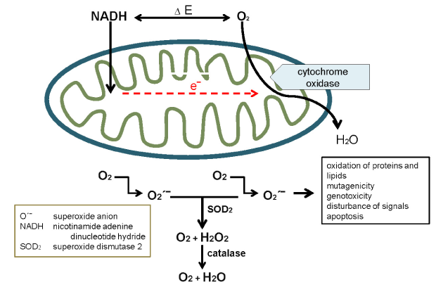
The respiratory electron transport chain consists of four independent complexes (I, II, III, and IV). Coenzyme Q (CoQ) transports the electrons from complexes I and II to complex III, cytochrome c from complex III to IV. NADH donates its electrons to complex I. From there electrons are transferred to CoQ to form QH2, which passes its electrons on to complex III. A proton translocation reaction is needed to provide the energy. Complex II (succinate dehydrogenase) transports electrons without providing energy for proton translocation or ATP formation [8,9,10]. The major sources of physiological oxygen radicals which are natural by-products of oxidative phosphorylation can be found between complex I and CoQ and between CoQ and complex III (Figure. 2).
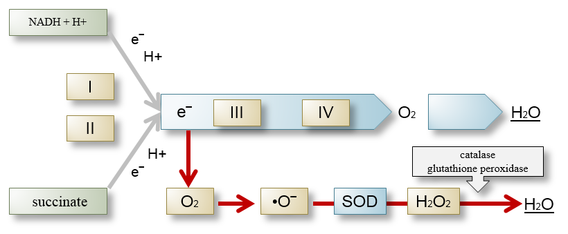
They account for about 1% of the total oxygen uptake. This situation changes completely during artificial respiration as an elevated level of electrons and O2 form high amounts and ultimately uncontrollably high amounts of superoxide anions reacting with nitric oxide to form peroxynitrate, which in turn forms free radicals and induces oxidative stress [5]. Mitochondrial DNA is 15 times more sensitive to oxidative damage than nuclear DNA [11].
The physiological process of ATP production is also the dominant source of ROS and consequently of oxidative stress. Oxygen is an element urgently needed but also an element causing risks when activated to form singlet oxygen (¹O2) by means of energy transfer or by electron transfer forming superoxide anion radicals (•O2‾) [10]. As long as ROS remain compensated in a homeostatic and physiological reaction or are used for controlled defense for a limited period of time, deleterious effects can be avoided. The most important enzymes to act on •O2‾ are superoxide dismutases (SODs) which exist in three forms: Mn-SOD2) in mitochondria as well as copper-zinc superoxide dismutase (Cu/Zn-SOD2) in cytosol membranes.
2.2 Superoxide
There are several metabolic steps leading to the production of superoxide. The most important in this context is the electron transport chain in the mitochondrion during ATP generation. Superoxide exists in two interconvertible forms. The more important one has an unpaired electron and a negative charge (٠O2‾ ), the other one is an acid (٠OH). The negative charge keeps superoxide in the cells where it serves as an intracellular signaling molecule. Higher levels are damaging to cells. Therefore enzymes, (SODs) are needed to degrade superoxide. They can be found in all organisms dependent on the presence of oxygen in the air [10]. There are three forms: the first form is involved in the energy metabolism of the mitochondria, the second one can be found in the cytoplasm of cells, and the third one outside of cells. Each SOD is encoded by its own gene and has a distinct structure. Superoxide is metabolized by Mn-SOD2 to H2O2. Both ٠O2‾ and H2O2 have low reactivity. In the presence of transition metals, however they develop high amounts of ROS.
2.3 Reactive Nitrogen Species
RNS include nitric oxide (NO٠) and nitrogen dioxide (٠NO‾2), both of which are free radicals, and peroxynitrite (ONOO‾), a nonradical. They all have high oxidizing potential. The toxic effect of these molecules is referred to as “nitrosative stress” [12, 13]. They are also classified as ROS and summarized as reactive oxygen and nitrogen species (RONS). ٠NO is a relatively stable free radical with a half-life of 1 s. It is produced by oxidation of L-arginine to L-citrulline by the enzyme nitric oxide synthase (NOS) which takes three forms: inducible NOS (iNOS), endothelial NOS (eNOS), and neuronal NOS (nNOS) [14, 15]. NO٠ is a soluble lipid that can diffuse through membranes and is thus critically important in neurogenic membranes containing a different structural correlation of lipids and proteins [16].
NO٠ has both physiological and pathophysiological properties [17]. Because of its regulatory function it is considered a cytokine, in spite of the fact that it is structurally not identical to cytokines [18]. It causes vasodilation to regulate blood flow and consequently directs immune cells towards the site of inflammation. It also activates natural killer cells (NK cells). This positive effect is blocked when high levels of superoxide activate NO٠ and ٠O‾2 to form ONOO‾, a highly potent inducer of oxidative stress. This is the strongest inducer of oxidative stress and switch for the activation of nuclear factor-kB (NF-kB), which in turn triggers the production of proinflammatory cytokines (Figure. 3).
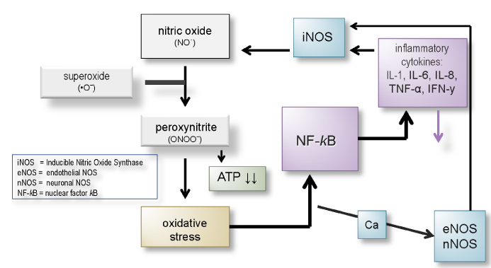
The blocking effect of OONO‾ acts more strongly on NK cells than the promoting effect of NO٠ [20, 21]. ONOO‾ can cause DNA fragmentation, lipid and protein oxidation, as well as nitration [22, 23].
All coronaviruses have the highest affinity for the respiratory tract among all organs and functional systems. Generally the inflammation involves the pharynx, trachea, bronchi, and bronchioli. However, in coronavirus infectious disease-19 (COVID-19) the inflammation often extends to the entire pulmonary tissue corresponding to an enormous expansion of the inflamed area. This provokes a strong antioxidative defense reaction by the activation of the NO/٠O2‾ /ONOO‾ cascade followed by the upregulation of proinflammatory cytokines such as IFN y, IL 1, IL 6, IL 8, TNF α and others by activating NF-kB and the sympathetic nervous system, which also interacts with the immune cells. This cascade is controlled by SODs, glutathione peroxidase (GPx), catalase, Treg cells, IL 10, cortisol, and the parasympathetic nervous system [24] The mitochondria are the regulators of those mechanisms. As shown before, the voltage difference between O2 and NADH (ΔΕ) is the driving force. The uptake of even physiological amounts of oxygen by artificial respiration with compressed air in particular will deregulate and escalate the whole process of inflammation. The more oxygen is used, the higher the production of electrons and consequently of superoxide will be. It is less reactive in physiological concentrations. It becomes very reactive however, if a high quantity is formed, producing ٠ONOO‾ by reacting with ٠NO. The oxidative and nitrosative stress may become so acute that it results in an oxidative burst which in turn leads to damage of granulocytes, especially neutrophils, and macrophages breaking down cellular defense mechanisms () [10].
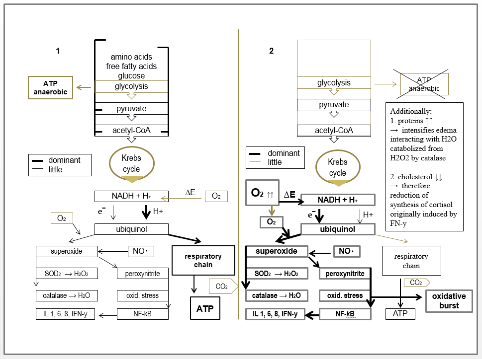
This may damage the sensitive mitochondrial DNA and immediately influence mitochondrial function. It has been demonstrated that the therapeutic treatment of various diseases with oxygen increased mortality without improving other patient-relevant outcomes [25]. The authors published a meta-analysis of 25 randomized studies with 16,037 patients treated with low and high-rate artificial respiration for various indications. Mortality was higher in all cases treated with high O2 flow than in the groups suffering from the same disease but treated with low levels of oxygen. In artificial treatment of COVID-19 patients, the oxygen reaches the inflamed tissue directly and is not distributed or attenuated through the blood stream. Therefore, its effect in COVID-19 is far more pronounced than in the cases referred to in the studies. A leakage of superoxide develops between complex I and CoQ and complex III, depending on the quantity and properties of CoQ. The overload of superoxide leads to an even stronger activation of inflammation by the vicious NO/ONOO‾ cascade and at the same increases the production of H2O2 and ultimately H2O by SOD and catalase. Latest findings demonstrate that SARS-CoV-2 in contrast to other coronaviruses does not only lead to elevated protein synthesis for viral reduplication but higher levels of protein synthesis in general [26]. This increased production of proteins is an important factor for H2O accumulation in the tissue causing a wet lung syndrome associated with the sensation of drowning. The same group showed that cholesterol levels are lowered, which inevitably leads to a reduced synthesis of sex hormones and steroid hormones. As a result, the anti-inflammatory effect of cortisol is blocked. The effect can be even more pronounced in patients who had been treated with statins. Additionally, statins can also lead to an increased NO٠ level. Consequently, insulin resistance can be expected. It always develops in acute inflammation due to an increase in circulating glucose and free fatty acids which are no longer absorbed by the adipose tissue, liver, or muscles in order to provide energy for the central nervous system and the immune system [24, 27].
Environmental factors influence the extent of oxidative stress. Transition metals with atomic numbers 21-30, 39-48, 57-80, and 89-112 are electron donors and catalyze the formation of superoxide and free radicals. It is also one of the reasons why COVID-19 takes a more severe course in pe, itople in polluted regions like China, Lombardy, and large cities all over the world. Furthermore also is one the contributing factors for the severe clinical course in elderly patients as their total body burden is generally higher. Metals are also inducers of cell adhesion molecules on the surface of the endothelium [28]. They are catalyzers of superoxide but may as well increase the risk of thromboembolic processes often seen in COVID-19 patients. It has been shown just recently that nitrogen dioxid (NO2) levels are a contributing factor to COVID-19 fatality [29]. The antioxidative capacity depends on the quality and quantity of micronutrients. The presence of CoQ might be particularly important. Although there is no current data on this aspect for COVID-19 as yet, it is a well-known fact that a mismatch of micronutrients causes mitochondrial dysfunction [7].
Oxygen is a promoter of oxidative stress as a defense reaction and as an activator of the innate immune system. As long as these reactions stay under control, the required shielding effect is guaranteed, but high levels of oxygen render them uncontrollable. Artificial respiration increases this risk considerably within a short period of time. So what could be the solution? In patients showing signs of oxidative and nitrosative stress spiraling out of control, high dosages of substances of the cellular antioxidant defense system should be administered, such as vitamin C and E, reduced glutathione, taurine, cysteine, methionine, s-adenosyl-L-methionine, melatonin, selenium, and polyamines [30]. They should be combined with anti-inflammatory substances and drugs. Ivermectin has antioxidative and anti-inflammatory properties and also blocks mRNA of COVID-19 [31]. In severe cases, corticosteroids should be taken into consideration in spite of the viremia. In case of symptoms of wet lung disease, hemofiltration should be started immediately to extract the fluid from the blood and consequently from the lung via osmotic pressure. This should alleviate the sensation of drowning within 2 to 3 hours. Additionally, a therapeutic concept to avoid pulmonary fibrosis is required until the radiograph has normalized.
Dresden International University (DIU). Dresden, Germany. Feilbergstrasse 32.
The author declares that he has no competing interest.
Dear Editorial Team, Clinical Medical Reviews and Reports. My experience with the journal was highly positive. The peer-review process was rigorous, constructive, and completed in a timely manner. The reviewers provided valuable comments that helped improve the quality and clarity of our manuscript. The editorial office was professional, responsive, and supportive throughout all stages of the publication process. Communication was clear and efficient, and any questions were addressed promptly. Overall, I found the journal to maintain high scientific standards and an excellent publication workflow. I would be pleased to consider submitting future work to this journal. Best wishes from, Elena Popa.

It was my pleasure to submit my testimonial concerning the Reviewer Board of our Scientific Journal “Brain and Neurological Disorders”. The Reviewers focused on some modifications and their contribution was helpful. The ladies of our Editorial Office were also supported my efforts. It was my honor to have such a co-operation and I am looking forward for more collaboration.

Dear Grace Pierce, Editorial Coordinator of Journal of Clinical Research and Reports, Thank you for the speedy and efficient peer review process. I appreciate the fact that your peer reviewers do not take months to respond like with some other journals. I would also like to thank the editorial office for responding quickly to my questions. It is an excellent journal. I plan to submit more manuscripts in the future. Best wishes from, Robert W. McGee

Dear Grace Pierce, Editorial Coordinator of Journal of Clinical Research and Reports, Working with you and your team on our recent publication in JCRR has been a truly wonderful and enjoyable experience. The responses were prompt, and the reviewers were patient, constructive, and highly professional. One reviewer in particular gave me the feeling that a professor was carefully reading and commenting on my coursework, which was deeply touching. The entire process was straightforward and hassle‑free, with no tedious online forms to complete. I highly recommend this journal. Best wishes from, DR Aibing Rao, Head of R&D

I Appreciate the Opportunity to Share my Experience with the Journal of Clinical Research and Reports. The peer review process was timely and constructive, and the feedback provided helped improve the quality of our manuscript. The editorial office was professional, responsive, and supportive throughout the process, ensuring smooth communication and efficient handling of the submission. Overall, it was a positive experience collaborating with your team.

Dear Mercy Grace, Editorial Coordinator of Obstetrics Gynecology and Reproductive Sciences, We would like to express our gratitude for your help at all stages of publishing and editing the article. The editors of the magazine answer all the necessary questions and help at every stage. We will definitely continue to cooperate and publish other works in the Obstetrics Gynecology and Reproductive Sciences! Best wishes from, Alla Konstantinovna Politova,
