
Case Report | DOI: https://doi.org/10.31579/2690-4861/666
1SPACIALIST ENT department of Guelmim’s REGIONAL HOSPITAL. Morocco.
2Maxillofacial Professor of department CHU hassane, Professor of Higher Education at the University Hospital IBN sina; hopital des spécialités de Rabat Morocco.
*Corresponding Author: N. Belhaj, Spacialist Ent department of Guelmim’s Regional Hospital. Morocco.
Citation: N. Belhaj, Z. Sayad, R. Bencheikh, M. A. Benbouzid, L. Houssini. Essakalli, (2025), Rare Location of a Large Intraoral Lipoma: About a Clinical Observation and a Literature Review, International Journal of Clinical Case Reports and Reviews, 23(5); DOI:10.31579/2690-4861/666
Copyright: © 2025, N. Belhaj. This is an open-access article distributed under the terms of the Creative Commons Attribution License, which permits unrestricted use, distribution, and reproduction in any medium, provided the original author and source are credited.
Received: 23 December 2024 | Accepted: 21 January 2025 | Published: 27 February 2025
Keywords: rectus sheath hematoma; trauma; bleeding
Rectus sheath hematoma (RSH) is a rare but notable cause of abdominal pain, commonly associated with trauma or anticoagulation therapy. It occurs due to the rupture of a branch of the inferior epigastric artery at its attachment to the rectus abdominis muscle, often without effective hemostasis. Diagnosing RSH can be challenging, as it frequently mimics other intra-abdominal conditions. Preoperative identification is uncommon, with most cases diagnosed intraoperatively, and only a limited number of cases reported in the literature. Here, we present the case of a 57-year-old male who developed RSH following blunt trauma to the left lower abdomen caused by a motorbike rim during repair work. The patient reported left lower paraumbilical pain, and intraperitoneal bleeding was confirmed via clinical evaluation and CT imaging. This case highlights the diagnostic complexities of RSH in acute abdominal presentations and emphasizes the importance of thorough examination and imaging for timely diagnosis and management.
The lipoma corresponds to a benign mesenchymal tumor made up of lipid tissue, which is slowly evolving and often discovered by chance (Kumaraswamy, 2009). This pathology represents 2.4% of all tumours of the oral cavity with an identical distribution between the two sexes. Its pathogenesis is poorly understood and its development is independent of metabolism and diet. Although this benign soft tissue tumor is the most common, intraoral locations are rare.
In this work, we report the observation of a 42-year-old patient who was admitted by her dentist for an intraoral jugal swelling interfering with dental care.
Patient was 42 YEARS without any notable pathological anecedents; who has been presenting for years an intraoral discomfort but given the age of this mass the patient neglects it. During dental care, the patient was admitted by her dentist because of the swelling that bothered the doctor during the treatment.
Clinical examination found a mass of four centimeters of major axis, mobile and soft on palpation, painless, homogeneous, circumscribed, oral mucosa in healthy view which occupied almost the entire right retromolar space. The patient also reported that the mass became annoying aesthetically and chewing. The orthopantomogram, except for carious lesions, is unremarkable.
Faced with this atypical location and its size, magnetic resonance imaging is recommended. The latter found clearly identifiable septa, hypo-intense in T1 and T2 weighting, rising very shortly after injection of Gadolinium.
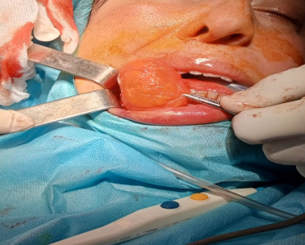
A complete excision is performed under general anesthesia. (Picture 1.2.3) .

Histological analysis of the operative specimen concludes that the lipoma is without cellular atypia or signs of malignancy, with limits of healthy excision. The search for the MDM2 and CDK4 cell markers did not was carried out immediately in front of the typical appearance of the lesion. The post-operative check-up at fifteen days is back good healing.
Lipoma is considered a benign mesenchymal neoplasm formed by mature fat cells with varying amounts of collagen bundles and blood vessels, most frequently affects the thorax, back and shoulders, while only 5% of tumors in the oral cavity [2]. In the intraoral region, it mainly occupies the jugal mucosa, lips, tongue and floor of the mouth [3].
According to the latest WHO classification, lipomas most commonly affect patients between the ages of 40 and 60 years. According to the same classification, localization in the intraoral region is found in a small number of cases in the literature [4]. The studies show a slight discrepancy with men, who are more affected [5]. Its pathogenesis remains relatively unknown, and its development is independent of lipid metabolism and diet [6]. For our patient no etiology was suspected (no history of trauma, infection, heredity...) Clinically it takes the form of a yellowish colored tumor of soft consistency, which can have different dimensions. When it is intraoral, it causes a lot of discomfort to the patient, in addition to aesthetic and phonetic problems, depending on the affected area. In our patient, only the aesthetic problem was present since the lipoma was developed mainly towards the outside and did not interfere with the oral cavity. The diagnosis is not always easy. In cases where the overlying mucosa is thin and the yellow color of the tumor can show through it, the diagnosis of lipoma is easily made. On the contrary, in cases of deep lipomas, the diagnosis is not easy. In this case, the tumor may be diagnosed as a cyst, an encapsulated abscess or another type of tumor. In this patient, the diagnosis of lipoma was not made in the first place, but the ultrasound guided the diagnosis.
Lipomas are benign tumors, rare in the mouth. As there are other lesions that can be confused with lipoma, it is important to make a correct diagnosis that allows for proper treatment. The treatment, in most cases, is simple and consists of surgical removal of the lesion, the prognosis is good
Dear Editorial Team, Clinical Medical Reviews and Reports. My experience with the journal was highly positive. The peer-review process was rigorous, constructive, and completed in a timely manner. The reviewers provided valuable comments that helped improve the quality and clarity of our manuscript. The editorial office was professional, responsive, and supportive throughout all stages of the publication process. Communication was clear and efficient, and any questions were addressed promptly. Overall, I found the journal to maintain high scientific standards and an excellent publication workflow. I would be pleased to consider submitting future work to this journal. Best wishes from, Elena Popa.

It was my pleasure to submit my testimonial concerning the Reviewer Board of our Scientific Journal “Brain and Neurological Disorders”. The Reviewers focused on some modifications and their contribution was helpful. The ladies of our Editorial Office were also supported my efforts. It was my honor to have such a co-operation and I am looking forward for more collaboration.

Dear Grace Pierce, Editorial Coordinator of Journal of Clinical Research and Reports, Thank you for the speedy and efficient peer review process. I appreciate the fact that your peer reviewers do not take months to respond like with some other journals. I would also like to thank the editorial office for responding quickly to my questions. It is an excellent journal. I plan to submit more manuscripts in the future. Best wishes from, Robert W. McGee

Dear Grace Pierce, Editorial Coordinator of Journal of Clinical Research and Reports, Working with you and your team on our recent publication in JCRR has been a truly wonderful and enjoyable experience. The responses were prompt, and the reviewers were patient, constructive, and highly professional. One reviewer in particular gave me the feeling that a professor was carefully reading and commenting on my coursework, which was deeply touching. The entire process was straightforward and hassle‑free, with no tedious online forms to complete. I highly recommend this journal. Best wishes from, DR Aibing Rao, Head of R&D

I Appreciate the Opportunity to Share my Experience with the Journal of Clinical Research and Reports. The peer review process was timely and constructive, and the feedback provided helped improve the quality of our manuscript. The editorial office was professional, responsive, and supportive throughout the process, ensuring smooth communication and efficient handling of the submission. Overall, it was a positive experience collaborating with your team.

Dear Mercy Grace, Editorial Coordinator of Obstetrics Gynecology and Reproductive Sciences, We would like to express our gratitude for your help at all stages of publishing and editing the article. The editors of the magazine answer all the necessary questions and help at every stage. We will definitely continue to cooperate and publish other works in the Obstetrics Gynecology and Reproductive Sciences! Best wishes from, Alla Konstantinovna Politova,
