
Research Article | DOI: https://doi.org/10.31579/2641-0419/046
1The Heart and Vascular Institute, Germantown, TN – USA
2Department of Cardiology, Drexel University College of Medicine, and ANSAR Medical Technologies, Inc., Philadelphia, PA - USA
*Corresponding Author: Gary L. Murray, The Heart and Vascular Institute, Germantown TN- USA
Citation: Gary L. Murray, Colombo J (2020) Ranolazine Preserves and Improves Left Ventricular Ejection Fraction and Autonomic Measures when wdded to Guideline-Driven Therapy in Chronic Heart Failure. J. Clinical Cardiology and Cardiovascular Interventions, 3(3); Doi:10.31579/2641-0419/046
Copyright: © 2020 Gary L. Murray. This is an open-access article distributed under the terms of the Creative Commons Attribution License, which permits unrestricted use, distribution, and reproduction in any medium, provided the original author and source are credited.
Received: 22 January 2020 | Accepted: 30 January 2020 | Published: 07 February 2020
Keywords: congestive heart failure; left ventricular ejection fraction; parasympathetic function; Patient outcomes; ranolazine; sympathetic function
Background: Ranolazine (RAN) reduces cardiac sodium channel 1.5’s late sodium current in congestive heart failure (CHF), reducing myocardial calcium overload, potentially improving left ventricular (LV) function. RAN blocks neuro- nal sodium channel 1.7, potentially altering parasympathetic and sympathetic (P&S) activity. The effects of RAN on LV ejection fraction (LVEF) and P&S function in CHF were studied.
Methods: Matched CHF patients were given open-label RAN (1000 mg po-bid) added to guideline-driven therapy (RANCHF, 41 systolic, 13 diastolic) or no adjuvant therapy (control, NORANCHF, 43 systolic, 12 diastolic). Echocar- diographic LVEF and P&S measures were obtained at baseline and follow-up (mean 23.7 months).
Results: LVEF increased in 70% of RANCHF patients, an average of 11.3 units. Mean LVEF remained unchanged in NORANCHF patients. P&S measures indicated cardiovascular autonomic neuropathy (P≤0.1 bpm2) in 20% of NORANCHF patients at baseline and in 29% at follow-up (increasing in both groups). At baseline, 28% of patients had high sympathovagal balance (SB), RAN normalized SB over 50% of these; in contrast, the NORANCHF group had a 20% increase in patients with high SB.
Conclusions: RAN preserves or improves LVEF and decreases high SB in CHF.
Despite advances in pharmacologic management [1-5] and device therapy [6], improvement in left ventricular (LV) function in congestive heart failure (CHF) patients, while statistically significant, remains relatively mild in many sub- jects. The late sodium current (INa) present in CHF cause an intramyocardial calcium (Ca++) overload that results in diastolic dysfunction and microvascular compression that can worsen LV function [7]. RAN binds to amino acid F1760 of the cardiac sodium channel 1.5 (Nav1.5), thereby reducing the late INa. In a therapeutic concentration (6 μmol), intra- myocardial Ca++ overload is reduced 50%. Additionally, RAN blocks neuronal sodium channel 1.7 (Nav1.7) in a strongly use-dependent manner via the local anesthetic receptor [8, 9]. Therefore, RAN may directly alter function of the parasympathetic and sympathetic (P&S) branches of the autonomic nervous system (ANS). We postulated these actions of RAN should result in favorable changes in LV func- tion and P&S measures in CHF.
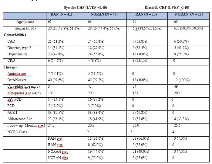
One hundred and nine systolic or diastolic, New York Heart Association (NYHA) class 2-4 CHF patients were included in this study. They were treated according to standard heart failure guidelines [10]. In an open-label fashion, patients were prescribed Ranolazine (RAN, 1000 mg po-bid) in addi- tion to standard heart failure therapy (RANCHF, 41 systolic, 13 diastolic) or no adjuvant therapy (control, NORANCHF, 43 systolic, 12 diastolic), in an unblinded fashion. Patients were matched for age, gender and history. Patient demographics are presented in Table I. Since patients were on maximally tolerated doses of beta-blocker and angiotensin-converting enzyme inhibitor (ACE-I) or angiotensin receptor blockers (ARBs), only the diuretic dose was adjusted as needed.
Diastolic CHF is defined as CHF with LV ejection fraction (LVEF) ≥0.40. Baseline 2D-echocardiograms were obtained and the LVEF calculated as the average of the apical 2 and 4 chamber Simpson’s method [11], and studies were repeated within 36 months (mean follow-up for RANCHF patients is 24.5 months and for NORANCHF 22.8 months, (Table. II). The accuracy of the initial echocardiographic LVEF was confirmed by being within 5 ejection fraction units (EFUs) of the LVEF as measured by nuclear multigated acquisition.
Serial changes in any patient of ≥±7 EFUs are consid- ered clinically significant [12]. Other measurements are per American Society of Echocardiography guidelines [13]. CHF is classified as systolic or diastolic, rather than CHF with pre- served (normal) LVEF or reduced LVEF, because the RANCHF group only had one subject with a normal LVEF.
P&S function in response to Ewing challenges [14] was as- sessed noninvasively using the ANSAR Medical Technologies, Inc., Philadelphia, PA, and ANX 3.0 Autonomic Function Monitor. P&S activity was computed simultaneously and independent- ly based on concurrent, continuous time-frequency analyses of respiratory activity (RA) and heart rate variability (HRV) [15-19]. Parasympathetic activity (measured as the respira- tory frequency area, RFa) is defined as the spectral power within a 0.12 Hz-wide window centered on the fundamen- tal respiratory frequency (FRF) in the HRV spectrum. FRF is identified as the peak spectral mode from time-frequency analysis of RA. Effectively, FRF is a measure of vagal outflow as it effects the heart (a measure of cardiovagal activity). Sym- pathetic activity (low-frequency area, LFa) is defined as the remaining spectral power, after computation of RFa, in the low-frequency window (0.04-0.15 Hz) of the HRV spectrum. High sympathovagal balance (SB = LFa/RFa) is defined as a resting LFa/RFa ratio >3.0 (established in our laboratory by evaluating 260 healthy volunteers) [11]. P&S activity was re- corded from a standard autonomic test, including 5 minutes rest, 1 minute paced breathing (6 breaths/min), a Valsalva challenge (including a 15-sec Valsalva maneuver) and a quick stand followed by 5 minutes of quiet stand. The average SB reported is the average of the ratios recorded during the sam- pling period, not a ratio of averages [11].
Cardiovascular autonomic neuropathy (CAN) was defined in standard fashion [20, 21], reflecting very low, resting RFa (<0.1 bpm2) (22). The P&S method is valid regardless of chal- lenge or patient state or history. Normal SB is 0.43.0) and CAN define a high mortality risk, including silent MI, sudden cardiac death and acute coronary syndrome (ACS) [15, 16, 23-25]. Records including high-quality arrhyth- mia are omitted. P&S and HRV measures are correlated with outcomes. While the patient population is underpowered to make final health outcome assessments, we determined the occurrence of major adverse cardiac events (MACE), defined as cardiac death (determined from hospital records or death certificates), heart failure hospitalization and ven- tricular tachycardia or fibrillation (as determined by defibrilla- tor therapy, or administration of intravenous amiodarone for arrhythmia termination) alone or as a composite endpoint. All subjects signed appropriate informed consent forms for the studies and treatments rendered.
Continuous data were assessed for normality with normally distributed data analyzed using Student t-tests and non-nor- mally distributed data assessed using a Mann-Whitney test. Dichotomous data were analyzed using the Chi-square test or Fischer’s Exact Test. A p-value of ≤0.05 was considered signifi- cant. We determined that we needed 50 patients per group to have a sufficient sample size using an alpha of 0.05, differ- ence of means of 6 units and expected standard deviation of 15 units with a power of 80%. All statistics are performed under SPSS v 1.4. Student t-tests are performed as two-tailed with equal variance. Significance values are determined on the null hypothesis that pre- and posttreatment values are equal.
Overall, 109 age-, gender- and history-matched CHF pa- tients already treated according to standard heart failure guidelines [10] were included in the study, with 54 patients re- ceiving RAN and 55 patients in the control group. Demographic comparisons are provided in Table I and are similar between groups: 93% of the patients are evenly divided between NYHA class 2 and 3; 98% are on a beta-blocker (NORANCHF sub- jects at a slightly higher dose). Slightly more diastolic RANCHF patients have hypertension and chronic renal insufficiency.
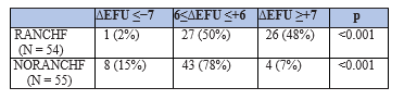
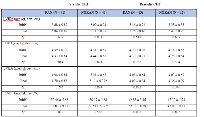
On follow-up, RANCHF patients had significantly higher LVEF (Table. II) systolic CHF: p<0.001, diastolic CHF: p = 0.003). Controls had no significant change in the mean LVEF. When viewed dichotomously (Tab. III), 26/54 (48%) RANCHF patients experienced a clinically significant increase in LVEF (≥+7 EFU) as compared to 4/55 controls (7%, p<0.001, Table. III). From the systolic RANCHF subgroup, 17/41 (41%) subjects experienced a clinically significant increase (>7 EFUs) in LVEF as compared to 9/13 (69%) diastolic RANCHF patients (p<0.001). Final LVEF in cohort patients experiencing MACE was significantly lower than in those who were MACE- free (Table. IV and V, p = 0.005). In the RANCHF group MACE subpopulation, the initial to final LVEF increase was less than in patients without MACE, 6 EFUs vs. 9 EFUs (Tab. IV, p<0.020). In control patients, insignificant changes in LVEF occurred regardless of MACE or not (p>0.050).
Systolic RANCHF patients demonstrated a decrease in left ventricular internal dimension in systole (LVIDs). Diastolic RANCHF patients demonstrated a slight increase in LVID- diastole (LVIDd) coupled with a slight decrease in LVIDs. Base- line LVID (Table. II) trended similar between groups (p>0.050). LVIDd averaged 5.88 and 6.09 cm for systolic RANCHF and NORANCHF patients, and 5.16 and 5.28 cm for diastolic RANCHF and NORANCHF patients, respectively. LVIDs aver- aged 4.94 and 5.21 cm for systolic RANCHF and NORANCHF patients, and 4.08 and 4.03 cm for diastolic RANCHF and NORANCHF patients, respectively. RANCHF vs. NORANCHF patients had significantly lower LVIDs at follow-up (>0.36 cm, p<0.001, Tab. II). No significant differences (p>0.050) in base- line or follow-up LVIDd or LAD occurred between experimen- tal groups, although LAD tended to decrease in the systolic RANCHF cohort (4.6 to 4.3 cm, Table. II, p = 0.084).
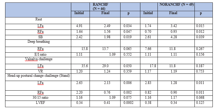
Arrhythmia-free, P&S studies were accomplished every 6 months for 95/109 (87%) patients; 13% of the patients (8 RANCHF and 6 NORANCHF) had arrhythmias precluding a complete assessment. While P&S measures are readable [26], HRV analyses are contraindicated for arrhythmia [27]. Autonomic measures of the RANCHF and control groups are presented in Table VI. The average RANCHF patient demon- strated significant P&S responses to RAN (p≤0.050), except for paced breathing RFa (a parasympathetic stimulus; p = 0.065). This included significant reductions in absolute and relative measures of sympathetic activity. None of the Time Domain Ratio responses to RAN were significant (p≥0.050). The abso- lute and relative resting sympathetic changes from baseline to follow-up in the control patients were also significant.
Sympathetic activity remained high for cohort patients with events (Table. IV and V), even though SB demonstrated a relative decrease from 6.25 to 4.86 (unitless). The high pre- RAN SB (higher than the ratio of the averages might suggest, (Table. IV) is due to two patients with severe CAN. Post-RAN, these patients were found to no longer be in CAN and dem- onstrated an increase of ≥7 EFUs, on average (p = 0.0002). The parasympathetic response to deep breathing is slight. The change in RFa is well correlated with the changes in LVEF (p<0.001). The exhalation to inhalation (E/I) ratio de- creases (not significant). The sympathetics (LFa) decrease with Valsalva challenge. The VR decreases (not significant). The Valsalva challenge responses are well correlated with the changes in LVEF (p<0.001). Sympathetic withdrawal (SW) was demonstrated by 9/15 RANCHF patients. These patients all demonstrated an abnormal BP response to standing. Upon follow-up, these patients demonstrated an average increase in sympathetic activity (a normalized response) as compared with rest, with improved standing BP. Only four RANCHF pa- tients continued to demonstrate SW after history of RAN. The stand responses are well correlated with changes in LVEF (p<0.001).
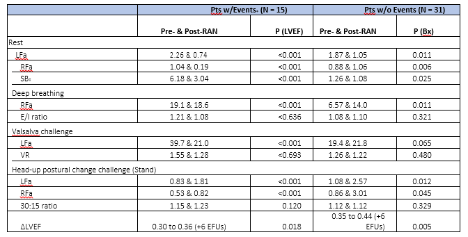
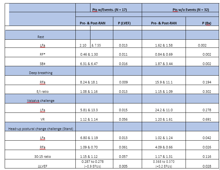
For NORANCHF cohort patients (Table. V), the relative sym- pathetic measure (SB) increased (p<0.05). In the RANCHF group without events (Table. IV), the relative measure (SB) de- creased. These SB changes are significantly associated with changes in LVEF (p<0.001). The associated average increase in LVEF is more than +9 EFUs. The patients without events started in balance (normal SB) and remained in balance. The resting changes are well correlated with the changes in LVEF (p<0.001). The pre- and post-RAN resting P&S responses in both the subpopulations with and without events are sig- nificant (p≤0.025). The pre- and post-RAN deep breathing parasympathetic measures (RFa) in both the subpopulations with and without events are significant (p≤0.011), but not the increases in E/I ratio (p>0.321). Nearly half (14/27) of the pre- RAN event patients demonstrated SW in response to stand, indicating orthostatic dysfunction. These findings are associ- ated with abnormal blood pressure responses to stand. Post- RAN, the average patient without events reversed their SW. This is a normalized response. Only six patients continued to demonstrate SW after history of RAN. The pre- and post-RAN autonomic responses to stand in both subpopulations are sig- nificant (p≤0.045).
Table V presents baseline and follow-up P&S measures and LVEF in the NORANCHF patients with and without events. P&S changes were significant (p≤0.050) for patients with events. Their SB started high and increased upon follow-up. The patients without events demonstrated opposite absolute changes upon follow-up. However, the net result was an in- crease in SB to above normal. Only the E/I ratio change for the patients with events was significant (p = 0.013).
The composite MACE endpoint occurred in 17/54 (31.5%) RANCHF patients and 21/55 (38.2%) control patients. When evaluated separately, each MACE endpoint was lower in the RANCHF patients.
In the past 30 years, improvements in LV function and out- comes in systolic CHF have been attributed to pharmacologic therapy addressing the neurohumoral paradigm, together with the advent of device therapy [1-6]. However, even more improvement is needed. This has triggered stem cell trials [28] and a search for new pharmacologic agents. To date, no therapy in diastolic CHF has shown improved survival. RAN is a first in class drug. It reduces the late sodium current (INa) resulting in a 50% reduction of the intramyocellular Ca++ overload caused by the late INa via the Na+/Ca++ exchanger [7]. This improves diastolic and microvascular dysfunction, and should result in improved LV systolic function [29]. Since LVEF is widely accepted as one of the most important prognostic indicators in CHF [30], we focused on its changes after RAN was added to guideline-driven therapy. In therapeutic concentrations (2-6 μmol), RAN also inhibits neuronal Nav1.7 via the local anesthetic receptor in a use-dependent fashion [8, 9]. Consequently, RAN potentially can alter ANS function directly, improving P&S measures. High sympathetic tone (high SB) with critically low parasympathetic activity (CAN) indicates high mortality risk, and has been associated with sudden cardiac death, CHF and ACS [15-19, 31]. This study is the first to correlate CHF outcomes with changes in both LVEF and P&S measures.
We found RAN significantly increased LVEF by 6.4 EFUs in systolic CHF patients and 9.5 EFUs in diastolic CHF (Table. II). In the NORANCHF group, final LVEF fell 1 EFU in the systolic CHF patients and 0.5 EFU in the diastolic CHF patients (Tab. II). These LVEF changes represent mean values of the cohort groups. In the systolic RANCHF patients, the increase in LVEF was solely due to a decrease in LVIDs (Table. II). In diastolic RANCHF pa- tients, the increase in LVEF was due to a slight increase in LVIDd (suggesting increased diastolic filling) coupled with a slight de- crease in LVIDs (suggesting improved systolic emptying; Table. II). Individually, only 1/54 (2%) RANCHF patients decreased LVEF by ≤−7 EFUs, and 26/54 (48%) RANCHF patients increased LVEF by ≥+7 EFUs, with the remaining 50% of patients showing little LVEF change (p<0.001, Table. III). Increases in the RANCHF pa- tients’ LVEF were sufficient to avoid defibrillator implantation in 10 subjects, resulting in substantial cost savings. In the control group, 8/55 (15%) decreased LVEF by ≤−7EFUs, and only 4/55 (7%) patients increased LVEF by ≥+7EFUs, with the remaining 43/55 (78%) demonstrating little change (Table. III). Therefore, LVEF is more than 6 times as likely to increase and 1/8th as likely to decrease following RAN therapy in CHF patients. LVEF can increase regardless of the initial LVEF. RAN increased LVEF by ≥+7 EFUs in 17/41 (41.5%) systolic CHF patients vs. 9/13 (69%) diastolic CHF patients (p<0.001). Furthermore, when RAN in- creased LVEF by ≥+7 EFUs, 9/26 (35%) patients had a history of CAD, whereas 17/26 (65%) did not (p<0.001). Since almost 80% of the CAD patients were revascularized, and only 14% had a positive stress test, we feel the smaller increases in LVEF in CAD patients were due to LV scarring secondary to remote myocardial infarctions. Finally, whether or not LVEF increased by ≥+7 EFUs did not depend upon the maximum tolerated dose of beta-blocker (94% took carvedilol), as the mean daily dose differed by only 0.5 mg.
Autonomic (P&S and HRV) measures have been docu- mented to be associated with LVEF and cardiovascular risk (32). Table VI presents the P&S and LVEF data without regard to clini- cal outcomes. RANCHF patients demonstrated a decrease in SB from 2.42 to 1.98 (p = 0.019) mainly resulting from a reduc- tion in LFa, for example, a sympatholytic effect. Sympatholytics, such as beta-blockers, are known to be cardioprotective. This protection is at least in part due to a decrease in SB (balance) toward 1.0 indicating less sympathetic activity and a relative increase in parasympathetic activity [33]. and it is associated with reduced CAN risk. NORANCHF patients almost doubled their initially high-normal SB as a result of a marked increase in LFa with only a small increase in RFa, increasing the risk for MACE. The ANS responses to standing were more normal af- ter RAN, indicating improved ANS function and reduced risk of orthostasis. Orthostasis not uncommonly limits the doses of beta-blockers and ACE-Is/ARBs CHF patients can tolerate. Conversely, NORANCHF patients on average displayed a more abnormal standing response during follow-up, resulting from a decrease in LFa (SW) consistent with worsening of ANS func- tion, increasing the risk for orthostasis. In contrast to the dra- matic LFa changes noted in both groups, RFa (parasympathetic) activity changes were very small, consistent with the lack of sig- nificant changes in the Time Domain Ratios, and CAN was not, on average, improved. The lack of a significant impact upon CAN means RAN’s reduction of SB might be an important miti- gating factor reducing the CV risk of CAN. Differences in ANS measures in patients with or without events are presented in Tables IV and V.
While this study was an open enrollment (nonrandom- ized) trial and underpowered to make final health outcome assessments, we found a qualitative reduction in the compos- ite endpoint of cardiac death, CHF admissions and therapies for Ventricular Tachycardia and Ventricular Fibrillation (VT/VF) in the RANCHF group. There was a 40% event reduction, with 57% fewer deaths, 60% fewer VT/VF therapies and 20% fewer CHF hospitalizations. The initial LVEF was lower in MACE pa- tients than in non-MACE patients (Tabs. V and VI). Only the RANCHF group increased LVEF during follow-up, and the in- crease was more in patients without events. The increase in MACE patients’ LVEF (Table. IV) was the same as the LVEF increase of the entire systolic RANCHF group (Table. II), yet RANCHF patients had 40% fewer events. Therefore, high sym- pathetic activity as indicated by high SB was more predictive of MACE than a change in LVEF. When SB was ≤2.5 or LVEF was ≥0.32, 81% or 79% of subjects, respectively, were MACE- free; when SB was >2.5, 59% of patients suffered MACE vs. 50% of patients when LVEF was <0.32.
This is a single-center study. Recently, it was proposed that diastolic CHF be defined as CHF with LVEF≥0.50 (10). Had we used this definition, only one of our diastolic RANCHF pa- tients would have remained, increasing the systolic RANCHF group to 50 patients. With a new definition of systolic CHF requiring an LVEF<0.50 (instead of ≤0.40), RAN would have increased LVEF ≥+7 EFUs in 26/53 (49%) systolic CHF patients, an increase from the 14/41 (34%) herein reported (p<0.001), with RAN being the last add-on therapy.
Using spectral analysis of HRV to estimate cardiac sympa- thetic activity in CHF has its limitations. The sinoatrial node be- comes less responsive to norepinephrine and acetylcholine, so HRV decreases despite high norepinephrine levels [34]. There- fore, absolute cardiac LFa is inversely related to sympathetic outflow to muscle. Spectral analysis measures the modulation of autonomic neural outflow to the heart. SB reflects this modu- lation, and an SB>2.5 has a positive predictive value of 61% for MACE. In comparison to123 Iodine, Metaiodobenzylguanidine (MIBG) imaging to assess cardiac sympathetic activity, only 29% of CHF patients with high MIBG washout suffered MACE within a mean follow-up of 31 months [35].
RAN preserved or improved LVEF during a 24 month follow-up period when added to guideline-driven therapy in CHF. Since LVEF has long been considered one of the most important prognostic indicators in CHF, and since RAN seems free of the potentially harmful side effects of some of the agents that increase LVEF (such as catecholamines and phos- phodiesterase inhibitors), RAN has the potential to improve CHF mortality and morbidity without significant adverse ef- fects. Reduced sympathetic tone (LFa) and SB were present in RANCHF patients; the lowest measures of both were in RAN- treated patients without MACE. When SB was ≤2.5, only 19% of subjects experienced MACE. High SB with low RFa (<0.1 bpm2, defined as CAN) is associated with increased mortality and morbidity risk. Therefore measuring P&S function should improve our ability to risk-stratify our patients and adjust their management accordingly. Periodic P&S measures have become just as a routine management tool in our CHF pa- tients as assessment of LVEF or measurement of (pro-) brain natriuretic peptide.
Dear Editorial Team, Clinical Medical Reviews and Reports. My experience with the journal was highly positive. The peer-review process was rigorous, constructive, and completed in a timely manner. The reviewers provided valuable comments that helped improve the quality and clarity of our manuscript. The editorial office was professional, responsive, and supportive throughout all stages of the publication process. Communication was clear and efficient, and any questions were addressed promptly. Overall, I found the journal to maintain high scientific standards and an excellent publication workflow. I would be pleased to consider submitting future work to this journal. Best wishes from, Elena Popa.

It was my pleasure to submit my testimonial concerning the Reviewer Board of our Scientific Journal “Brain and Neurological Disorders”. The Reviewers focused on some modifications and their contribution was helpful. The ladies of our Editorial Office were also supported my efforts. It was my honor to have such a co-operation and I am looking forward for more collaboration.

Dear Grace Pierce, Editorial Coordinator of Journal of Clinical Research and Reports, Thank you for the speedy and efficient peer review process. I appreciate the fact that your peer reviewers do not take months to respond like with some other journals. I would also like to thank the editorial office for responding quickly to my questions. It is an excellent journal. I plan to submit more manuscripts in the future. Best wishes from, Robert W. McGee

Dear Grace Pierce, Editorial Coordinator of Journal of Clinical Research and Reports, Working with you and your team on our recent publication in JCRR has been a truly wonderful and enjoyable experience. The responses were prompt, and the reviewers were patient, constructive, and highly professional. One reviewer in particular gave me the feeling that a professor was carefully reading and commenting on my coursework, which was deeply touching. The entire process was straightforward and hassle‑free, with no tedious online forms to complete. I highly recommend this journal. Best wishes from, DR Aibing Rao, Head of R&D

I Appreciate the Opportunity to Share my Experience with the Journal of Clinical Research and Reports. The peer review process was timely and constructive, and the feedback provided helped improve the quality of our manuscript. The editorial office was professional, responsive, and supportive throughout the process, ensuring smooth communication and efficient handling of the submission. Overall, it was a positive experience collaborating with your team.

Dear Mercy Grace, Editorial Coordinator of Obstetrics Gynecology and Reproductive Sciences, We would like to express our gratitude for your help at all stages of publishing and editing the article. The editors of the magazine answer all the necessary questions and help at every stage. We will definitely continue to cooperate and publish other works in the Obstetrics Gynecology and Reproductive Sciences! Best wishes from, Alla Konstantinovna Politova,
