
Case Report | DOI: https://doi.org/10.31579/2690-1919/270
1 Department of Radiodiagnosis, Jawaharlal Nehru Medical College and Hospital, Aligarh Muslim University, Aligarh 202002.
2 Department of Radiodiagnosis, JNMCH, AMU, Aligarh 202002.
*Corresponding Author: Md. Khalaf Saba, Department of Radiodiagnosis, JNMCH, AMU, Aligarh 202002.
Citation: Aniket Verma, Khalaf Saba, Syed Yusuf Masood, Saifullah Khalid. (2022). Pseudo-Subdural Collection on Ultrasound: A Mirror Image Artefact, J. Journal of Clinical Research and Reports, 11 (4) DOI: 10.31579/2690-1919/270
Copyright: © 2022 Khalaf Saba. This is an open-access article distributed under the terms of The Creative Commons Attribution License, which permits unrestricted use, distribution, and reproduction in any medium, provided the original author and source are credited.
Received: 23 September 2022 | Accepted: 30 September 2022 | Published: 05 October 2022
Keywords: contrast enhanced computer tomography (CECT); radio-diagnosis; low frequency; high frequency
Teaching point: -Extra-calvarial subcutaneous or subgaleal collection can mimic as subdural collection on ultrasound due to mirror image artefacts.
A 2 months old female infant was referred by paediatrician to radio-diagnosis department for ultrasonography of a left sided retro-auricular swelling for past 5 days. This was associated with mild off and fever. There was no significant clinical history apart from this.
High frequency and low frequency ultrasound probe image (Figure 1 and 2) show a well-defined heterogeneous predominantly hypoechoic extracalvarial collection in layers of scalp in left retroauricular region with no internal vascularity. Also noted similar subdural collection underlying this extracalvarial collection. But the infant had no neurological symptoms present.
To confirm the presence of subdural collection, further evaluation was done by contrast enhanced computer tomography (CECT) of the brain. Axial CECT image at the level of interest shows extracalvarial peripherally enhancing collection without any underlying bony erosions or intracalvarial extension of extra axial collection. Abscess was aspirated under USG guidance and culture revealed staphylococcus aureus.
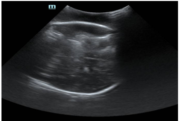
Figure 1: Low frequency ultrasound image of collection.
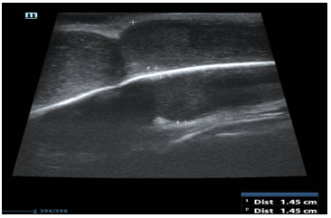
Figure 2: High frequency ultrasound image of the collection
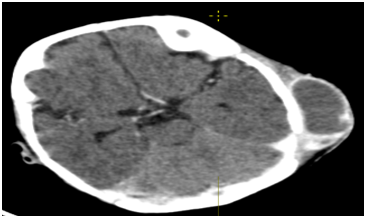
Figure3: CEC Taxial section
The ultrasonographic findings of subdural collection was due to the phenomenon of a mirror-image artifact.
Mirror-image artefacts happen when the primary beam hits a highly reflective surface that is obliquely oriented, is reflected by that surface, but then runs into another structure on the way to the transducer. The mirror-image artefact is produced on the opposite side of the reflective surface when the transducer interprets the delayed echo as reflecting from a structure deeper down (Figure 4).
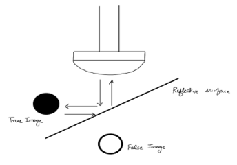
In this instance, the skull bone reflects the ultrasound beam, creating an artefact that resembles an epidural/subdural collection on the side opposite the extracalvarial collection.
Other typical mirror-image aberrations include reflections of the amniotic sac and ascites across the diaphragm that simulate heterotopic pregnancy and pleural effusion respectively.
The mirror-image artefact may closely resemble the original structure, but it also has the potential to be weaker, to distort the original structure's picture, or to appear on photos when the original structure is not simultaneously visible.
In order to avoid needless further investigations and patient worry, this is crucial to distinguish such mirror-image abnormalities and not consider them as pathology.
By adjusting the primary beam's angle or, in the instance of an
extracalvarial collection, by application of graded compression, which demonstrates compression of the false mirror picture simultaneously with the graded compression of genuine image, so it is possible to assess the presence of mirror-image artefacts. It is also possible to utilise doppler ultrasound to demonstrate the normal cerebral blood flow within the area of misleading mirror image [1].
Dear Editorial Team, Clinical Medical Reviews and Reports. My experience with the journal was highly positive. The peer-review process was rigorous, constructive, and completed in a timely manner. The reviewers provided valuable comments that helped improve the quality and clarity of our manuscript. The editorial office was professional, responsive, and supportive throughout all stages of the publication process. Communication was clear and efficient, and any questions were addressed promptly. Overall, I found the journal to maintain high scientific standards and an excellent publication workflow. I would be pleased to consider submitting future work to this journal. Best wishes from, Elena Popa.

It was my pleasure to submit my testimonial concerning the Reviewer Board of our Scientific Journal “Brain and Neurological Disorders”. The Reviewers focused on some modifications and their contribution was helpful. The ladies of our Editorial Office were also supported my efforts. It was my honor to have such a co-operation and I am looking forward for more collaboration.

Dear Grace Pierce, Editorial Coordinator of Journal of Clinical Research and Reports, Thank you for the speedy and efficient peer review process. I appreciate the fact that your peer reviewers do not take months to respond like with some other journals. I would also like to thank the editorial office for responding quickly to my questions. It is an excellent journal. I plan to submit more manuscripts in the future. Best wishes from, Robert W. McGee

Dear Grace Pierce, Editorial Coordinator of Journal of Clinical Research and Reports, Working with you and your team on our recent publication in JCRR has been a truly wonderful and enjoyable experience. The responses were prompt, and the reviewers were patient, constructive, and highly professional. One reviewer in particular gave me the feeling that a professor was carefully reading and commenting on my coursework, which was deeply touching. The entire process was straightforward and hassle‑free, with no tedious online forms to complete. I highly recommend this journal. Best wishes from, DR Aibing Rao, Head of R&D

I Appreciate the Opportunity to Share my Experience with the Journal of Clinical Research and Reports. The peer review process was timely and constructive, and the feedback provided helped improve the quality of our manuscript. The editorial office was professional, responsive, and supportive throughout the process, ensuring smooth communication and efficient handling of the submission. Overall, it was a positive experience collaborating with your team.

Dear Mercy Grace, Editorial Coordinator of Obstetrics Gynecology and Reproductive Sciences, We would like to express our gratitude for your help at all stages of publishing and editing the article. The editors of the magazine answer all the necessary questions and help at every stage. We will definitely continue to cooperate and publish other works in the Obstetrics Gynecology and Reproductive Sciences! Best wishes from, Alla Konstantinovna Politova,
