AUCTORES
Globalize your Research
Research Article | DOI: https://doi.org/10.31579/2690-4861/407
Department of Neurosurgery, The Affiliated Changzhou No. 2 People’s Hospital of Nanjing Medical University, Changzhou, Jiangsu, China
*Corresponding Author: Fang Liu, MD Department of Neurosurgery, The Affiliated Changzhou No. 2 People’s Hospital of Nanjing Medical University, 68 Gehu Road, Changzhou 213000, Jiangsu, China.
Citation: He-Qi Qu, Yu-Wei Zhang, Yi-Zhuo Guo, Fang Liu, (2024), Prognostic value Analysis of Intraoperative Transcranial Electrical Stimulation motor evoked Potential Monitoring in Postoperative limb Function changes of Neurosurgical Patient, International Journal of Clinical Case Reports and Reviews, 17(2); DOI:10.31579/2690-4861/407
Copyright: © 2024, Fang Liu. This is an open-access article distributed under the terms of the Creative Commons Attribution License, which permits unrestricted use, distribution, and reproduction in any medium, provided the original author and source are credited.
Received: 23 February 2024 | Accepted: 22 March 2024 | Published: 30 April 2024
Keywords: intraoperative electrophysiology; motor evoked potential; glioma; intracranial aneurysm
Objective: To explore the predictive value of intraoperative transcranial electrical stimulation motor evoked potential (TES-MEP) monitoring for postoperative muscle strength change in patients undergoing craniocerebral surgery.
Methods: In this study, 166 patients who underwent intraoperative motor evoked potential (MEP) monitoring were retrospectively analyzed. Univariate analysis and binary Logistic regression were used to analyze the influencing factors of postoperative muscle strength changes. Receiver operating characteristic (ROC) curve analysis was performed to evaluate the predictive value of abnormal changes of MEP amplitude in postoperative muscle strength changes.
Results: Binary Logistic regression analysis showed that the abnormal amplitude of MEP during operation was an independent risk factor for short-term and long-term muscle strength decline after operation (P < 0.05). ROC curve analysis showed that the area under the curve (AUC) for abnormal changes of MEP amplitude to predict short-term postoperative muscle strength decline was 0.754, with a sensitivity of 0.516, with a specificity of 0.993, and the AUC for long-term postoperative was 0.782, with a sensitivity of 0.591 and specificity of 0.972.
Conclusions: The decrease of MEP amplitude more than 50% as a warning standard has a good predictive value for the change of limb function after operation, and the abnormal changes of MEP amplitude indicate that the muscle strength of patients after operation may be lower than that before operation.
In neurosurgery, the purpose of surgery is to remove the lesion as much as possible, and to avoid new neurological defects related to the operation as far as possible, so as to improve the quality of life of patients. In recent years, transcranial electrical stimulation motor evoked potential (TES-MEP) monitoring has received increasing attention in neurosurgery, which can monitor the disorder of the pyramidal tracts in brain and spinal surgery , thereby increase the safety of surgery and reducing the incidence of postoperative neurologic deficits[1-3]. However, there are different views on the warning standard of intraoperative MEP. The common views of MEP warning standard are as follows: the latency of MEP is prolonged by 10%, the amplitude is decreased by more than 50% or 80%, all or none, and the stimulation threshold is increased[4-6].
In this paper, 166 patients who underwent intraoperative MEP monitoring in the Department of Neurosurgery of the Affiliated Changzhou No.2 people's Hospital of Nanjing Medical University from January 2019 to December 2022 were included. The intraoperative MEP amplitude changes were recorded and the decrease of MEP amplitude more than 50% was used as the early warning standard to explore the predictive value of abnormal changes of MEP amplitude for postoperative muscle strength changes.
Patient Selection and Study Design
The inclusion criteria of patients were as follows: (1) craniotomy involving functional and non-functional areas. (2) surgery involving the pyramidal tract and its surroundings. (3) endarterectomy for carotid stenosis. (4) intracranial vascular surgery such as clipping of intracranial aneurysms and resection of vascular malformations. (5) surgery for brain stem or peri-brainstem lesions. Exclusion criteria: (1) patients and their families refused intraoperative monitoring. (2) patients with intracranial implants or history of epilepsy can not be monitored by MEP. The study was approved by the Ethics Committee of the Affiliated Changzhou No.2 people's Hospital of Nanjing Medical University. Before operation, we informed the patients or their families of the purpose of this study, and the patients signed the informed consent form after understanding the situation.
Anesthesia
All patients were induced with propofol (150mg) and sufentanil (25ug). Propofol (4-6mg/kg/h) and remifentanil (0.1-0.2ug/kg/min) were used to maintain intraoperative anesthesia. Muscle relaxants were only used during induction anesthesia and not used during other periods of anesthesia.
Neurophysiologic monitoring
According to the International EEG Electrode Placement 10–20 System Standard, the limb stimulation electrodes were placed at C1 and C2 points, and the recording electrode was connected to the abductor pollicis brevis of the upper limb and the abductor muscle of the lower limb. MEPs were generally obtained by 5-8 pulse trains, with a stimulus interval of 1-2ms, a stimulus intensity of 100–400 V, and a stimulus frequency of 250–500 Hz. The band-pass filter range was 30–3000 Hz, with a notch filter at 50 Hz, and the analysis time was 100 ms. When the anesthesia is stable and the dura mater is not opened, the MEP amplitude is recorded as the baseline amplitude, and the MEP amplitude decrease of more than 50% of the baseline amplitude as warning standard. Pay close attention to the changes of MEP amplitude during the operation. When the MEP amplitude decreases more than 50% of the baseline during the operation, report immediately to the surgeon, who will stop the operation and take measures to restore the MEP amplitude as much as possible.
According to the change of MEP amplitude during operation, the change of MEP amplitude can be divided into MEP amplitude without abnormal change and MEP amplitude abnormal change. The abnormal change of MEP amplitude includes the following two situations: 1)the decrease of MEP amplitude is always less than or equal to 50% of the baseline amplitude, which is called no deterioration of MEP amplitude. 2)the MEP amplitude decreases by more than 50% of the baseline amplitude and then returns to more than 50% of the baseline amplitude, which is called the reversible deterioration of MEP amplitude. The abnormal amplitude of MEP shows that the decrease of MEP amplitude is more than 50% of the baseline amplitude and has not recovered to more than 50% of the baseline amplitude at the end of the operation, which is also called irreversible deterioration.
Clinical follow-up evaluation
The changes of muscle strength at 1 week and 1 month after operation were evaluated. The grading standard of muscle strength was the six-stage grading method of 0-5 grade. According to the change of muscle strength after operation, the selected patients were divided into two groups: 1) no decrease group: the muscle strength of the patients remained unchanged or improved by at least 1 grade compared with that before operation; 2) decrease group: the muscle strength of the patients decreased by at least 1 grade compared with that before operation. The short-term muscle strength decline was defined as the decrease in muscle strength at 1 week after operation as compared with that before operation. The long-term decline of motor function was defined as the decrease of muscle strength 1 month after operation as compared with that before operation.
SPSS22.0 (IBM, Chicago, Illinoi, USA) and R (Ver.3.6.3) were used for statistical analysis. The classification variables were expressed as frequency or percentage and analyzed by Pearson chi-square test, continuous correction chi-square test or Fisher exact test. Determining whether a continuous variable is normally distributed using the Shapiro-wilk test. The data of normal distribution were expressed by mean ± standard deviation (SD), and the independent sample t-test was used for comparison. The continuous variables of non-normal distribution were expressed by median and inter-quartile range (IQR), and compared by Mann-whitney U test. Binary Logistic regression analysis was used to evaluate the independent risk factors of postoperative muscle strength. Receiver operating characteristic (ROC) curve was used to analyze the ability of abnormal changes of MEP amplitude to predict the change of muscle strength after operation. P < 0>
Patient Characteristics
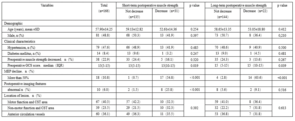
A total of 166 patients who underwent intraoperative MEP monitoring were included. There were 81 males and 85 females with an average age of 57.90 ±14.25. The preoperative GCS score was 5-15, with a median score was 15. There were 87 cases of Patients with intracranial tumor or function, 40 cases of aneurysm, 20 cases of severe stenosis of internal carotid artery, 19 cases of intracranial vascular malformation. Preoperative MRI showed that the lesions in 67 patients were located in the motor function area and corticospinal tract (CST) .39 patients had lesions located in non-motor functional areas and CST regions, and CTA showed lesions in the vessels in 60 patients(Table1).
MEP monitoring and postoperative muscle strength
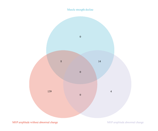
Short-term follow-up results showed that among the 166 patients included, 31(19%) patients had decreased muscle strength and 135(81%) patients had no decrease in muscle strength. Among the 31 patients with decreased muscle strength, the MEP amplitude without abnormal change in 14 (45.2%) cases (11 with no deterioration, 3 with reversible deterioration) and MEP amplitude abnormal change in 17 (54.8%) cases. Among the 135 patients with no decrease in muscle strength, the MEP amplitude without abnormal change in 134(99.3%) cases (125with no deterioration, 9 with reversible deterioration), and the MEP amplitude abnormal change in 1(2.0%) case (Table1, Figure1).
Long-term follow-up result showed that among the 166 patients included, 22 (13.3%) patients showed decreased muscle strength, while 144 (86.7%) patients did not. Of the 22 patients with muscle strength decline, 3 (13.6%) showed MEP amplitude without abnormal change (no deterioration in 1 case, reversible deterioration in 2 cases) and 19(86.4%) showed MEP amplitude abnormal change. Of the 144 patients with muscle strength did not decrease, the MEP amplitude without abnormal change in 140 (97.2%) cases (no deterioration in 130 cases, reversible deterioration in 10 cases) and the MEP amplitude abnormal change in 4(2.8%) case. (Table 1, Figure 1).

Figure 1: Venn diagram of the relationship between MEP amplitude change and postoperative muscle strength decline. (A) Venn diagram of changes in MEP amplitude and short-term postoperatively reduced muscle strength. (B) Venn diagram of changes in MEP amplitude and long-term decline in postoperative muscle strength. MEP: motor evoked potential.
The abnormal changes of MEP amplitude were an independent risk factor for postoperative muscle strength decline
Univariate analysis showed that there were significant differences in preoperative GCS score, postoperative imaging features and changes of MEP amplitude between short-term decreased muscle strength group and non-decreased group (p < 0>
preoperative GCS scores and MEP amplitude changes between long-term decreased muscle strength group and non-decreased group (P<0>0.05). Gender, age, hypertension, diabetes mellitus, preoperative changes of muscle strength had no statistical significance on postoperative changes of muscle strength (P > 0.05) (Table 1)

SD. standard deviations. GCS: Glasgow Coma Scale. IQR, interquartile range. MEP: motor evoked potential. CST: Corticospinal tract. Comparison of postoperative muscle strength between non-decreased group and decreased group.SD. standard deviations. GCS: Glasgow Coma Scale. IQR, interquartile range. MEP: motor evoked potential. CST: Corticospinal tract. Postoperative imaging abnormalities include intracerebral hemorrhage or cerebral infarction. Asterisks indicate significant differences.
Table 1: Comparison of postoperative muscle strength between non-decreased group and decreased group
The factors with statistical significance in the univariate analysis were subjected to binary logistic regression analysis to determine the independent predictors of postoperative muscle strength changes. The results showed that the abnormal change of MEP amplitude was an independent risk factor for short-term (OR:218.145, 95% confidence interval [CI] 22.491-2115.828, P<0>% CI20.685-606.127, P<0>

OR, odds ratio. CI, confidence interval. GCS, Glasgow Coma Scale. MEP, motor evoked potential. Postoperative imaging abnormalities include intracerebral hemorrhage or cerebral infarction.
Table 2: Analysis of multiple factors affecting the changes of postoperative muscle strength.
Using ROC curve analysis to assess the ability of MEP amplitude abnormal change to predict the change of muscle strength. The short-term postoperative AUC was 0.754 (95% CI 0.639-0.870), the sensitivity was 0.516, the specificity was 0.993. The long-term postoperative AUC was 0.782 (95% CI 0.652-0.911), the sensitivity was 0.591 and specificity was 0.972. (Figure 2).


Figure 2: Univariate analysis of potential factors affecting postoperative muscle strength changes.(A)Univariate analysis of potential factors affecting short-term postoperative muscle strength changes. (B) Univariate analysis of potential factors affecting long-term muscle strength changes after operation. GCS: Glasgow Coma Scale. CST: Corticospinal tract
MEP reversible deterioration and postoperative muscle strength changes
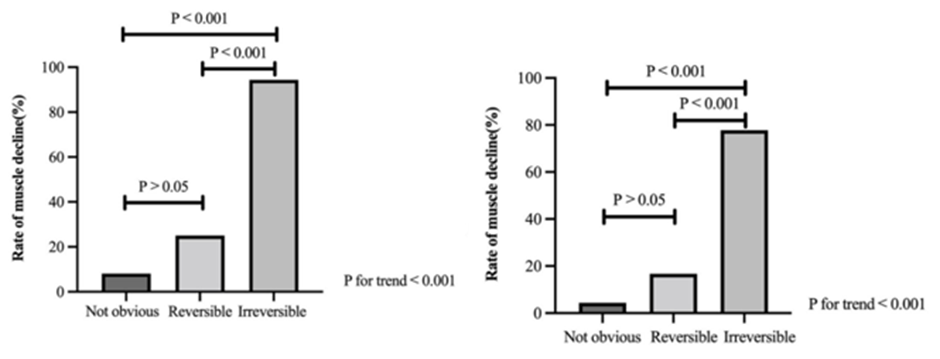
Further study on the relationship between the change of MEP amplitude and the change of postoperative muscle strength showed that there were significant differences in no deterioration, reversible deterioration and irreversible deterioration of MEP between the postoperative muscle strength decrease group and the non-decrease group (p<0.01) (Table3), and there was a linear correlation among the three groups in the incidence of postoperative muscle strength decline (p<0.001) (Figure 3). There was a significant difference between MEP reversible deterioration and irreversible deterioration in the incidence of long-term postoperative muscle strength decline (p<0.001) (Figure 3). Binary Logistic regression analysis showed that compared with MEP irreversible deterioration, MEP reversible deterioration decreased the risk of short-term (OR:0.020, 95% CI 0.002-0.217, p=0.001) and long-term (OR:0.057, 95% CI 0.009-0.375, p=0.003) muscle strength decline after operation (Table 4).

MEP, motor evoked potential. Fisher exact test for data analysis
Table 3: Comparison of the changes of MEP amplitude between the decreased muscle strength group and the non-decreased group after operation.

MEP,motor evoked potential. OR, odds ratio. CI, confidence interval.
Table 4:Odd ratios of factors affecting postoperative muscle strength changes.

Figure 4: The histogram shows the relationship between the non-deterioration, reversible deterioration and irreversible deterioration of MEP and the decrease of muscle strength after operation. (A) Short-term after operation. There is a significant difference between no deterioration and irreversible deterioration of MEP. (p<0.01) (B) Long-term after operation. There were significant differences between MEP non-deterioration (p<0.001) and reversible deterioration (p<0.05) with irreversible deterioration of MEP. The change of MEP amplitude in three groups was linearly correlated with the decrease of muscle strength after operation (p<0.001). P: The p value after bonferroni's adjustment.
This research found that a reduction in MEP amplitude by more than 50% independently predicts a higher likelihood of postoperative muscle strength decline. Patients experiencing this level of MEP reduction faced a greater risk of losing muscle strength after surgery compared to those with less than a 50% decrease. Furthermore, it was observed that reversible deterioration in MEP was associated with a lower risk of muscle strength reduction post-surgery, in contrast to irreversible MEP damage. Thus, a decline in MEP amplitude exceeding 50% serves as a reliable indicator for forecasting changes in muscle strength following surgery. In brain tumor surgery, when the tumor is very close to the anatomical structure or arterial branches of the motor cortex or CST, the incidence of new postoperative neurological dysfunction is very high, especially pure motor sequelae (no sensory disturbance). Therefore, continuous evaluation of the function of motor cortex or CST is very important to reliably detect and prevent motor cortex or CST injury[7-10]. TES-MEP monitoring technique is a neuroelectrophysiological method for monitoring motor conduction function by transcranial electrical stimulation of the cerebral cortex, activating cortical motor neurons and recording action potentials on limb muscles. It can be used to monitor the integrity of the motor system during operation[11]. At the same time, MEP has a relatively high sensitivity in monitoring cortex, subcortical ischemia and brain function damage. When monitoring cerebral vascular operations such as aneurysm clipping and carotid endarterectomy, the amplitude change of MEP can reflect cerebral ischemia earlier[12, 13]. In neurosurgery, the use of MEP monitoring to protect limb motor function has been paid more and more attention, and has been widely used in aneurysms[14, 15], gliomas[16, 17], carotid endarterectomy[18, 19] and other diseases.
The importance of intraoperative TES-MEP monitoring in protecting motor function is self-evident, and the selection of warning standards in monitoring directly affects the monitoring results. The main warning standards of the MEP mainly include prolonged latency, increased stimulus threshold, amplitude decrease of more than 50% or 80%, all or none[4-6]. The changes of MEP latency showed great differences[10, 20], and the prolongation of latency led to lower sensitivity and specificity[21]. Amplitude Standard and threshold standard are the more commonly used alarm standards of MEP[22]. Compared with threshold standard, amplitude variation is more commonly used and practical as the warning standard of MEP[23], and the usefulness of threshold criteria in the process of brain lesion clearance has not been fully demonstrated[6]. In supratentorial tumor surgery, the MEP amplitude is less than 50% of the baseline value, which is better than the standard of all or no loss[8, 24-26]. Therefore, during intraoperative electrophysiological monitoring, we adopted the MEP amplitude drop of more than 50% as the warning standard.
It has been widely reported that the amplitude reduction of MEP is more than 50% as the alarm standard of intraoperative electrophysiological monitoring in the study of postoperative motor function, but its evaluation has not been consistent. In the study of Umemura et al[27], the 50% change of MEP as warning standard has high sensitivity and specificity for the prediction of postoperative motor function. While Yamashita et al[28] suggest that it has high specificity and low sensitivity. In order to further clarify the clinical application value of the 50% change of MEP amplitude as an early warning standard, we included a variety of diseases to study the relationship between the change of MEP amplitude and the change of muscle strength after operation. Our results are consistent with those of Asimakido et al[29]: in most studies, MEP shows high specificity and low or moderate sensitivity. Intraoperative MEP amplitude reduction of more than 50% as a warning standard has a good predictive value for postoperative muscle strength changes.
In the study, we found that some patients with MEP reversible deterioration showed decreased muscle strength after operation, which aroused our concern: will MEP reversible deterioration increase the risk of postoperative muscle strength decline? Therefore, we divided MEP amplitude changes in more detail, and further studied the relationship between MEP non-deterioration, reversible deterioration, irreversible deterioration and postoperative muscle strength changes.
We found a linear correlation between MEP no deterioration, reversible deterioration and irreversible deterioration in the incidence of postoperative muscle strength decline. Compared with the irreversible deterioration of MEP, the reversible deterioration of MEP reduces the risk of decreased muscle strength after operation. Holdefer et al [30]reported that compared with the irreversible deterioration of MEP, the deterioration of MEP reversibility was negatively correlated with new postoperative motor dysfunction, and our results were similar. However, Li et al[31] reported that the occurrence of postoperative motor dysfunction was associated with the duration of MEP deterioration, with reversible MEP deterioration lasting more than 13 minutes and an increased risk of new postoperative motor dysfunction, while the cutoff points reported by Guo et al[32] and Kameda et al[33] were 8.5min and 5min, respectively. In this study, we did not study the relationship between the duration of MEP deterioration and postoperative muscle strength changes. Although our purpose and results are not the same as those of Li et al, but our results all suggest that when MEP is significantly decreased or disappeared during surgery, the surgeon should intervene in time to restore the MEP amplitude to normal level, which can reduce the risk of new postoperative motor dysfunction.
Reversing the deterioration of MEP can reduce the risk of new motor dysfunction after surgery, but not all MEP deterioration can be reversed. Compared with vascular surgery, tumor surgery has a higher incidence of irreversible deterioration of MEP, which may be related to the type of surgical injury. Most injuries caused by tumor surgery are mechanical injuries[29], such as direct damage to the motor cortex or corticospinal tract, these injuries are irreversible. Vascular injury often causes ischemic changes. Hokari et al[34] reported that when the cerebral blood flow is lower than 16mL/min/100g, the amplitude of cortical evoked potential decreases gradually, and when it is lower than 12mL/min/100g, the amplitude of cortical evoked potential disappears. Timely recovery of blood supply (such as raising high blood pressure., adjustment of temporary clamp, etc.) can correct the state of cerebral ischemia and restore the amplitude shape of MEP.
The emergence of false negatives (there was no abnormal change in the amplitude of MEP during the operation, but the muscle strength decreased after operation) and false positives (the amplitude of MEP changed abnormally during the operation, but the muscle strength did not decrease after operation) may mislead surgeons in surgical procedures. In this study, 14 patients showed intraoperative MEP amplitude without abnormal change, but postoperative muscle strength decreased. 6 cases had short-term muscle strength decline and 8 cases had long-term muscle strength decline. The short-term decrease of muscle strength may be caused by the decrease of local brain function, or the compression of motor pathway caused by local brain hemorrhage and edema after operation. After the edema or hematoma was relieved, the function or pressure of the brain group returned to normal, and the limb function of the patients improved. In addition, the injury of the functional connection from the auxiliary motor area to the primary motor cortex during the operation will also lead to temporary postoperative dyskinesia, but there is no significant change in MEP during the operation[35].
Among the eight patients who experienced prolonged muscle strength loss, one displayed evidence of new post-surgical bleeding in the surgical area on an MRI scan. This suggests that the muscle strength decrease in this individual might have been caused by the new hemorrhage, indicating that it might not truly be a false negative case. Another patient, who suffered a long-term decline in muscle strength, was diagnosed with an aneurysm. During surgery, this patient's MEP amplitude was impaired for over 30 minutes. Although the amplitude recovered to normal levels after treatment during the operation, the patient later developed a cerebral infarction and a decline in muscle strength. It's plausible that the cerebral infarction occurred during the surgery, making this instance a false negative. The rest of the patients showed no significant changes in MEP amplitude during surgery or notable post-operative imaging abnormalities, yet they experienced a decline in muscle strength, classifying these instances as false negatives as well. According to research reports, excessive intraoperative electrical stimulation may lead to false negative. Because the activation site of the electrical stimulation threshold is the subcortical superficial white matter, when the stimulation is too large, the maximum current can cross the infarcted area and activate the subcortical or brainstem area[36-38], so even if the infarction occurs during the operation, it cannot be detected. In addition, Rothwell et al[39] pointed out that strong stimulation current can even activate CST in the foramen magnum, resulting in false negative. Therefore, intraoperative stimulation close to the motor threshold is used to avoid the excitation of the corticospinal tract fibers deep in the white matter and even at the brainstem level, thus reducing the occurrence of false negatives[6, 8, 40].
In this study, there was a patient, whose intraoperative MEP monitoring was false positive. According to the available data, we could not find the possible cause of the false positive. Chung et al[4] suggested that the false positives were caused by the enlargement of the gap between the cerebral cortex and the skull, which may be caused by the contraction of brain tissue caused by excessive drainage of cerebrospinal fluid. In addition, elderly patients with motor dysfunction may be more likely to detect unstable MEP, so they are more likely to have false positives[21].
There are limitations to our study. First, the sample size included in our study is relatively small, and the follow-up time is relatively short. Therefore, we will further collect more samples and follow up for a longer period of time for the following experimental study. Second, our study is a retrospective study, and we need further prospective studies to verify our results.
Our study shows that the decrease of MEP amplitude more than 50% as an alarm standard has a good predictive value for postoperative muscle strength changes. Abnormal changes in MEP amplitude is an independent risk factor for postoperative muscle strength decline, and intraoperative MEP amplitude abnormal changes indicate that patients may have muscle strength decline after operation. In addition, reversible MEP deterioration can reduce the risk of postoperative muscle strength decline compared with irreversible deterioration.
The authors declare that they have no known competing financial interests or personal relationships that could have appeared to influence the work reported in this paper.
This work was supported by the international cooperation program of Changzhou (grant number CZ20200039), the "333" talent project of Jiangsu Province (grant number BRA2020151) and the Key Research and Development Program of Jiangsu Province (grant number BE2019652).
This study was approved by the Ethical Review Boards of The Affiliated Changzhou No. 2 People’s Hospital of Nanjing Medical University. The ethics committee waived the need to obtain informed consent from eligible patients, because of the retrospective design of this study.