
Research Article | DOI: https://doi.org/10.31579/2578-8868/175
1,2Paediatric Surgery Unit, Department of Surgery, Enugu State University Teaching Hospital, Park lane, Enugu, Enugu State, Nigeria
*Corresponding Author: KE Chukwubuike, Paediatric Surgery Unit, Department of Surgery, Enugu State University Teaching Hospital, Park lane, Enugu, Enugu State, Nigeria
Citation: Ozor I. Ikemefuna., Chukwubuike K. Emeka., Enyi Nnenna., (2021) Profile of Neurological Congenital Anomalies in the Two Teaching Hospitals in Enugu, Nigeria. J. Neuroscience and Neurological Surgery. 9(1); DOI:10.31579/2578-8868/175
Copyright: © 2021 KE Chukwubuike, This is an open-access article distributed under the terms of The Creative Commons Attribution License, which permits unrestricted use, distribution, and reproduction in any medium, provided the original author and source are credited
Received: 29 March 2021 | Accepted: 12 April 2021 | Published: 16 April 2021
Keywords: central nervous system; congenital anomalies; neurological; teaching hospital
Background: Congenital anomalies of the central nervous system (CACNS) are birth defects of the physical structure of the brain or spinal cord that occur during intrauterine growth. The purpose of study was to obtain the incidence, types and risk factors of congenital anomalies of the central nervous system in the 2 teaching hospitals in Enugu, Nigeria.
Materials and Methods: This was a hospital based observational study carried out on infants delivered at University of Nigeria Teaching Hospital (UNTH) and Enugu State University Teaching Hospital (ESUTH), Enugu during the periods of January 2013 and December 2018. Diagnosis of neurological congenital anomaly was made through clinical examination by a pediatrician and a neurosurgeon. Stillborns were excluded.
Results: During the study period, 15,820 were delivered in the 2 teaching hospitals, out of which 79 infants had CACNS, which gave an incidence of 0.5%. Neural tube defect was the most common neurological anomaly. A significant number of the mothers took herbal concoctions during pregnancy. About one-fifth of the neurological anomalies were diagnosed prenatally.
Conclusion: This study showed an incidence of CACNS of 0.5% in the two teaching hospitals in Enugu, South East Nigeria. Neural tube defect was the most common anomaly.
Short title: Neurological Congenital Anomalies
Congenital anomalies of the central nervous system (CACNS) are birth defects of the physical structure of the brain or spinal cord that occur during intrauterine growth. Other terms synonymous with CACNS are central nervous system abnormalities/anomalies; brain and spinal cord birth defects. They refer to any morphological abnormality of the central nervous system that dates to the embryonic or fetal period, regardless of the mechanism of its origin [1]. CACNS is a common cause of emotional, economic and physical stress for both the child and the family. It encompasses a wide range of anomalies from minor to major abnormalities [2]. Neural tube defects results from failure of neural tube to close between the 3rd and 4th week of embryonic development and is one of the most common type of CACNS [3]. Advances in investigations such as amniocentesis, fetoscopy, ultrasound, computerized tomography and magnetic resonance imaging have improved the diagnostic accuracy of CACNS [2]. A combination of maternal serum alpha fetoprotein and maternal sonography has almost 100 percent accuracy with regards to prenatal diagnosis [4]. CACNS may be associated with anomalies of other systems such as cardiac malformation [1]. The incidence of CACNS may vary over time or with geographical location [5,6]. The exact etiology of CACNS is unknown but is most likely multifactorial involving genetic and environmental factors. The timing of an insult to the fetus is more important than the nature of the insult in determining the type of resulting anomaly. Maternal diabetes mellitus, maternal age over 35 years, multiple pregnancy, oligohydramnion, maternal hyperthermia, use of valproate by epileptic women during pregnancy are some of the risk factors associated with the development of CACNS [7]. The aim of study was to obtain the incidence, types and risk factors of congenital anomalies of the central nervous system in the 2 teaching hospitals in Enugu, Nigeria.
This was an observational study carried out at the two teaching hospitals in Enugu, namely: University of Nigeria Teaching Hospital (UNTH) Enugu and Enugu State University Teaching Hospital (ESUTH), Enugu, South East, Nigeria. The hospital serves the whole of Enugu State, which according to the 2016 estimates of the National Population Commission and Nigerian National Bureau of Statistics, has a population of about 4 million people and a population density of 616.0/km2. The hospitals also receive referrals from its neighboring states. Ethical approval was obtained from the ethics and research committees of the 2 teaching hospitals and informed consent was obtained from the patients’ mothers. For the purposes of this study, our interest was on clinically obvious and observable abnormality of structure of the brain or spinal cord which was present at birth or noticed a few days/weeks after birth. All the live babies born in UNTH and ESUTH during the period of this study were included. Stillborns were excluded from this study. This study covered a period of 5 years, from January 2013 to December 2018.
Protocol
The protocol conforms to the declaration of Helsinki. All the consecutive children who were born in both teaching hospitals during the study period had a thorough physical examination (general and systemic) performed by a pediatrician and a neurosurgeon at birth and at 6 weeks postnatal visit respectively. This 2-stage physical examination minimized the error of missing any CACNS. Diagnosis of CACNS was based only on clinical evaluation of the babies by the pediatrician and neurosurgeon. Investigations such as radiography, ultrasonography, and computed tomography scan and magnetic resonance imaging of the spine and spinal cord were not performed. For each patient, the following data were collected: sex, age of the baby at the time of diagnosis, maternal age, gestational age of the pregnancy before delivery (term/preterm), baby’s birth weight and mode of delivery. Baby’s birth weight greater or equal to 2.5 kilograms (kg) were considered to be normal while birth weight less than 2.5 kg were considered as low birth weight. Babies born at less than 37 completed weeks, calculated from the first day of last menstrual period, were considered preterm while babies born at or after 37 completed weeks were considered term. Other data collected include family history of congenital anomaly, maternal comorbidities and use of drugs and herbs.
Data Analysis
Statistical Package for Social Science (SPSS) for Windows version 23 (IBM Corp., Armonk, NY) was used for data entry and analysis. Data were expressed as percentages, medians and means.
3.1 Patents’ demography
There were 15,820 babies were born in the 2 teaching hospitals during the study period. 10,547 babies were delivered in ESUTH (55 babies had CACNS) while 5,273 babies were delivered in UNTH (24 babies had CACNS). 284 babies had one congenital anomaly or the other which gave an overall incidence of all congenital anomalies of 1.8% or 18 babies per 1000 live births. Out of the 15,820 babies delivered during the study period, 79 babies had CACNS. This gave an incidence of 0.5% or 5 babies per 1000 live births. Considering the congenital anomalies system by system, CACNS accounted for 27.8% of all the systems. Other demographic features are shown in Table 1.

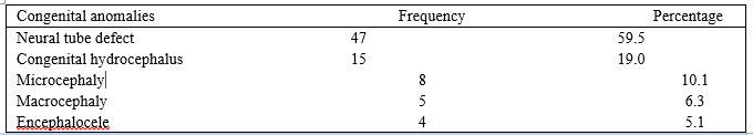
3.2. Distribution of Central nervous system congenital anomalies
These are shown in Table 2.
3.3. Associated risk factors of CACNS
Possible associated risk factors are depicted in Table 3.

3.4. Prenatal diagnosis of CACNS
Seventeen (21.5%) patients had their central nervous system anomaly diagnosed prenatally using maternal ultrasound scan. Neural tube defect was the most commonly diagnosed, prenatally.
In utero, normal development of the central nervous system involves the following stages: Induction; neural tube formation; regionalization and specification; proliferation and migration; connection and selection [8]. CACNS can occur at any stage of this development and represent an important factor of morbidity and mortality. Autopsy reports have shown that CACNS are among the most common birth defects [9]. CACNS can occur singly (isolated) or as part of syndromes and can be associated with other congenital anomalies [8].
In the present study, 0.5% incidence of CACNS recorded is similar to the reports of other authors [1,7]. The incidences of CACNS vary widely and may depend on geographical variations, cohort of patients studied such as livebirths, stillbirths or autopsy series [9,10,11]. Prevalence of CACNS is higher in stillbirths [12]. CACNS in the present study accounted for 27% of all the anomalies which is lower than what was reported by other researchers [13,14]. The reason for this discrepancy could lie in the thorough search for congenital anomalies through investigations such as computed tomography scan and magnetic resonance imaging which were not performed in the present study.
The male predominance reported in the current study is consistent with the report of Eke et al [10]. However, there is a report of female predominance [15]. The reason for the gender difference is not known. The median age at diagnosis of CACNS may be related to the degree of severity of the anomaly. For instance, encephalocele is obvious at first glance but spina bifida occulta may not be instantly obvious. There were more CACNS in preterm babies than in term babies in present study. Brown in his research reported a strong association between preterm birth and brain malformation. He stated that the brain defects themselves or the underlying cause of the defects may be inducing preterm birth [16]. In the present study, majority of the babies who had CACNS had a low birth weight. Other researchers also found low birth weight as a significant risk factor for congenital anomalies [7,17]. Most of the mothers whose babies had CACNS were aged 20 years to 35 years. Howbeit, other series reported maternal age of above 35 years [1,10]. Guardiola documented young maternal age as a risk factor for CACNS [7]. These differences are difficult to explain. In the current study, majority of the babies were delivered vaginally. Guardiola et al also reported vaginal delivery in most of their patients [7]. Some researchers have reservations about vaginal delivery of these babies with CACNS because of the risk of neurological trauma [10]. Birth injury can occur through any route of delivery and caesarian section should be based on obstetric indications [18].
Neural tube defect is consistently the most common congenital anomaly of the central nervous system as attested to by several studies [1,7,10,11]. Neural tube defects, which include spina bifida and anencephaly, result from failure of complete closure of the neural tube during embryonic development. Macrocephaly and microcephaly are birth defects where the baby’s head circumference is bigger (greater than 2 standard deviations) or smaller (less than 2 standard deviations) than expected when compared to babies of the same sex and age respectively [19]. Problems of macrocephaly/microcephaly include seizures, delayed developmental milestone, intellectual disability, balance, vision and hearing problems [20].
Two (2.5%) mothers had a history of previous birth of a child that had congenital anomaly. One (1.3%) mother accepted marrying her relative (consanguinity). Mosayebi and Movahedian reported that congenital anomalies are 3.5 times more common in consanguineous marriages [21]. In the index study, about one-twentieth of our patients were diabetic or hypertensives. Maternal diabetes mellitus and hypertension predisposes the baby to congenital anomalies with up to 9-fold increase in congenital anomalies when compared with the rate seen in non-diabetic, non-hypertensive mothers [22]
A significant number of mothers whose children had CACNS accepted taking native medications (herbal concoction) during pregnancy. Other studies on CACNS in Nigeria also reported the use of herbs by the mothers during pregnancy [10,13]. Folic acid is required for the synthesis, repair and methylation of DNA. Herbal concoctions are anti-folate and interfere with the functions of folic acid [10]. In the current study, about half of the mothers were not regular with the intake of the folic acid prescribed at the antenatal clinics. Periconceptional folic acid supplementation has been shown to decrease the incidence of neural tube defects [23].
Diagnosis of CACNS can be made prenatally at about 20 weeks gestation. However, some CACNS may be detected at the first trimester or early second trimester of pregnancy [24]. About one-fifth of the mothers had prenatal diagnosis of the CACNS in the present study. Poverty and lack of expertise required for interpretation of the results may be responsible for this low prenatal detection rate of CACNS.
Limitations of this study
This study was limited by the small number of birth defects. A larger number of cases would have availed better analysis.
Only obvious and observable birth defects were assessed for this study. Investigations were not performed. Stillborns were excluded from this study.
Conclusion: This study showed an incidence of CACNS of 0.5% in the two teaching hospitals in Enugu, South East Nigeria. Neural tube defect was the most common anomaly. Future studies should community based so that babies delivered in the maternity homes and private hospitals can be captured.
Our gratitude goes to the resident doctors who assisted with logistics.
Conflict of interest:None
Dear Editorial Team, Clinical Medical Reviews and Reports. My experience with the journal was highly positive. The peer-review process was rigorous, constructive, and completed in a timely manner. The reviewers provided valuable comments that helped improve the quality and clarity of our manuscript. The editorial office was professional, responsive, and supportive throughout all stages of the publication process. Communication was clear and efficient, and any questions were addressed promptly. Overall, I found the journal to maintain high scientific standards and an excellent publication workflow. I would be pleased to consider submitting future work to this journal. Best wishes from, Elena Popa.

It was my pleasure to submit my testimonial concerning the Reviewer Board of our Scientific Journal “Brain and Neurological Disorders”. The Reviewers focused on some modifications and their contribution was helpful. The ladies of our Editorial Office were also supported my efforts. It was my honor to have such a co-operation and I am looking forward for more collaboration.

Dear Grace Pierce, Editorial Coordinator of Journal of Clinical Research and Reports, Thank you for the speedy and efficient peer review process. I appreciate the fact that your peer reviewers do not take months to respond like with some other journals. I would also like to thank the editorial office for responding quickly to my questions. It is an excellent journal. I plan to submit more manuscripts in the future. Best wishes from, Robert W. McGee

Dear Grace Pierce, Editorial Coordinator of Journal of Clinical Research and Reports, Working with you and your team on our recent publication in JCRR has been a truly wonderful and enjoyable experience. The responses were prompt, and the reviewers were patient, constructive, and highly professional. One reviewer in particular gave me the feeling that a professor was carefully reading and commenting on my coursework, which was deeply touching. The entire process was straightforward and hassle‑free, with no tedious online forms to complete. I highly recommend this journal. Best wishes from, DR Aibing Rao, Head of R&D

I Appreciate the Opportunity to Share my Experience with the Journal of Clinical Research and Reports. The peer review process was timely and constructive, and the feedback provided helped improve the quality of our manuscript. The editorial office was professional, responsive, and supportive throughout the process, ensuring smooth communication and efficient handling of the submission. Overall, it was a positive experience collaborating with your team.

Dear Mercy Grace, Editorial Coordinator of Obstetrics Gynecology and Reproductive Sciences, We would like to express our gratitude for your help at all stages of publishing and editing the article. The editors of the magazine answer all the necessary questions and help at every stage. We will definitely continue to cooperate and publish other works in the Obstetrics Gynecology and Reproductive Sciences! Best wishes from, Alla Konstantinovna Politova,
