
Research Article | DOI: https://doi.org/10.31579/2766-2314/071
*Corresponding Author: Loutfy H. Madkour, Chemistry Department, Faculty of Science Tanta University, 31527, Tanta, Egypt
Citation: Loutfy H. Madkour (2022). Pharmacogenomics-synergistic strategies using a chimerical peptide for enhanced chemotherapy based on ROS and DNA nanosystem. Biotechnology and Bioprocessing. 3(2): DOI: 10.31579/2766-2314/071
Copyright: ©2022, Loutfy H. Madkour, This is an open access article distributed under the Creative Commons Attribution License, which permits unrestricted use, distribution, and reproduction in any medium, provided the original work is properly cited.
Received: 03 August 2021 | Accepted: 15 September 2021 | Published: 06 January 2022
Keywords: peptide; positive feedback strategy; ROS-responsive; tumor therapy; mesoporous silica nanoparticle; drug delivery; synergistic therapy; p53; DOX
Co-delivery of gene and drug for synergistic therapy has provided a promising strategy to cure devastating diseases. The use of genomic predictors of response to cisplatin and pemetrexed can be incorporated into strategies to optimize therapy for advanced solid tumors.
Here, a positive feedback strategy was utilized to amplify the concentration of intracellular reactive oxygen species (ROS) and a ROS-triggered self-accelerating drug release nanosystem (defined as T/D@RSMSNs) was demonstrated for enhanced tumor chemotherapy. It was found that in human breast cancer (MCF-7) cells, T/D@RSMSNs could not only release DOX and a-TOS initiatively, but also lead to increased concentration of intracellular ROS, which could be used as new trigger to cut away TK linkage and then in turn facilitate the further release of DOX for enhanced chemotherapy. Standard treatment for advanced non–small-cell lung cancer (NSCLC) includes the use of a platinum-based chemotherapy regimen. This novel ROS triggered self-accelerating drug release nanosystem with remarkably improved therapeutic effects could provide a general strategy to branch out the applications of existing ROS-responsive drug delivery systems (DDSs).
Here, an amphiphilic chimeric peptide (Fmoc)2KH7-TAT with pH-responsibility for gene and drug delivery was designed and fabricated. As a drug carrier, the micelles self-assembled from the peptide exhibited a much faster doxorubicin (DOX) release rate at pH 5.0 than that at pH 7.4. As a nonviral gene vector, (Fmoc)2KH7-TAT peptide could satisfactorily mediate transfection of pGL-3 reporter plasmid with or without the existence of serum in both 293T and HeLa cell-lines. Besides, the endosome escape capability of peptide/DNA complexes was investigated by confocal laser scanning microscopy (CLSM). To evaluate the co-delivery efficiency and the synergistic anti-tumor effect of gene and drug, p53 plasmid and DOX were simultaneously loaded in the peptide micelles to form micelleplexes during the self-assembly of the peptide. Cellular uptake and intracellular delivery of gene and drug were studied by CLSM and flow cytometry respectively. And p53 protein expression was determined via Western blot analysis. The in vitro cytotoxicity and in vivo tumor inhibition effect were also studied. The results [1] suggest that the co-delivery of gene and drug from peptide micelles resulted in effective cell growth inhibition in vitro and significant tumor growth restraining in vivo. The chimeric peptide-based gene and drug codelivery system will find great potential for tumor therapy.
The mesoporous silica nanoparticles (MSNs) based nanocarriers were gated by β-cyclodextrin (β-CD) through the ROS-cleavable thioketal (TK) linker to encapsulate the anticancer drug doxorubicin hydrochloride (DOX) and ROS producing agent α-tocopheryl succinate (α-TOS), whose surface was further anchored with adamantane conjugated poly(-ethylene glycol) chain (AD-PEG) via host-guest interaction. Both in vitro and in vivo experiments demonstrated that T/D@RSMSNs exhibited more significant antitumor activity in the human breast cancer than the traditional single-DOX loaded ROS-responsive nanocarrier.
Chemotherapy as a dominant approach for the treatment of tumors has been widely used in clinical trials [2, 3]. However, the curative effect of chemotherapy is severely dampened due to severe systemic toxicity, low bioavailability of anti-tumor drug as well as the emerging of drug resistance of tumor cells after repeated administration of anti-tumor drug [4]. Gene therapy, which focuses on the modulation and repair of particular gene defect, provides a new avenue for tumor therapy in gene level. Unfortunately, the biosecurity concerns of viral vectors and the low transfection efficiency of non-viral vector greatly limit its application. Recently, synergistic therapy based on co-administration of gene and drug has emerged rapidly as a promising modality for tumor treatment. Decreased drug dosage, negligible side effect and high inhibition efficacy are hopeful to achieve, mostly because of the synergistic effect [5] or reversal of drug resistance to some extent via specific gene [6] by the co-delivery systems.
The key point of synergistic therapy was to transport gene and drug to the same cells and tissues. Considerable efforts have been devoted to developing delivery systems and various carriers based on dendrimers [7], liposomes [8], polymers [9, 10] and nano-scaled inorganic particles [11] were proposed. But the barriers such as unsatisfactory cellular uptake, endosome escape and vectorinduced toxicity [12] are still fatal problems for these delivery systems. Especially in gene delivery process, the major DNA-loaded complex internalized into cells through endocytosis is believed to traffic from endosome to lysosome. The milieu in lysosome is hostile to DNA due to the existence of various enzyme systems, which may lead to the degradation of DNA/vector and failure in gene transfection [13]. Strategies based on the proton-sponge hypothesis are widely used to overcome the endosome escape barrier [14]. Generally, carriers with satisfied buffering ability always accompany with severe cytotoxicity, such as PEI and chloroquine.
Amphiphilic peptide-based micelles with a typical coreeshell structure are an important class of promising co-delivery systems for gene and drug [15, 16]. The peptide-based hydrophobic core can be used to encapsulate hydrophobic drugs, while the cationic peptide shells can be utilized to condense DNA. Additionally, benefiting from the blossom of modern molecular biology, peptides with tremendously appealing bioactivity have been exploited. For instance, TAT peptide (YGRKKRRQRRR), which derives from the transactivating transcriptional activator protein of HIV-1, has been confirmed to possess ability of autonomous and receptor independent cell membrane translocation [17, 18]. Besides, many viruses utilized pH-sensitive peptide to accelerate the endosome escape [19], pH-sensitive peptides such as GALA or histidine-rich peptide [20, 21] were fabricated as promising candidates to overcome the endosome escape barrier. Since peptide is originated from organism and mainly composed of nature amino acids, peptidebased biomaterials can always perform undeniable biocompatibility and biodegradability.
In this chapter, we reported an amphiphilic chimeric peptide, (Fmoc)2KH7-TAT with the chemical structure indicated in Scheme. 1, which can self-assemble into micelleplex with DNA and drug simultaneously.

As illustrated in Scheme. 2, at the physiological pH, the hydrophobic drug DOX was encapsulated in the hydrophobic micellar core through hydrophobic interaction and π-π stacking interaction, while the DNAwas combined with cationic shell of TAT peptide. After cell internalization, the complexes could escape from the endosome due to the protonation of heptahistidine domain of (Fmoc)2KH7-TAT at low pH milieu, resulting in the swelling of complexes, accelerated drug release, and the delivery of therapeutic gene to nucleus subsequently. The properties of peptide micelles were characterized in terms of drug/DNA loading ability, drug release behavior in vitro, gene transfection ability as well as codelivery of gene/drug etc. Furthermore, the synergistic anti-tumor effect both in vitro and in vivo was evaluated in detail [1]

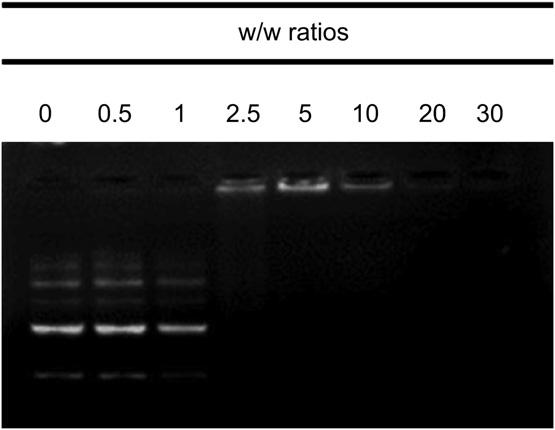
The condensation of DNA is crucial for gene transfection. To demonstrate the DNA binding ability of the micelles, agarose gel electrophoresis assay was studied. As shown in Fig. 1, (Fmoc)2KH7-TAT could completely retard the mobility of pGL-3 DNA at w/w of 5. The good DNA binding ability of (Fmoc)2KH7-TAT was probably due to the formation of micelle structure, since the DNA binding ability of TAT alone was relatively poor according to our previous work [22].
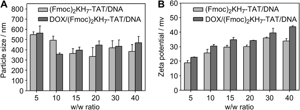
Besides, the particle size and zeta potential of (Fmoc)2KH7-TAT/pGL-3 DNA complexes were also determined as shown in Fig. 2. The size decreased and the zeta potential increased with increasing w/w ratio, indicating the more compact complexes formed. Furthermore, size and zeta potential of DOX/ (Fmoc)2KH7-TAT/pGL-3 DNA micelleplexes were performed to investigate the influence of loaded drug. It was found that loading of DOX brought nearly negligible influence in zeta potential, while the loaded DOX increased the size slightly as compared with that of the (Fmoc)2KH7-TAT/pGL-3 DNA complexes, indicating that the loaded DOX swelled the micellar hydrophobic core to some extent.
In order to get direct insight into fabricated (Fmoc)2KH7-TAT micelles, and evaluate the effect of DNA and/or drug loading on peptide micelle, four corresponding morphologies were observed by TEM. As shown in Fig. 3A2-D2, all the samples were welldispersed with uniform structure. The loaded DOX increased the size of both peptide and peptide/pGL-3 DNA complexes. No matter drug was loaded or not, the condensation of DNA decreased the size of peptide micelles. The decreased size in complexes was attributed to the introducing of electrostatic interaction between cationic peptide and DNA. Furthermore, the hydrodynamic size of the four types of nanoparticles at the same peptide concentration was studied by dynamic light scattering (DLS). As shown in Fig. 3A1-D1, the tendency in hydrodynamic size was consistent with the TEM results. Moreover, the size observed by TEM was smaller than that by DLS and the discrepancy was ascribed to the shrinking of complexes during the preparation of TEM samples [23].

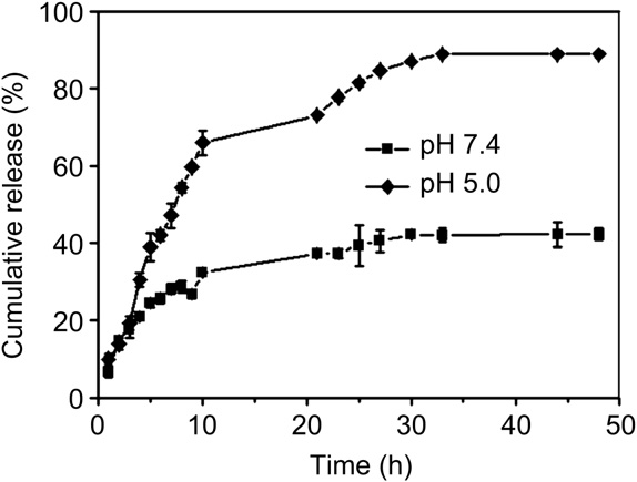
DOX was chosen [1] as the model drug to assess the drug loading behavior, and the EE and DLE values of peptide micelles were 21.8% and 11% respectively at pH 7.4. The peptide could efficiently entrap the DOX due to the π-π stacking interaction between Fmoc group and DOX as well as hydrophobic interaction between deprotonated histidine and DOX at neutral pH. To demonstrate the endosome-pH sensitivity of (Fmoc)2KH7-TAT, the drug release behaviors under different pHs were also evaluated, and pH 5.0 was chosen to imitate the endosome pH. As reflected in Fig. 4, the cumulative release of DOX at pH 5.0 was significantly faster than that at pH 7.4. This finding was attributed to the increased hydrophilicity of the peptide at pH 5.0. Since the pKa of imidazole group of histidine was about 6.0, the protonation of imidazole under slightly acid environment would vanish and destroy the hydrophobic interaction between H7 and DOX, leading to the decrease in compactness of micelles and rapid release of drugs at pH 5.0.
Translocating through plasma membrane mediated by TAT peptide was confirmed involving the classical endocytosis-uptake pathway [14]. As a result, complexes are always trafficked into endosome vesicles. Successfully conquering the endosome barrier is vital to co-delivery delivery system. Here, CLSM observation was performed to estimate the endosome escaping ability of (Fmoc)2KH7-TAT/pGL-3 DNA complexes. The complexes were labeled with FITC,while LysosomeRedwas specially used to stain the acidic organelles lysosome. (Fmoc)2K-TAT/pGL-3 DNA was used as a negative control, and the corresponding ESI-MS of (Fmoc)2K-TATwas studied [1]. As shown in Fig. 5, only a small amount of (Fmoc)2KH7-TAT/pGL-3 DNA complexes was trapped in lysosome, and the majority of complexes escaped from the endosome successfully, which was confirmed by the isolated distribution of red fluorescence and green fluorescence in Fig. 5D2. In contrast, the existence of large area of yellow fluorescence in Fig. 5D1 suggested that the majority of (Fmoc)2K-TAT/pGL-3 DNA complexes was localized in lysosome.

\
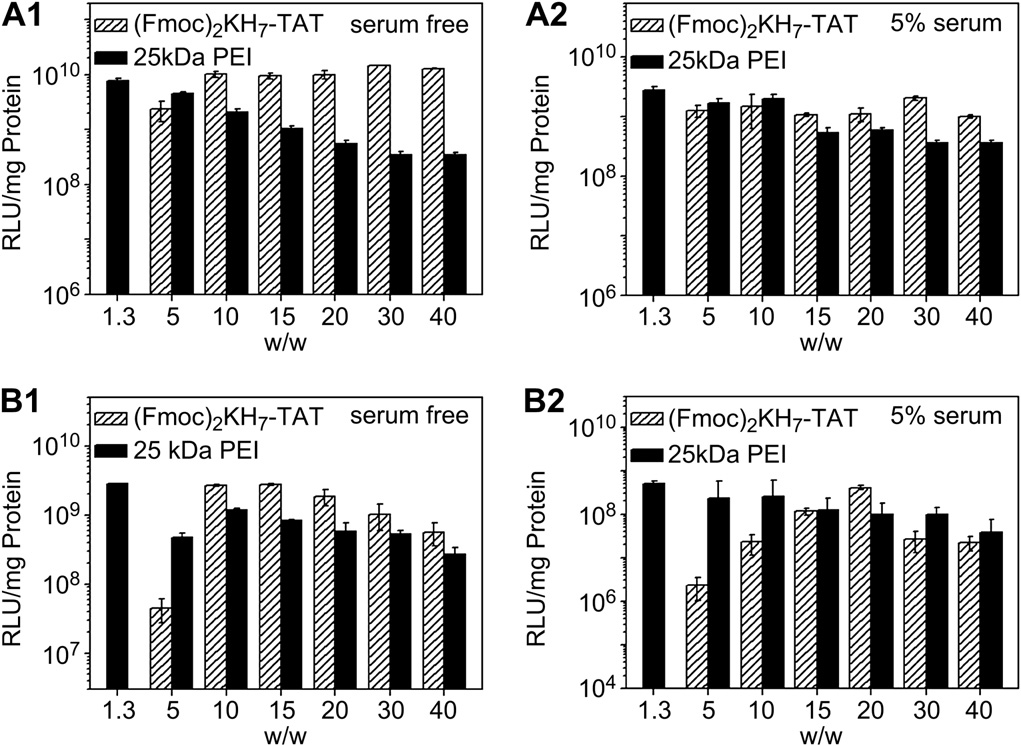
The luciferase expression of pGL-3 DNA mediated by (Fmoc)2KH7-TAT was investigated in 293T and Hela cell-lines. Herein, branched 25 kDa PEI was used as a positive control. Fig. 6A1 and B1 revealed that (Fmoc)2KH7-TAT performed comparable transfection efficiency to 25 kDa PEI at an optimal w/w ratio of 1.3 in both cell-lines under serum-free condition. Results demonstrated that the peptide could mediate satisfactory gene transfection. The multifunctional peptide with low cytotoxicity could form micelles through self-assembly which could enhance the stability of the complexes greatly. Moreover, excellent cellular uptake and endosome escape ability of the complexes could extremely improve their bioavailability.
The transfection ability under 5% serum-containing condition was further assessed as shown in Fig. 6A2 and B2. Due to the negative influence of serum to cationic gene carrier, the transfection efficiency mediated by PEI or (Fmoc)2KH7-TAT decreased to some extent in both cell-lines. However, the transfection efficiency of peptide was still comparable to that of 25 kDa PEI.
As a good gene carrier, low cytotoxicity of vector itself was essential for practical applications. Cytotoxicity of (Fmoc)2KH7-TAT in vitro was estimated against HeLa cells via MTT assay. 25 kDa PEI was used as a negative control. As shown in Fig. 7A, the cell viability was still above 90% when the concentration of peptidewas 250 mg/L. On the contrary, the PEI showed serious cytotoxicity. Obviously, the peptide presented negligible toxicity, originating from the inherent biocompatibility of peptide.
The toxicity of doxorubicin hydrochloride (DOX.HCl) and DOX loaded
peptide system is also shown in Fig. 7B. The IC50 (the concentration required for 50% inhibition of cellular growth) of free DOX was 0.17 mg/L, while the one of DOX encapsulated in peptide micelles was 1.45 mg/L. Since the peptide was biocompatibility, the cytotoxicity was mainly induced by the release of loaded DOX. It was also found that at low DOX concentration, free DOX showed significantly higher cytotoxicity than DOX loaded peptide. With the increasing concentration of DOX, the difference in cytotoxicity between them was decreased, since free DOX can be readily transported into cytoplasm and nuclei by passive diffusion, while the DOX release from micelles was time-consuming [24].

In HeLa cells, the nucleus was stained with Hoechst 33258 (blue) and pGL-3 DNA was labeled with YOYO-1 (green). The w/w ratio of (Fmoc)2KH7-TAT/pGL-3 DNA was 15, while the final concentration of DOX was 0.638 mg/L because the toxicity at this concentration was negligible. Cellular uptake at different time (1 h and 4 h) was performed as shown in Fig. 8. With the increasing incubation time, the green and red fluorescence intensity increased significantly, indicating that the uptake of micelleplexes was time-dependent. Besides, the green and red fluorescence got well overlapped after internalization for 4 h (Fig. 8D2). In the light of this finding, peptide micelles could successfully load pGL-3 DNA and DOX simultaneously. The excellent co-delivery behavior may benefit from the core-shell structure of the cationic peptide micelles. Meanwhile, image of merged2 at 4 h revealed that the majority of DOX/peptide/pGL-3 DNA micelleplexes entered the nucleus or were around the nucleus periphery (Fig. 8E2).

To further quantify the simultaneous delivery of gene and drug, two-color flow cytometry was performed as shown in Fig. 9. The (Fmoc)2KH7-TAT micelle was used as a blank control. Here, YOYO-1 stained (Fmoc)2KH7-TAT/pGL-3 DNA complexes were green fluorescence positive and DOX loaded (Fmoc)2KH7-TAT were red fluorescence positive. As expected, double-positive cells presented an overwhelming majority of the cells. This result demonstrated the excellent ability of the peptide in co-delivery of DNA and DOX to the same cells.

The key point of co-administration of gene and drug was to achieve synergistic effect. Herein, the cell viability against HeLa cells via MTT assay was employed to evaluate the synergistic effect in vitro. p53 gene was chosen as a therapy gene to suppress the growth of tumor. As shown in the Fig. 22.7C, (Fmoc)2KH7-TAT/pGL-3 DNA exhibited negligible toxicity in HeLa cells, while (Fmoc)2KH7-
TAT/p53 DNA complexes showed certain cytotoxicity. The DOX/(Fmoc)2KH7-TAT/p53 DNA micelleplexes presented significantly higher cytotoxicity than that of (Fmoc)2KH7-TAT/p53 DNA complexes. Since the concentration of loaded DOX in micelleplexes was 0.5 mg/L, the loaded DOX showed negligible cytotoxicity according to Fig. 7B, the much increased cytotoxicity was attributed to the synergistic effect of DOX and p53 gene. Clearly, co-administration of gene and drug exceedingly decreased the dose of DOX in peptide micelles and resulted in the enhanced cell growth inhibition rate.
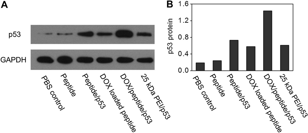
To further confirm the synergistic effect, Western blot analysis was used to determine the p53 protein expression in different transfected cells. GAPDH was used as the internal control. According to Fig. 10A, peptide induced very low expression level of p53 protein. Additionally, (Fmoc)2KH7-TAT/p53 DNA could mediate higher expression level of p53 protein than that of 25 kDa PEI/p53 DNA (w/w 1.3), demonstrating the excellent gene transfection efficacy of (Fmoc)2KH7-TAT in vitro once again. Remarkably, DOX/(Fmoc)2KH7-TAT/p53 micelleplexes mediated considerably higher expression level of p53 protein than either (Fmoc)2KH7-TAT/p53 DNA complexes or DOX loaded (Fmoc)2KH7-TAT. This result was consistent with that in cytotoxicity. Besides, the relative p53 protein expression was also provided in Fig. 10B. The relative p53 protein expression was assessed as the light intensity ratio of p53 to GAPDH from Western blot results in Fig. 10A.
The anti-tumor effect in vivo of PEI/p53 complexes, DOX loaded (Fmoc)2KH7-TAT, (Fmoc)2KH7-TAT/p53 DNA complexes and Dox/(Fmoc)2KH7-TAT/p53 DNA micelleplexes was tested, and PBS was used as the control. Fig. 11 revealed that all testing groups showed certain anti-tumor effect compared to PBS control. In detail, (Fmoc)2KH7-TAT/p53 DNA complexes exhibited better tumor inhibition than 25 kDa PEI/p53 DNA complexes, indicating the superiority of (Fmoc)2KH7-TAT in gene delivery in vivo. Notably, DOX/(Fmoc)2KH7-TAT/p53 DNA micelleplexes performed remarkably higher efficiency in tumor inhibition than either (Fmoc)2KH7-TAT/p53 DNA or DOX loaded (Fmoc)2KH7-TAT, attributed to the synergistic effect of co-delivery of gene and drug. Similar results were also found from the tumor separated from mice at the 11th day after treatment.
The representative tumor imaging was shown in Fig. 12. Besides, the body weight of the group that injected with DOX/(Fmoc)2KH7-TAT/p53 DNA micelleplexes was relatively stable, suggesting the decreased side effect of this treatment by the use of peptide as well as the satisfactory anti-tumor effect, since the malignant growth of tumor increased the body weight to some extent.

Over the past decades, plenty of smart drug delivery systems (DDSs) have been proposed to overcome unwanted side effects and maximize therapeutic efficacy of tumor chemotherapy through achieving “on demand” drug release at tumor-targeted site under unique external or internal stimuli [25,26]. Besides the additional trigger of light and magnetic field, taking advantage of tumorrelated intrinsic stimuli, including overexpressed proteases, low pH value, hyperthermia and over secreted glutathione (GSH), stimuli-responsive DDSs could further mediate tumor microenvironment targeted therapy [27-32]. Recently, the relatively high level of intracellular reactive oxygen species (ROS) in tumor environment [33-35] was also exploited as a biochemical basis for researchers to propose innovative strategies for tumor-targeted treatment [36-38]. For example, Xia et al. developed a ROS sensitive cationic polymer (PATK) for targeted gene delivery. The containing thioketal cross-linkers could be cut away under abundant ROS environment, leading to the degradation of DNA/PATK polyplexes in PC3 cells, resulting in the release of DNA [39]. Although there are a variety of ROS cleavable bonds that have been utilized to endow the materials with ROS-responsibility, very few of them exhibit sufficient sensitivity to control the drug release efficiently at biological concentration of ROS, which is too low to trigger reaction in vivo [40].
To cope with the problem mentioned above, improving the sensitivity of materials might open up appealing possibilities. A good case in point is that Sung et al. reasonably utilized the H2O2 at biologically relevant concentration, which could diffuse into hollow microsphere carrier to generate CO2 gas and disrupt the shell by reacting with a series of the encapsulated molecules, resulting in the release of loaded drug [41]. However, this strategy involved complicated design and syntheses. Besides constructing the ultrasensitive ROS-responsive carriers, another prevalent approach critically relies on the generated ROS during the processes of photodynamic therapy (PDT) to expand the utility of ROSresponsive reservoirs [42-45]. Nevertheless, precisely controlling of the irradiation time and intensity for extra laser source remains a hurdle. In addition, non-selectivity of ROS elevated in both normal cell and cancer cell under the irradiation scope could also cause lethal damage to healthy tissues.
Here, we proposed a ROS-triggered self-accelerating drug release nanosystem (defined as T/D@RSMSNs) based on a positive feedback strategy to overcome insufficient ROS generation of traditional DDSs through amplifying the intracellular ROS concentration, resulting in adequate drug release selectively in ROS abundant cancer cells in vivo. Firstly, as one of the most classic and primary nanocarriers, mesoporous silica nanoparticles (MSNs) were selected as drug carriers, owing to their distinct advantages, including large pore volume for loading drugs, easy surface functionalization and great biocompatibility [46-49]. Then, α-tocopheryl succinate (α-TOS), a vitamin E analogue, which could rapidly generate ROS in cells after interacting with mitochondrial respiratory complex II and interfering the electron transportation chain in mitochondria [50-52], and anticancer drug doxorubicin (DOX) were co-encapsulated in the pores of MSNs. The gatekeeper β-CD was anchored on the surface of MSNs through the ROS-cleavable thioketal linker (TK) for stimuli-responsive drug release [53]. Furthermore, adamantane conjugated poly(ethylene glycol) chain (AD-PEG) was introduced via host-guest interaction to enhance the stability of nanoparticles and prolong the circulation time in vivo. As shown in Scheme 3, after accumulation in tumor tissues through the enhanced permeability and retention (EPR) effect, T/D@RSMSN would be uptaken by tumor cell efficiently. At very beginning, only limited pores were open because of the existent but insufficient intracellular ROS, resulting in the simultaneous release of loaded DOX and α-TOS. Then, released α-TOS interacted with mitochondria in tumor cells to generate additional ROS. In other words, the intracellular ROS would be self-regenerated and amplified, which in turn facilitated the cutting of TK linkage to remove the gatekeeper β-CD and led to more release of α-TOS as well as DOX in MSNs.
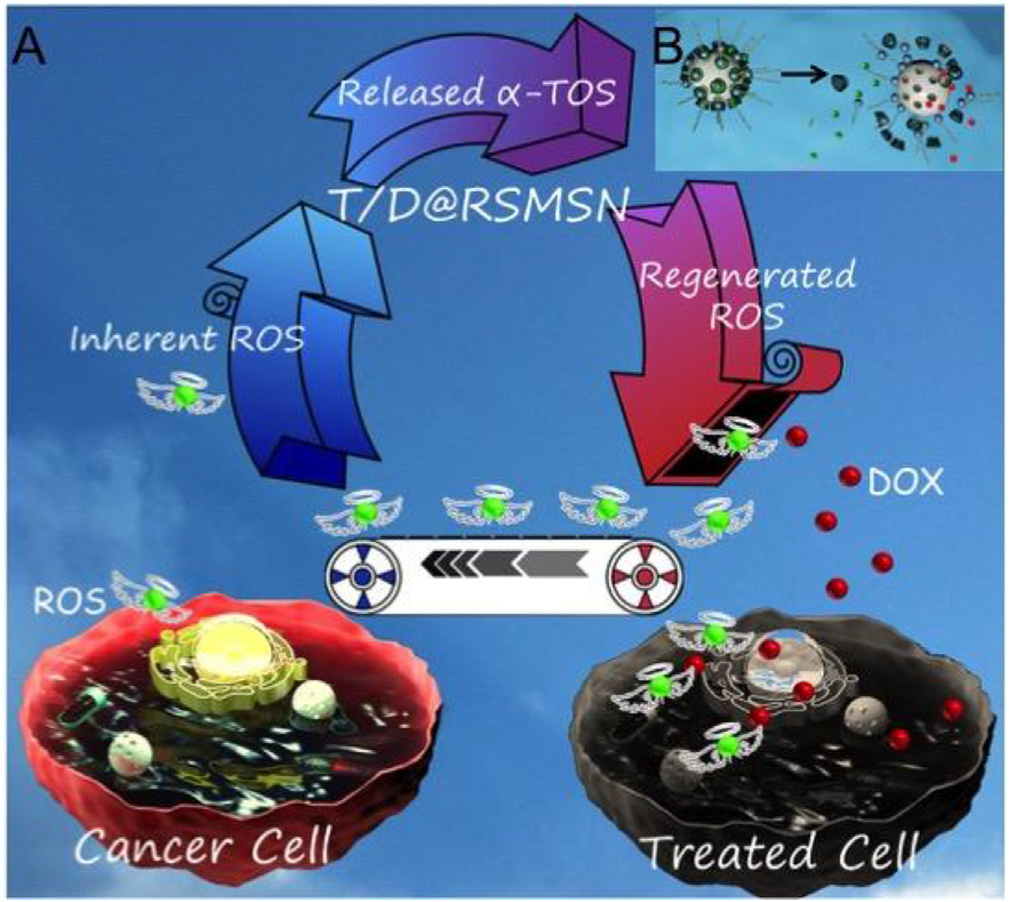
The detailed positive feedback effect with ROS-triggered self-accelerating drug release of this nanosystem was illustrated in Scheme 4. This novel nanosystem could not only achieve remarkable therapeutic effects, but also provide a general and vital strategy to surmount the restrictions of existing ROS-responsive DDSs.

MSN was synthesized by using the classic basecatalyzed co-condensation methods with several modifications according to a previous report [54]. As shown in scanning electron microcopy (SEM) (Fig. 13A) and transmission electron microscopy (TEM) (Fig. 13B) images, well-distributed and uniform MSN with mesoporous structure and a mean diameter of ~65 nm was prepared successfully. The hydrodynamic diameter of MSN measured by dynamic light scattering (DLS) [55] was 116.1 nm (PDI = 0.029) and relatively larger than TEM size because of the hydrated layer surrounding the nanoparticles. The step-by-step functionalization on the surface of MSNs was monitored by zeta potential measurements (Fig.13C) and Fourier transforms infrared (FT-IR) spectra. As demonstrated in [55], the MSN was firstly reacted with 3-aminopropyltrimethoxysilane (APTMS) to obtain MSN-NH2. After the surface modification of amino groups, the zeta potential of MSN-NH2 increased significantly from negatively charge of MSN (-17.73 mV) to positively charge (23.36 mV). Then the surfactant template (CTAC) in the pores of MSN was removed completely, and which was confirmed by the disappearance of characteristic absorption peak of CeH stretching vibrations at 2926 and 2854 cm-1 in FT-IR spectra. Subsequently, the ROScleavable thioketal linker (TK) [55] was introduced to the surface of MSN-NH2 based on amide condensation reaction, giving rise to the negatively charged MSN-TK nanoparticles (-14.13 mV). DOX and a-TOS were co-loaded into the pores of MSNs after stirring the mixture solution of MSN-TK nanoparticles, DOX and α-TOS vigorously. Finally, the resultant nanoparticle was modified with the gatekeeper β-CD through another amide condensation reaction and long-circulating AD-PEG5000 was further introduced via host-guest interaction to obtain T/D@MSN-TK-CD/AD-PEG5000 (defined as T/D@RSMSN). As shown in Fig. 13D, in comparison with the distinct mesoporous structure of blank MSN, the mesostructure of T/D@RSMSN nanoparticles in TEM image was unclear and fuzzy after the cargoes loading and surface modification. In addition, the hydrodynamic diameter of MSNs measured by dynamic light scattering (DLS) increased with the continuous modification [55]. And the hydrodynamic diameter of T/D@RSMSN (Fig. 13E) was 159.2 nm (PDI = 0.110) with a monodisperse distribution, which was suitable for achieving the passive targeting to tumor tissue through EPR effect. To further explore the stability of nanosystems, hydrodynamic diameter changes of T/D@RSMSN in PBS and PBS with 10% serum after incubation for 24 h were measured. As indicated in [55], there was no significant aggregation during 24 h incubation, demonstrating well stability of T/D@RSMSN under physiological condition. Furthermore, thermogravimetric analysis (TGA) was employed to detect the weight loss of the nanoparticles after the stepwise decoration. As illustrated in Fig. 13F, when raising their temperature to 800 oC, the weight loss of MSN-NH2, MSN-TK and T/D@RSMSN were 11%, 20% and 35%, respectively, which further confirmed the successful functionalization. Moreover, the mean number of CD molecules per nanoparticle was calculated to be 530 and the number of AD-PEG5000 conjugated on T/D@RSMSN could be calculated to be 431, according to the calculation method in a previous report [56]. Besides, as measured by the RF-5301PC spectrofluorophotometer and high performance liquid chromatography (HPLC), the loading efficiency (LE) of T/D@RSMSN was 5.1% for DOX and 2.5% for α-TOS. As to the control material D@MSNTK-CD/AD-PEG5000 (D@RSMSN), which only encapsulated DOX, the LE was 6.7%.

In this study, the T/D@RSMSN was capped by b-CD/AD through the ROS-cleavable TK linkers. After stimulating of ROS, the TK linkers would be broken, leading to the release of the loaded drug from the pores of nanoparticles. To investigate the ROS responsibility of this nanosystem, H2O2 was used as a typically ROS stimulus in in vitro experiments [42, 57], and the release profiles of the encapsulated DOX from T/D@RSMSN in PBS with different H2O2 concentration were monitored by spectrofluorophotometer. As shown in Fig. 14, without adding extra H2O2, T/D@RSMSN was gated by β-CD/AD effectively and less than 20% of drug was leaked even after 72 h incubation, proving the efficiency of anchoring of nanovalves through the TK linkers. In contrast, 60% DOX was released after the incubation with 100 µM H2O2 for 108 h. And when the nanoparticles were incubated with 1 mM H2O2, the release percentage of DOX reached 83% over 108 h. Apparently, the concentration of H2O2 had a remarkable and positive influence on the release rate and cumulative release amount of DOX, which was the basement of our self-accelerating drug release in cellular environments. Moreover, the release amount of a-TOS from T/D@RSMSN after incubating with different concentrations of H2O2 for 24 h was monitored by HPLC. As shown in [55], after incubating with different concentrations of H2O2 for 24 h, the accumulative release amount of α-TOS from T/D@RSMSN was enhanced markedly with the increased concentration of H2O2, confirming the successful ROS-responsible release of α-TOS, which was the prerequisite for ROS generation. The results above demonstrated that the β-CD/AD could act as an effective nanovalve and open the pores after the stimulating of ROS expectedly.

First of all, in order to better explore the feasibility of positive feedback strategy to overcome the obstacles in ROS-responsive DDSs, the frequently used ROS abundant human breast cancer cells (MCF-7) were chosen. And the prevailing intracellular ROS sensitive probe 2́,7-dichlorofluoresceindiacetate (DCFH-DA) was utilized to confirm the ROS generation. Cell permeable nonfluorescent DCFH-DA could be rapidly oxidized to dichlorofluorescein (DCF) with green fluorescence by the intracellular ROS [58]. As shown in Fig. 15A1 and B1, the conspicuous green fluorescence of DCF in MCF-7 cells clearly proved the inherently existence of intracellular ROS. Further, compared to the MCF-7 cells without staining (control), the fluorescence signal in MCF-7 cells incubated with DCFH-DA increased approximately 10 folds measured by the corresponding flow cytometry analysis (Fig. 15C).
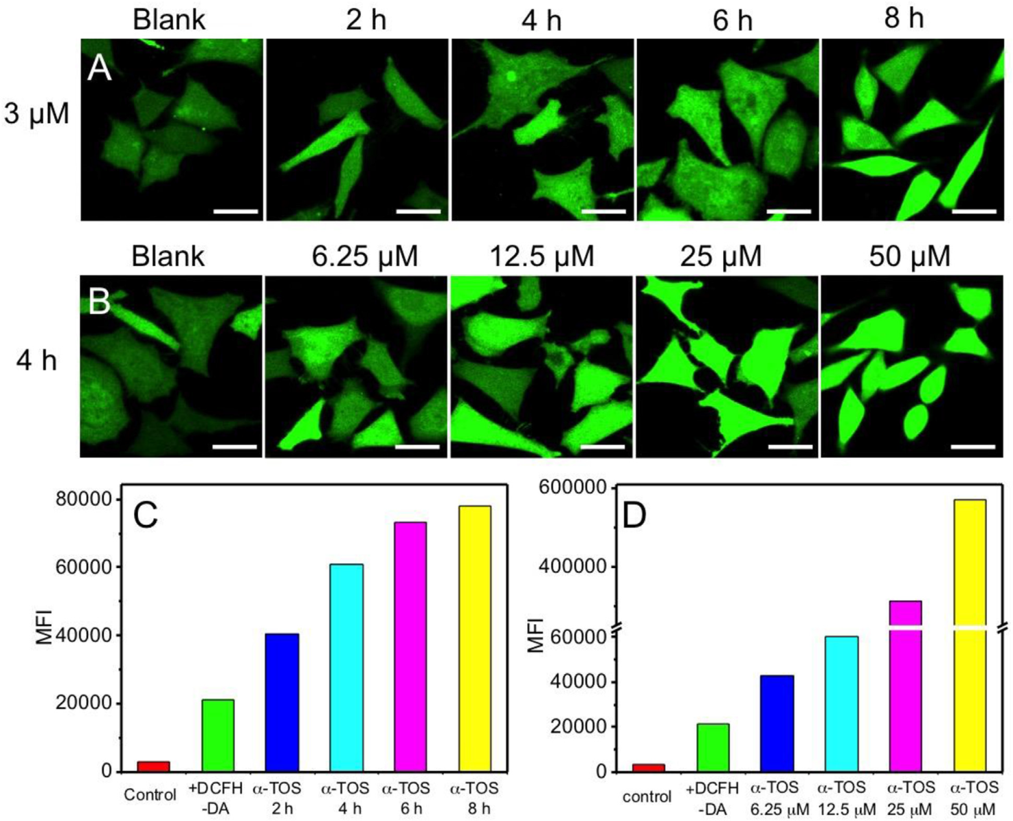
Afterwards, since the ROS generation of α-TOS was the key point in this positive feedback strategy based DDS, the intracellular ROS augmentation capability of α-TOS was also evaluated by the same ROS sensitive probe through confocal laser scanning microscopy (CLSM) and flow cytometry analysis. As shown in Fig. 15A and B, when the MCF-7 cells were incubated with α-TOS, the green fluorescent signal was noticeably stronger than that without α-TOS (blank), owing to the ROS generating capability of α-TOS, which could restrain the bioactivity of mitochondrial respiratory complex II, resulting in the electron transfer to produce ROS from oxygen. In addition, after prolonging the incubation time, the green fluorescence in MCF-7 cells incubated with a specific concentration of α-TOS (3 µM) became stronger (Fig. 15A). Besides the co-incubation time, the concentration of a-TOS is another factor to influence the levels of intracellular ROS. As demonstrated in Fig.15B, the fluorescent signal was enhanced markedly with the increased concentration of α-TOS. Thereafter, to quantitative investigate ROS producing ability of α-TOS, flow cytometry analysis was also conducted. As coincident with the CLSM results above, in the initial 2 h, the mean fluorescence intensity (MFI) values in MCF-7 cells increased to 1.5-fold, and up to 3.7-fold after prolonging the treated time to 8 h (Fig. 15C). Additionally, there was 2.8-fold and 26-fold increases of the levels of ROS in MCF-7 cells treated with 12.5 µM and 50 µM for 4 h, respectively (Fig. 15D). Moreover, the ROS producing capability of α-TOS loaded in RSMSNs (T@RSMSNs) was also evaluated by CLSM. As shown in [55], with the increasing incubation time, the green fluorescence became stronger significantly, demonstrating the releasing of α-TOS from ROSresponsive nanocarriers. Further, no obviously change of green fluorescence indicated that the possible interfering of blank MSNs was excluded based on the same incubation conditions. These CLSM and flow cytometry analysis results above confirmed the intracellular ROS producing ability of α-TOS and also revealed the possibility of loading α-TOS for positive feedback strategy based DDS.
Encouraged by the satisfying ROS generating capability of α-TOS, cellular co-incubation experiments were further performed to estimate the intracellular ROS-triggered ROS signals amplification and self-accelerating drug release efficiency of T/D@RSMSN in vitro. To highlight the important positive feedback effect induced by ROS producing agent, a single-DOX loaded nanoparticle with same modification, which was designed to like a traditional ROSresponsible DDS, was employed as a control group (defined as D@RSMSN). As shown in Fig. 16B1-B4, green fluorescencewas found in the MCF-7 treated with D@RSMSN, due to inherent ROS in MCF-7 cells. Although the green fluorescence was continuously existent, no obviously change was observed, even with increasing the incubation time from 4 h to 36 h, demonstrating that without α-TOS, the intrinsic intracellular ROS was barely growing. In addition, the red fluorescence of DOX, which was released from the pores of D@RSMSN after being uptaken, was clearly detected, but not enhanced significantly after prolonging the incubation time (Fig. 16C1- C4). It was understandable that as a result of limited intrinsic ROS, only a part of TK linkers of D@RSMSN could be cut away, leading to the partial release of DOX within the time allotted.

Compared to the unsatisfying drug release situation of control group, the T/D@RSMSN nanoparticles have shown the high efficiency of ROS-triggered ROS signals amplification and selfaccelerating drug release in vitro. As illustrated in Fig. 17B1- B4, the green fluorescence was stronger than that in MCF-7 cells incubated with D@RSMSN (Fig. 17B1-B4) at each time point, indicating that besides the inherent intracellular ROS, the α-TOS also took effect on producing ROS. And with increasing incubation time, the green fluorescence was markedly increased, confirming that with the initial intracellular ROS triggering, the encapsulated α-TOS was released from the pores of T/D@RSMSN after the TK linkers were cleaved, resulting in the ROS regeneration and replenishment. What's more, augmenting levels of intracellular ROS would in turn assist in cutting away more TK linkers and produce the positive feedback effect. As expected, during this ROS triggered ROS amplification process, the co-loaded drug DOX showed a selfaccelerating release situation, which was proved by significantly increasing of red fluorescence signal with the extension of incubation time (Fig. 17C1-C4). Moreover, compared to the relatively weak red fluorescence in MCF-7 cells treated with D@RSMSN was mainly located in cytoplasm after incubating for 36 h, MCF-7 cells treated with T/D@RSMSN exhibited the strong red fluorescence in cytoplasm at 4 h and 10 h, stronger red fluorescence in cytoplasm and weak fluorescence in the nucleoplasm at 24 h, and after 36 h incubation, the red fluorescence of the released DOX spread throughout the whole MCF-7 cells (Fig. 17E). Further, quantitative fluorescence-intensity analysis by ImageJ software of red fluorescence (DOX) in MCF-7 cells showed that the released DOX amplified by α-TOS after incubation for 36 h was 1.9-fold higher than initial released DOX. These CLSM results demonstrated that the high efficiency of drug release in T/D@RSMSN based on ROS triggered positive feedback effect could improve the discontenting efficacy of omnipresent ROS-responsible DDS.

To further evaluate the ROS microenvironment selective ability of T/D@RSMSN, human embryonic kidney (293T) normal cells with relatively negligible ROS were utilized as a control group. As shown in Fig. 18, compared to the obviously fluorescence in MCF-7 cells incubated with T/D@RSMSN or D@RSMSN, both negligible green fluorescence and red fluorescence were detected in 293T cells incubated with T/D@RSMSN or D@RSMSN, illustrating that without the abundant intracellular ROS, the pores were still capped and the loaded α-TOS and DOX could avoid being premature released from the nanoparticles, which was coincident with the cargoes release profiles above (Fig. 3). These results further confirmed that owing to the ROS cleavable TK linkers, the T/D@RSMSN could target to the tumor cells with high levels of ROS selectively, without affecting the healthy cells.

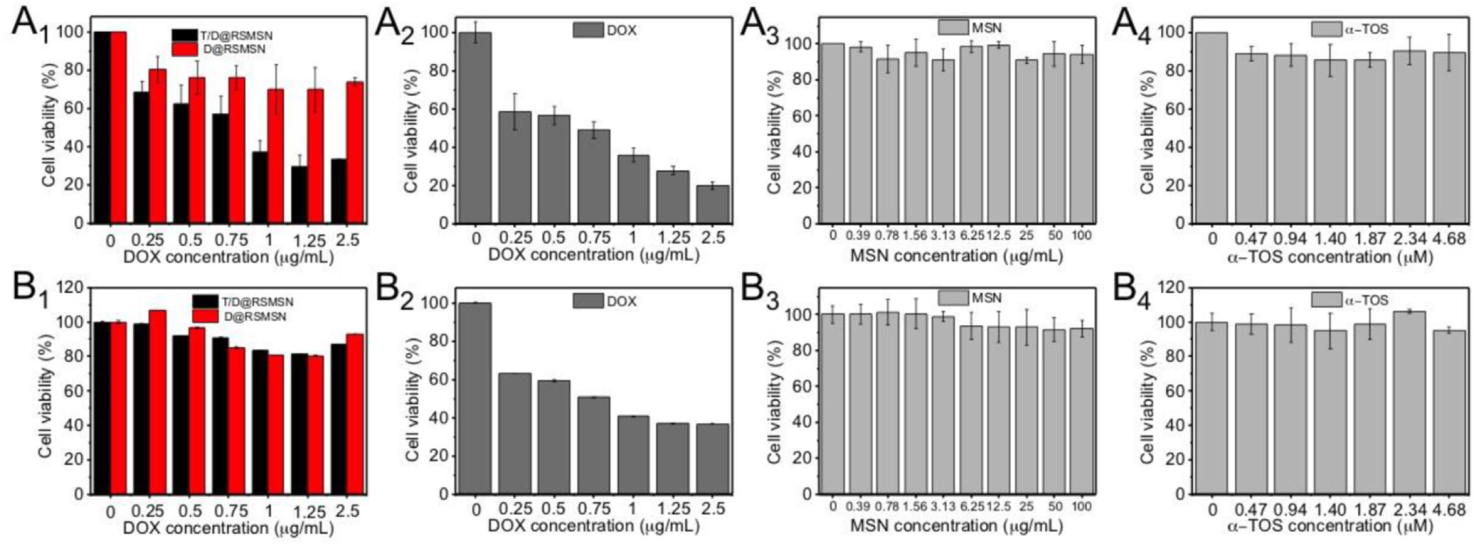
To testify the advantages of the ROS-triggered self-accelerating drug release nanosystem, the cell viability of MCF-7 cells and 293T cells treated with T/D@RSMSN and D@RSMSN was investigated. Firstly, the cytotoxicity of blank MSNs was evaluated. The cell viability of blank MSN in two cells was higher than 90%, even up to 100 µg/mL (Fig. 19), exhibiting low toxicity and suitability for drug delivery. Then, the cell viability of T/D@RSMSN or D@RSMSN in 293T cells had no obvious difference and was higher than 80% (Fig. 19A1 and B1), due to the nanoparticles were capped tightly without drug leakage under the really low concentration of ROS in normal cells. In comparison, the obviously cell viability was both observed in the MCF-7 cells incubated with T/D@RSMSN and D@RSMSN (Fig. 19A1). Compared to the cell viability in 293T cells, the toxicity in MCF-7 cells was noticeable, because the gatekeepers of nanoparticles could be removed after being uptaken by tumor cells with relatively high levels of ROS, leading to the diffusion of DOX. Also, therewas a significant difference in cell toxicity between MCF-7 cells incubated with T/D@RSMSN and D@RSMSN. The cell viability of D@RSMSN in MCF-7 cells was 69%, due to the limited DOX release caused by nonsufficient ROS during the incubation time. In comparison with MCF-7 cells treated with D@RSMSN, the cell viability of T/D@RSMSN in MCF-7 cells was much lower (29%), because after the inherent ROS triggering, the intracellular ROS was regenerated by the released part of α-TOS, utilized as new triggers to release much more DOX during the incubation period, which was well matched with above CLSM results (Fig. 19A2 and B2). IC50 value (half inhibitory concentration) of T/D@RSMSN was 0.84 mg/L (DOX concentration), whichwas much lower than D@RSMSN (>2.5 mg/L) for MCF-7 cells, indicating advantages of positive feedback strategy of T/D@RSMSN. And the IC50 of free DOX was calculated to be 0.71 mg/L for MCF-7 cells and 0.76 mg/L for 293T cells. Furthermore, the cell viability of 293T cells or MCF-7 cells incubated with free α-TOS (Fig. 19A4 and B4) was supplied for reference. Therefore, this designed nanosystem could deliver the drug targeting to the ROS abundant tumor cells and produce the cell toxicity selectively, particularly achieve enhanced chemotherapy compared to the traditional ROS-responsive nanocarriers.
Encouraged by the well performance in in vitro study, the feasibility of T/D@RSMSN in antitumor study in vivo was further evaluated through intravenous injection. The MCF-7 tumor-bearing nude mice were divided into four groups randomly (n > 5) and treated with PBS, free DOX, D@RSMSN and T/D@RSMSN, respectively. No obvious body weight variation was found in T/D@RSMSN group, confirming its well biocompatibility and nontoxicity (Fig. 20). The continuous tumor changes of MCF-7 tumor-bearing mice in each group during the 14 days treatment were exhibited through the digital photos (Fig. 20A). The tumor growth in the group injected with T/D@RSMSN was noticeably inhibited, in contrast with the other three groups. As displayed in Fig. 20C, the tumor volume in PBS group increased rapidly and grew to 10-fold of the initial volume after 14 days treatment. Moreover, the tumor treated with D@RSMSN could be inhibited to some extent, owing to the partial release of DOX triggered by the intracellular ROS. Compared to the free DOX, the main material T/D@RSMSN could inhibit the growth of tumor significantly, which illustrated the superiority of T/D@RSMSN in maximizing the antitumor efficiency, caused by positive feedback strategy. When all the mice were sacrificed after 14 days treatment, the tumors in each group were collected, average tumor weights (Fig. 20D) and the representative tumor photos (Fig. 20E) were provided to further confirm the therapeutic efficacy of T/D@RSMSN. Moreover, the therapeutic mechanism of T/D@RSMSN was studied by the terminal deoxynucleoitidyltransferaseddUTP nick end labeling (TUNEL) assay (Fig. 20F). An obvious large area of green fluorescence was displayed in T/D@RSMSN group, in comparison with none or a weak green fluorescent signal in other control groups, which was attributed to the cytotoxicity of the released DOX, leading to more apoptotic cells. In addition, hematoxylin and eosin (H&E) staining of tumor tissues was employed to evaluate the morphology and confirm the therapeutic efficiency of T/D@RSMSN. As exhibited in Fig. 20G, compared to abundant and compact tumor cells in PBS group and partial destroyed tumor cells in D@RSMSN or DOX group, the tumor cells in T/D@RSMSN group displayed that a more serious damage and many tumor cells were dead. These H&E staining and TUNEL staining assays results were coincident with the in vivo data of antitumor study above. Additionally, the low systemic toxicity of T/D@RSMSN was proved by no obviously pathological abnormalities in the heart, liver, spleen, lung, and kidney [55]. Furthermore, the normal myocardial five-enzyme parameters of the mice after treatment with T/D@RSMSN confirmed the T/D@RSMSN had no obvious cardiotoxicity [55]. These results above indicated that T/D@RSMSN displayed no systemic side effects and showed great potential in biomedical applications.

Multiple randomized controlled trials during the last two decades have established combination chemotherapy (cisplatin-based) to be the standard of care as first-line treatment in recurrent or metastatic NSCLC [59,60]. Despite these advances, response rates to first-line platinum based therapy is approximately 30%, with median survival between 24 to 31 months [61]. Those who develop resistance to cisplatin receive pemetrexed, docetaxel, or erlotinib in the second-line setting, but response rates are only 7% to 10%. Improving our ability to manage the disease by optimizing the use of existing drugs and/or developing new agents is essential in this endeavor [62]. To this end, individualizing treatments by identifying patients who will or will not respond to specific agents will potentially increase the overall effectiveness of these drugs and limit the incidence and severity of toxicities that impair the functional status of patients and their ability to tolerate further therapies.
Standard treatment for advanced non–small-cell lung cancer (NSCLC) includes the use of a platinum-based chemotherapy regimen. However, response rates are highly variable. Newer agents, such as pemetrexed, have shown significant activity as second-line therapy and are currently being evaluated in the front-line setting. It has been utilized a genomic strategy [62] to develop signatures predictive of chemotherapeutic response to both cisplatin and pemetrexed to provide a rational approach to effective individualized medicine. Using in vitro drug sensitivity data, coupled with microarray data, it has been developed gene expression signatures predicting sensitivity to cisplatin and pemetrexed. Signatures were validated with response data from 32 independent ovarian and lung cancer cell lines as well as 59 samples from patients previously treated with cisplatin.
Since the advent of chemotherapy to treat cancer, there have been numerous advances in the development, selection, and application of these agents, sometimes with remarkable successes. In several instances, particularly in early-stage disease, combination chemotherapy in the adjuvant setting has been found to be curative. However, most patients with clinically or pathologically advanced solid tumors will eventually relapse and die as a result of their disease. For example, in advanced non–small-cell lung cancer (NSCLC), third-generation regimens consisting of a platinum analog in combination with a second agent increases overall response and survival when compared with older regimens [59,63,64]. However, overall response is still only 20% to 30%, [64] suggesting that a majority of the patients do not respond to a platinum analog. Subsequently, those patients who experience treatment failure with platinum-based therapy typically receive pemetrexed, docetaxel, or targeted therapies as secondline treatment, with response rates of approximately 7% to 10% [65-67].
New technologies offer the potential to measure genome-wide gene activity that may serve as a powerful adjunct to currently available clinical and biochemical markers. Such complementary approaches may better characterize the complexity of the disease and identify discrete clinical and biologically relevant phenotypes [68-70]. The ability to find structure in the data, in the form of patterns of gene expression, provides snapshots of gene activity in a cell or tissue sample that can then be used to describe a phenotype [71, 72]. This transforms biology from an observational molecular science to a data-intensive quantitative genomic science [73-75]. The dimension and complexity of such data provide an opportunity to uncover patterns and trends that can distinguish subtle phenotypes in ways that traditional methods cannot.
Recently, we have described the use of gene expression profiling to develop signatures of drug sensitivity to individual chemotherapeutic drugs [76]. These signatures also reliably predicted in vitro and in vivo response to individual cytotoxic drugs. Thus, development of gene expression profiles that can predict response to commonly used cytotoxic agents may provide a unique opportunity to better utilize drugs previously shown to be effective in first- or second-line therapy. Here, we describe a novel approach to rationalized drug therapy in NSCLC, by developing predictors of cisplatin (a first-line agent) and pemetrexed (a second-line agent) sensitivity and demonstrating the clinical value of identifying the most appropriate drug on the basis of sensitivity profile for the treatment regimen of each individual patient, thus moving beyond empirical therapeutic choices that are now in current practice. Such an approach is likely to maximize response to chemotherapeutic drugs and may change the current paradigm of cancer therapy, particularly in NSCLC, and possibly in other advanced cancers.
17.1 Cell and RNA preparation
Full details of the methods used for RNA extraction and development of gene expression data from lung and ovarian tumors have been described previously [70, 76].
Briefly, total RNA was extracted using the Qiashredder and Qiagen RNeasy Mini kit (Qiagen, Hilden, Germany) and the quality of RNA was checked by an Agilent 2100 Bioanalyzer (Agilent Technologies, Palo Alto, CA). The targets for Affymetrix DNA microarray analysis were prepared according to the manufacturer’s instructions. Biotin-labeled cRNA, produced by in vitro transcription, was fragmented and hybridized to the Affymetrix U133A Gene- Chip arrays at 45°C for 16 hours and then washed and stained using the GeneChip Fluidics (Affymetrix). The arrays were scanned by a GeneArray Scanner (Affymetrix) and patterns of hybridization were detected as light emitted from the fluorescent reporter groups incorporated into the target and hybridized to oligonucleotide probes. All analyses [62] were performed in a MIAME (minimal information about a microarray experiment)-compliant fashion, as defined in the guidelines established by Microarray Gene Expression Data (MGED).
17.2 Classification of platinum response in ovarian tumors
Using Affymetrix U133A GeneChips, it has been measured [62] gene expression in 59 patients with advanced (International Federation of Gynecology and Obstetrics stage III/IV) serous epithelial ovarian carcinomas who received cisplatin therapy (Gene Expression Omnibus [GEO] accession number: GSE3149). All ovarian cancer specimens were obtained at initial cytoreductive surgery from patients and collected under the respective (Duke University Medical Center, Durham, NC, and H. Lee Moffitt Cancer Center, Tampa, FL) institutional review board protocols involving written informed consent.
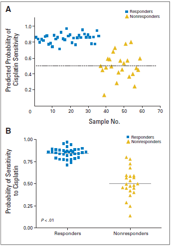
Response to therapy was evaluated using standard criteria for patients with measurable disease, based on WHO guidelines [77]. CA-125 was used to classify responses only in the absence of a measurable lesion and based on established guidelines [78]. A complete response (CR) was defined as a complete disappearance of all measurable and assessable disease or, in the absence of measurable lesions, a normalization of the CA-125 level after salvage therapy. A partial response (PR) was considered a 50% or greater reduction in the product obtained from measurement of each bidimensional lesion for at least 4 weeks or a drop in the CA-125 by at least 50%for at least 4 weeks. Progressive disease (PD) was defined as a 50% or greater increase in the product from any lesion documented within 8 weeks of initiation of therapy, appearance of any new lesion within 8 weeks of initiation of therapy, or a doubling of CA-125 from baseline. For the purposes of our analysis, a clinically beneficial response (ie, “responder”) included CRor PR.Apatient who did not demonstrate aCR or PR was considered a “nonresponder.”
17.3 Cross-Platform affymetrix Gene Chip Comparison
To map the probe sets across various generations of Affymetrix Gene- Chip arrays, we utilized Chip Comparer (http://tenero.duhs.duke.edu/genearray/perl/chip/chipcomparer.pl) as described previously [70,76].
17.4 Cell Proliferation and Drug Sensitivity Assays
Optimal cell number and linear range of drug concentration were determined [62] for each cell line and drug as described previously [70, 76]. For drug sensitivity assay, cells were plated in non–drug-containing media in 96-well plates. After incubation for 24 hours at 37°C, drugs were added to each well at a specific concentration. Cells were grown in the presence of drugs for an additional 96 hours, and sensitivity to cisplatin, docetaxel, paclitaxel, and pemetrexed in the cell lines was determined by quantifying the percent reduction in growth (v dimethyl sulfoxide [DMSO] controls) at 96 hours using a standard MTT colorimetric assay (CellTiter 96 Aqueous One 23 Solution Cell Proliferation Assay Kit; Promega, Madison, WI) [79, 80].
The experimental strategy for analysis employed in that study [62] is similar to that used for the development of oncogenic pathway and chemotherapy sensitivity signatures as described previously [70,76]. Samples representing extreme cases are used to train the expression data to develop a genomic signature that can predict drug sensitivity. A predictor of cisplatin sensitivity was developed by analyzing cell lines described by Györffy et al [81].
Using Bayesian binary regression analysis, genes highly correlated with drug sensitivity were identified and used to develop a model that could differentiate between cisplatin sensitivity and resistance. The developed model consisting of 45 genes based on cisplatin sensitivity (Fig. 21A) was validated in a leave-one-out cross-validation. The cisplatin sensitivity predictor includes DNA repair genes such as ERCC1 and ERCC4, among others, that had altered expression in the list of cisplatin sensitivity predictor genes. Interestingly, one previously described mechanism of resistance to cisplatin therapy results from the increased capacity of cancer cells to repair DNA damage incurred, by activation of DNA repair genes [82, 83].
18.1 Developing a gene expression–based predictor of pemetrexed sensitivity
In NSCLC, where platinum-based therapy is the standard of care, response rates are only 30%. One approach to identifying potential drugs effective in cisplatin-resistant patients is to examine the NCI-60 data set for agents whose IC50 profile showed an inverse relationship with cisplatin, focusing on those known to be effective in NSCLC. Of these drugs, an inverse correlation with cisplatin sensitivity was identified with docetaxel, abraxane, and pemetrexed. The strongest inverse correlation was found between cisplatin and pemetrexed sensitivity (P<.001; Pearson r value, 0.1; α = 0.05).

Using methods previously described [76], a predictor of pemetrexed sensitivity was developed by identifying NCI-60 cell lines that were most resistant or sensitive to pemetrexed. Using Bayesian binary regression analysis, genes whose expression was most highly correlated with drug sensitivity were used to develop a predictive model that could differentiate between pemetrexed sensitivity and resistance. The developed model consisting of 85 genes based on pemetrexed sensitivity (Fig. 21B) was validated in a leave-one-out cross-validation. Interestingly, multiple genes involved in nucleotide and cellular metabolism constituted the pemetrexed sensitivity predictor and is biologically consistent with the known mechanism of pemetrexed sensitivity, which involves interference with cell-cycle progression by reducing the pool of substrates necessary for DNA replication [84].
In addition to initial leave-one-out cross-validation, the true value of a
predictor lies in its ability to predict sensitivity in independent in vitro and in vivo settings. In the present study [62], the predictor of cisplatin sensitivity was independently validated in a panel of 32 (lung and ovarian cancer) cell lines, using cell proliferation assays and concurrent gene expression data. As shown in Figure 22A, the correlation between the predicted probability of sensitivity to cisplatin (in both lung and ovarian cell lines) and the respective IC50 for cisplatin confirmed the capacity of the cisplatin predictor to accurately predict sensitivity to the drug in cancer cell lines.
Similar to the independent validation of the cisplatin sensitivity predictor, the pemetrexed predictor was validated using gene expression data from an independent cohort of 17 NSCLC cell lines with respective in vitro drug sensitivity assays. As shown in Figure 22B, the correlation between the predicted probability of sensitivity to pemetrexed in the 17 NSCLC cell lines and the respective IC50 for pemetrexed validated the ability of the pemetrexed predictor to predict sensitivity to the drug in an independent cohort of cancer cell lines.

19.1 In Vivo validation of the cisplatin sensitivity Predictor
Although the ability of the cisplatin signature to predict sensitivity in independent samples validates the performance of the signature, it is the ability to predict response in patients that is obviously most critical. Using data from a previously published study that linked gene expression data with clinical response to cisplatin in an ovarian data set [70] (GEO accession number: GSE3149), it has been tested the ability of the in vitro cisplatin sensitivity predictor to accurately identify those patients who experience disease response with cisplatin. Using a predicted probability of response of 0.50 as the cutoff for predicting cisplatin sensitivity, the accuracy of the in vitro gene expression–based predictor of cisplatin sensitivity, based on available clinical data, was 83.1% (sensitivity, 100%; specificity, 57%; positive predictive value [PPV], 78%; negative predictive value [NPV], 100%; Fig.23). Furthermore, a Mann-WhitneyUtest revealed a significant difference in the predicted probabilities of cisplatin sensitivity between the resistant and sensitive cohorts of patients (P<.01; Fig. 23).
The cisplatin and pemetrexed predictors were utilized to profile the potential options of using these two drugs in a collection of 91 NSCLC described previously [85] (GEO accession number: GSE3141). These samples were first sorted according to the patterns of predicted sensitivity to cisplatin (Fig. 24A, left panel). The pattern observed indicated that those patients resistant to cisplatin (red) weremoresensitive to pemetrexed (blue). Although the data points in the scatter plot do not appear to be perfectly correlated, this analysis suggests that the relationship was statistically significant (P = .004, log-rank; Fig. 24A, right panel). A similar relationship was also demonstrated in the independent cohort of NSCLC cell lines (Fig.24B), suggesting the possibility of an alternative therapy for treatment of advanced or metastatic NSCLC patients who would be predicted to be platinum resistant. As a comparison, the pemetrexed signature was also applied to the ovarian cancer patient data set. In this analysis [62], however, only two (< 4>

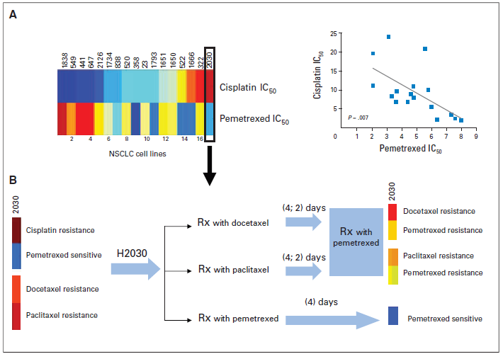
Currently, first-line treatment with a platinum-based regimen is the standard of care for advanced NSCLC. Those patients developing resistance to cisplatin are treated with a taxane, pemetrexed, or erlotinib as second-line options. To explore the effect of cisplatin resistance, as well as prior treatment with potentially ineffective therapies, the IC50 of various lung cancer cell lines to cisplatin and pemetrexed were analyzed and revealed an inverse relationship (Fig. 25A). Thereafter, one NSCLC cell line (H2030) that is resistant to cisplatin, paclitaxel, and docetaxel, but sensitive to pemetrexed on the basis of cell proliferation assays (IC50), was treated with pemetrexed, docetaxel, or paclitaxel in a systematic fashion. Interestingly, when H2030 was first treated for 4days with a taxane (docetaxel or paclitaxel), resistance to subsequent pemetrexed exposure was induced (Fig. 25B). In contrast, when H2030 was first treated with pemetrexed, H2030 was sensitive, as expected (Fig. 25B). Although these in vitro observations are only hypothesis generating at this time, this proof of principle experiment [62] suggests that the sequence of second-line chemotherapy in NSCLCmayprove to be important in determining clinical outcomes. Specifically, in tumors from cisplatin-refractory patients who are also predicted to be resistant to a taxane, treatment with a taxane (docetaxel or paclitaxel) before pemetrexed therapy may induce resistance to subsequent pemetrexed therapy. This preliminary observation, pending further validation, suggests the importance of including genomic-based, disease-specific, treatment prioritization in clinical practice.
In this study [62], the patterns of cisplatin sensitivity observed in our cohort of 91 NSCLC tumors suggests that not all patients may initially respond to first-line cisplatin-based therapy. As described herein, response rates to first-line platinum based therapy is approximately 30%, with median survival between 24 to 31 months [61].We have made use of in vitro drug sensitivity data in cancer cell lines, coupled with Affymetrix expression data, to develop gene expression signatures reflecting sensitivity to cisplatin and pemetrexed. The capacity of these signatures to predict response in independent sets of cell lines and patient studies begins to define a strategy that addresses the potential to identify cytotoxic agents that best match individual patients with advanced NSCLC and other advanced cancers (ovarian cancer). In addition, it can potentially be applied to patients with early-stage NSCLC to predict who may benefit from adjuvant cisplatin-based therapy. However, as promising as these approaches may seem, these strategies need to be first validated in a prospective clinical trial that would evaluate the performance of a genomic signature-based selection as an initial step in the individualized treatment strategy for patients with advanced NSCLC (Fig. 26).

In conclusion, the development of signatures of drug sensitivity provide an opportunity to optimize therapy for patients with NSCLC and perhaps other patients with advanced cancer where cisplatinbased therapy is considered the standard of care.
In this review, an amphiphilic chimeric peptide (Fmoc)2KH7-TAT was designed and synthesized. (Fmoc)2KH7-TAT peptide possessed good capacity of loading DNA and DOX simultaneously. The existence of H7 sequence in (Fmoc)2KH7-TAT peptide led to a much higher DOX release rate from(Fmoc)2KH7-TAT peptide at pH 5.0 than that at pH 7.4. At the same time, H7 sequence endowed the peptide/DNA complexes with well endosome escaping ability. (Fmoc)2KH7-TAT peptide mediated excellent transfection efficacy both in 293T and HeLa cell-lines under serum-free and serum-containing conditions. Besides, (Fmoc)2KH7-TAT micelle exhibited much superiority in transporting gene and drug to the same cells simultaneously and exhibited satisfactory synergistic effect both in vitro and in vivo. The results demonstrated a promising peptide-based micelle nanoplatform for efficient delivery [86-89] of gene and drug simultaneously and synergistic therapy in the realm of tumor treatment.
In summary, a novel ROS-triggered self-accelerating drug release nanosystem by amplifying the intracellular ROS concentration based on the positive feedback strategy was designed for enhanced tumor chemotherapy. It was found that owing to the ROS cleavable TK linkers, this designed nanosystem could deliver the drug to the ROS abundant tumor cells and produce the cell toxicity selectively, without affecting the normal cells. Moreover, both in vitro and in vivo studies proved that in MCF-7 cells, T/D@RSMSNs could not only release DOX and a-TOS initiatively, but also lead to the augmented concentration of intracellular ROS by α-TOS and accelerating release of DOX, displaying more remarkably antitumor activity than the traditional ROS-responsive nanocarriers. This novel ROS-triggered self-accelerating drug release nanosystem with enhanced tumor therapeutic efficiency could provide a general strategy to branch out the applications of existing ROSresponsible DDSs.
Dear Editorial Team, Clinical Medical Reviews and Reports. My experience with the journal was highly positive. The peer-review process was rigorous, constructive, and completed in a timely manner. The reviewers provided valuable comments that helped improve the quality and clarity of our manuscript. The editorial office was professional, responsive, and supportive throughout all stages of the publication process. Communication was clear and efficient, and any questions were addressed promptly. Overall, I found the journal to maintain high scientific standards and an excellent publication workflow. I would be pleased to consider submitting future work to this journal. Best wishes from, Elena Popa.

It was my pleasure to submit my testimonial concerning the Reviewer Board of our Scientific Journal “Brain and Neurological Disorders”. The Reviewers focused on some modifications and their contribution was helpful. The ladies of our Editorial Office were also supported my efforts. It was my honor to have such a co-operation and I am looking forward for more collaboration.

Dear Grace Pierce, Editorial Coordinator of Journal of Clinical Research and Reports, Thank you for the speedy and efficient peer review process. I appreciate the fact that your peer reviewers do not take months to respond like with some other journals. I would also like to thank the editorial office for responding quickly to my questions. It is an excellent journal. I plan to submit more manuscripts in the future. Best wishes from, Robert W. McGee

Dear Grace Pierce, Editorial Coordinator of Journal of Clinical Research and Reports, Working with you and your team on our recent publication in JCRR has been a truly wonderful and enjoyable experience. The responses were prompt, and the reviewers were patient, constructive, and highly professional. One reviewer in particular gave me the feeling that a professor was carefully reading and commenting on my coursework, which was deeply touching. The entire process was straightforward and hassle‑free, with no tedious online forms to complete. I highly recommend this journal. Best wishes from, DR Aibing Rao, Head of R&D

I Appreciate the Opportunity to Share my Experience with the Journal of Clinical Research and Reports. The peer review process was timely and constructive, and the feedback provided helped improve the quality of our manuscript. The editorial office was professional, responsive, and supportive throughout the process, ensuring smooth communication and efficient handling of the submission. Overall, it was a positive experience collaborating with your team.

Dear Mercy Grace, Editorial Coordinator of Obstetrics Gynecology and Reproductive Sciences, We would like to express our gratitude for your help at all stages of publishing and editing the article. The editors of the magazine answer all the necessary questions and help at every stage. We will definitely continue to cooperate and publish other works in the Obstetrics Gynecology and Reproductive Sciences! Best wishes from, Alla Konstantinovna Politova,
