
Case Report | DOI: https://doi.org/10.31579/2690-4861/348
1 Clinical Hospital Centre Sestre Milosrdnice, Clinic for Surgery, Zagreb, Croatia.
2 Private Orthopaedic Surgery Clinic Marinko Erceg Pty. Ltd., Split, Croatia.
3 General Hospital Šibenik, Šibenik, Croatia.
4 Clinical Hospital Center Split, Surgery Clinics, School of Medicine, University of Split, Croatia.
*Corresponding Author: Marinko Erceg, Private Orthopaedic Surgery Clinic, Marinko Erceg Pty. Ltd., Split, Croatia.
Citation: Erceg D., Erceg M., Bečić K., Bekavac J., (2023), Aloarthroplasty of the Hip After Pathological Fracture of the Right Femoral Neck and Right Femoral Dyaphysis and Osteosynthesis after Fracture of the Left Femoral Dyaphysis in 41-Year-Old Female Patient with Polyostotic Fibrous Dysplasia? A Case Report, International Journal of Clinical Case Reports and Reviews, 16(1); DOI:10.31579/2690-4861/348
Copyright: © 2023, Marinko Erceg. This is an open-access article distributed under the terms of the Creative Commons Attribution License, which permits unrestricted use, distribution, and reproduction in any medium, provided the original author and source are credited.
Received: 23 October 2023 | Accepted: 20 November 2023 | Published: 28 November 2023
Keywords: hip arthroplasty; periprosthetic femoral fractures; bone fragments
A 40 years old male patient had surgery on his right hip in 2010. when primary total cementless hip prosthesis was implanted because of posttraumatic arthrosis. Three months later he had a car accident and he got dislocation of the hip prosthesis with multiple femoral fractures of the proximal part of the femur around and below the femoral stem. The stem of the prosthesis was loose while acetaulum was stable. The patient had also fracture of the left acetabulum and ishial bone, without big dislocations. He underwent a new surgery on the right hip. Revisional long uncemented femoral stem was implanted, with cerclages and screws on multiple bone fragments. We met patient 8 years later with a very good function of his right hip. He has a problem with his left hip because of pseudarthrosis of the upper and lower part of the ischial bone. We think that this is an interesting case of periprosthetic femoral fracture, because of so many bone fragments around and below the femoral stem of the hip prosthesis, resulting in a very good function.
After many years of successful total hip arthroplasties (THA), today we have a substantial increase in the incidence of periprosthetic femoral fractures after hip replacement. Postoperative periprosthetic fractures of the femur are mostly located around femoral stem, typically in elderly patients with osteoporosis [1]. These fractures usually occur within days to several years after the procedure. The mean time from primary THA to the fracture is 7.4-8.1 years, and 3.9 years from revisional THA to the fracture. The prevalence of postoperative periprosthetic femoral fractures ranges from 0.1-4% [2]. The treatment of these fractures are mostly surgical. The surgical procedure depends of the site of fracture, number of bone fragments, age of patient, quality of bone, and the fact whether femoral stem is loose or well-fixed?. Today, the most used guide how to treat periprosthetic fractures is according to Vancouver classification [3,4]. Our patient had multiple fractures around and below the femoral stem with complete loose stem; group B3. This type of fracture needs to be treated with revisional long stem prosthesis and reposition and osteosynthesis of all bone fragments [4,5].
Male patient B. B., born 1970. had a traumatic dislocation of the right hip in car accident in 1990. After transosal traction clossed reduction of the right hip was done, but paresis of peroneal nerve existed till today. In March 2010. patient underwent surgery procedure for uncemented THA of the right hip, because of secondary posttraumatic hip arthrosis. Three months after hip arthroplasty the patient had a new car accident and he had traumatic dislocation of the hip prosthesis with periprosthetic comminuted fractures around and below the femoral stem. The stem was completely loose, while the acetabulum was well-fixed (Figure 1).
The patient also had fracture of the left acetabulum and ischial bone (body of ischium and ischial ramus), but without great dislocations of fragments (Figure 2).

Figure 1: X ray of the right hip and proximal femur shows comminuted periprosthetic femoral fractures 3 months after total hip replacement. Figure 2: X ray of the left hip with fractures of acetabulum and ischial bone.
The patient underwent surgery on his right hip and thighbone. Loosened femoral stem of prosthesis was removed. We found many femoral bone pieces, some of them were free of soft tissues and some were very small. Such peaces of bone have to be throwed out. First we implanted revisional uncemented long femoral stem in distal femoral fragment, and all other fragments were placed around femoral stem, keeping them together with seven cerclages and two screws. Some parts of femoral stem were not covered with bone, because some fragments were so small that we could not fixed them (Figure 3,4). Femoral stem was firm in distal fragment and after reposition, prothesis was stabile, but the leg became shorter. In postoperative time everything was going fine, only weight bearing was delayed. Later on we did not see the patient for seven years, because he lives and works outside of Croatia. When we met him again he gave us X rays of his both hips (Figure. 5,6). The patient uses orthopaedic shoes with 2 cm elevation on his right leg. He has paresis of peroneal nerve from first right hip dislocation, 20 years ago. The patient walks good. X ray of the right hip and femur (fig. 5) shows unchanged and good position of hip prosthesis and healed femoral fragments. One part of femoral stem is still uncovered with bone, but patient has not any problem with his right hip. Range of motion and function of the right hip and knee are good (Figure 7,8). The patient has some problems (pain) with his left hip, more in last few months while walking on longer distances and while sitting. X ray of the left hip (figure. 6) shows pseudoarthrosis on the proximal part of ischial bone below acetabulum and ischial ramus. The surgery for the ischial bone is not excluded in the future. Finally, the result after treatment of periprosthetic fractures in this patient is better than we expected. We think that this is an interesting case, with too much bone fragments, which resulted with good function.
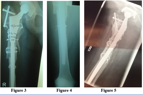
Figure 3: X ray of the right hip and femur bone 4 months after rearthroplasty and osteosynthesis. Figure 4: X ray; long cementless femoral stem. Figure 5: X ray 7 years after rearthroplasty.

Figure 6: X ray of the pelvis 7 years after rearthroplasty of right hip. Pseud. Figure 7: Good function of the right hip and knee joint. oarthrosis of the left ischial bone (ischial body and ischial ramus is seen). Figure 8: Good function of the right hip and knee joint.
The author declares no competing interests.
Dear Editorial Team, Clinical Medical Reviews and Reports. My experience with the journal was highly positive. The peer-review process was rigorous, constructive, and completed in a timely manner. The reviewers provided valuable comments that helped improve the quality and clarity of our manuscript. The editorial office was professional, responsive, and supportive throughout all stages of the publication process. Communication was clear and efficient, and any questions were addressed promptly. Overall, I found the journal to maintain high scientific standards and an excellent publication workflow. I would be pleased to consider submitting future work to this journal. Best wishes from, Elena Popa.

It was my pleasure to submit my testimonial concerning the Reviewer Board of our Scientific Journal “Brain and Neurological Disorders”. The Reviewers focused on some modifications and their contribution was helpful. The ladies of our Editorial Office were also supported my efforts. It was my honor to have such a co-operation and I am looking forward for more collaboration.

Dear Grace Pierce, Editorial Coordinator of Journal of Clinical Research and Reports, Thank you for the speedy and efficient peer review process. I appreciate the fact that your peer reviewers do not take months to respond like with some other journals. I would also like to thank the editorial office for responding quickly to my questions. It is an excellent journal. I plan to submit more manuscripts in the future. Best wishes from, Robert W. McGee

Dear Grace Pierce, Editorial Coordinator of Journal of Clinical Research and Reports, Working with you and your team on our recent publication in JCRR has been a truly wonderful and enjoyable experience. The responses were prompt, and the reviewers were patient, constructive, and highly professional. One reviewer in particular gave me the feeling that a professor was carefully reading and commenting on my coursework, which was deeply touching. The entire process was straightforward and hassle‑free, with no tedious online forms to complete. I highly recommend this journal. Best wishes from, DR Aibing Rao, Head of R&D

I Appreciate the Opportunity to Share my Experience with the Journal of Clinical Research and Reports. The peer review process was timely and constructive, and the feedback provided helped improve the quality of our manuscript. The editorial office was professional, responsive, and supportive throughout the process, ensuring smooth communication and efficient handling of the submission. Overall, it was a positive experience collaborating with your team.

Dear Mercy Grace, Editorial Coordinator of Obstetrics Gynecology and Reproductive Sciences, We would like to express our gratitude for your help at all stages of publishing and editing the article. The editors of the magazine answer all the necessary questions and help at every stage. We will definitely continue to cooperate and publish other works in the Obstetrics Gynecology and Reproductive Sciences! Best wishes from, Alla Konstantinovna Politova,
