
Case Report | DOI: https://doi.org/10.31579/2690-8794/074
1 Radiology Department, Usmanu Danfodiyo University, Sokoto.
2 Radiology Department, Gombe State University, Gombe.
3 Radiology Department, Usmanu Danfodiyo University Teaching Hospital, Sokoto
*Corresponding Author: Sule Muhammad Baba, Department of Radiology, Usmanu Danfodiyo University, Sokoto.
Citation: Sule M.B. Umar AU., Gele I.H., Umar F.K., Uzoma Gu. and Aliyu A.Z. (2021) Pericardial calcification with plain radiographic features of tuberculosis in a child: incidental finding and a case report. Clinical Medical Reviews and Reports 3(4); DOI: 10.31579/2690-8794/074
Copyright: © 2021 Sule Muhammad Baba, This is an open access article distributed under the Creative Commons Attribution License, which permits unrestricted use, distribution, and reproduction in any medium, provided the original work is properly cited.
Received: 02 March 2021 | Accepted: 08 March 2021 | Published: 22 March 2021
Keywords: tuberculosis; pericarditis; constrictive; calcification
Tuberculous pericarditis is frequently reported as the primary cause of pericardial calcification and occurs in about 1-2% of individuals with pulmonary tuberculosis, this however is a rare finding in the Western world. This is a 12-year-old male child that was referred from a peripheral health care center for plain radiograph of the chest on account of cough, easy fatiguability, night sweats, loss of weight, loss of appetite and dyspnea most times on excersion for more than a month duration.
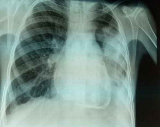
The plain chest radiograph demonstrated normal cardiac size with a cardiothoracic ratio of about 55/120, there is circumferential radio-opacity of calcic density around the peripheral walls of the heart; the pericardial calcification. The vascular pedicle appears slightly widened. The lung fields show extensive streaky opacities with cystic lung changes bilaterally more marked on the left lung field where consolidation, loss of lung volume and pleural effusion are also demonstrated. A two-dimensional echocardiography showed mild-moderate pericardial effusion, thickening of both visceral and parietal pericardium, and echogenic fond-like structures protruding in to the pericardial cavity. A diagnosis of pulmonary tuberculosis with features of tuberculous pericarditis in a 12-year-old male child was established. The patient has been placed on anti-tuberculous drugs, hematinic, and parents advised on good and adequate diet with adequate rehydration and strict drug compliance. Screening of the siblings and members of the family with close contact have also been emphasized. We report the radiographic features of pulmonary tuberculosis and pericardial calcification in a 12-year-old male child due to its peculiar presentation.
Pericardial calcification is present in less than 25% of all cases of constrictive pericarditis, with the etiology of pericardial calcification been uncertain most times [1, 2]. The true incidence of PC happens to be unknown [3, 4 and 5]. Tuberculosis is regarded a leading cause of pericarditis in some non-industrialized countries, in the United States however, it accounts for about 10% of cases of chronic constrictive pericarditis [6-8].
Tuberculous pericarditis presents clinically in three forms, these are: pericardial effusion, constrictive pericarditis, and a combination of constriction and effusion [9, 10]. Tuberculous involvement of the heart as form of extrapulmonary tuberculosis is regarded as the second commonest from the central nervous involvement especially with respect to morbidity and mortality [11].
Constrictive pericarditis (CP) is a relatively rare condition that is often caused by pericardial dysfunction following varying etiologies, and results in impaired cardiac function and associated with diastolic heart failure [11,12]. The presence of calcification often supports the diagnosis of CP, but not all the patients with CP have pericardial calcification [13, 14 and15]. Previously the most common cause of CP is tuberculosis, however in modern terms, the common causes are open heart surgery and mediastinal irradiation [16]. Constrictive pericarditis is often caused by fibrosis and calcification of the pericardium, leading to loss of normal elasticity of the pericardium, these causes decreased diastolic filling of the heart, and right heart failure [13, 14].
Pericardial calcification as a lone entity is often asymptomatic, however, signs and symptoms can evolve due to underlying disease processes such as CP, it however may (PC) occur in the absence of any constrictive physiology [17].
Pericardial calcifications are regarded as important sign of constrictive pericarditis which can be diagnosed and also confirmed following imaging, among which are computed tomography (CT), echocardiography and cardiac magnetic resonance imaging, however CT is a useful guide in the management and diagnosis of CP [13-15,18]. Pericardial calcification on plain chest radiograph is highly suggestive of CP, the presence of PC on chest radiographs frequently implicates CP as the cause of symptoms irrespective of the extent of pericardial thickening [13, 19 and 20].
This is a 12-year-old male child that was referred from a peripheral health care center for plain radiograph of the chest on account of cough, easy fatiguability, night sweats, loss of weight, and loss of appetite, weakness, occasional fever, chest pain and dyspnea most times on excersion for more than a month duration of onset.
The patient is the 6th child in a non-nuclear family of 14 children, the father is a farmer while the mother a housewife and not gainfully employed.
The patient is conscious and alert, mildly pale, mild dehydration, mildly icteric, acyanosed, mild respiratory difficulty, no significant pedal and facial edema, no finger clubbing, but has mild elevation of the jugular venous pulse.
The blood pressure is about125/90mmHg, pulse rate is about 86beats per minute, and respiratory rate is also about 28cycles per minute. Reduced heart sound intensity, audible pericardial rub with reduced breath sounds bilaterally more marked on the left lung field distally.
The sputum gram stain showed gram-negative Mycobacterium tuberculosis, the erythrocyte sedimentation rate (ESR) was also elevated; 17mm/hr.
The plain chest radiograph demonstrated normal cardiac size with a cardiothoracic ratio of about 55/120, there is circumferential radio-opacity of calcic density around the peripheral walls of the heart; the pericardial calcification. The vascular pedicle appears slightly widened; see figure1a.
The lung fields show extensive streaky opacities with cystic lung changes bilaterally more marked on the left lung field where consolidation with increased opacification is demonstrated on the mid and lower zones with non-demonstration of the left costophrenic sulci in keeping with loss of lung volume and effusion. Compensatory hyperinflation noted on the left upper zone and the right lung field. The bony thorax and overlying soft tissue outline show normal appearances; see figure 1b.
A two-dimensional echocardiography showed mild-moderate pericardial effusion, thickening of both visceral and parietal pericardium, and echogenic fond-like structures protruding in to the pericardial cavity.
A diagnosis of pulmonary tuberculosis with features of tuberculous pericarditis in a 12-year-old male child was established.
The patient has been placed on anti-tuberculous drugs (rifampicin, isoniazid, ethambutol and pyrazinamide for a minimum of six months), corticosteroid for prevention of CP, hematinic, and parents advised on good and adequate diet with adequate rehydration and strict drug compliance. Screening of the siblings and members of the family with close contact have also been emphasized.

Tuberculous involvement of the heart as form of extrapulmonary tuberculosis is regarded as the second commonest from the central nervous involvement especially with respect to morbidity and mortality11. The index case had pulmonary tuberculosis with involvement of the heart, no feature to suggest central nervous involvement, thereby conforming to this literature.
Pericardial tuberculosis is regarded as a chronic fibrinous pericarditis characterized by granulomatous inflammation and caseous necrosis [9, 21]. The index case was confirmed of having the gram-negative Mycobacterium tuberculosis, which is a granulomatous inflammation, thereby conforming to these literatures.
Tuberculous pericarditis presents clinically in three forms, these are: pericardial effusion, constrictive pericarditis, and a combination of constriction and effusion [9, 10]. The case under review had pericardial effusion and features of constrictive pericarditis (CP), thereby conforming to these literatures.
Pericardial calcification as a lone entity is often asymptomatic, however, signs and symptoms can evolve due to underlying disease processes such as CP, it however may (PC) occur in the absence of any constrictive physiology [17]. The index case had features of CP and not asymptomatic.
Pericardial calcifications are regarded as important sign of constrictive pericarditis which can be diagnosed and also confirmed following imaging, among which are computed tomography (CT), echocardiography and cardiac magnetic resonance imaging, however CT is a useful guide in the management and diagnosis of CP [13-15,18]. The case under review was not an exception, he had chest radiograph and echocardiography, thereby conforming to these literatures.
Pericardial calcification can be detected on chest radiograph in almost 50% of cases of CP, though extensive PC can be present without signs and symptoms of CP, there are varying causes of PC, tuberculosis has historically been the leading cause worldwide [22]. The index case was a confirmed case of tuberculosis, presented with plain chest radiographic features of PC and clinical signs and symptoms of tuberculosis and tuberculous pericarditis thereby conforming to this literature.
Echocardiography has been regarded as a sensitive diagnostic imaging modality for detecting pericardial effusion and intrapericardial abnormalities [23], the index case also benefitted from echocardiography, this demonstrated pericardial effusion, thickening and calcification of the pericardium, thereby conforming to these literatures.
Constrictive pericarditis results from fibrous thickening of the pericardium, this is most often accompanied with calcification thereby resulting in affectation of diastolic filling of the heart [24, 25], and the index case had pericardial calcification with features of reduced diastolic filling of the heart, thereby conforming to these literatures.
In tuberculous pericarditis, treatment is aimed at achieving three goals, these are eradicating the active Mycobacterium, relief of cardiac compression with adverse hemodynamic sequelae, and thirdly prevention of complications of maladaptive remodeling and healing, CP inclusive26. Similar treatment modalities which include institution of antibacterial agents, corticosteroid for CP, institution of hematinic, adequate rehydration and close monitoring of patient and contact tracing were employed in the index case, thereby conforming to this literature.
Tuberculosis is regarded endemic in developing countries, patients presenting with features of pulmonary tuberculosis should be investigated by imaging and adequately monitored to prevent occurrence of extrapulmonary affectation like pericardial involvement, and this will reduce morbidity and mortality associated with such complications.
Dear Editorial Team, Clinical Medical Reviews and Reports. My experience with the journal was highly positive. The peer-review process was rigorous, constructive, and completed in a timely manner. The reviewers provided valuable comments that helped improve the quality and clarity of our manuscript. The editorial office was professional, responsive, and supportive throughout all stages of the publication process. Communication was clear and efficient, and any questions were addressed promptly. Overall, I found the journal to maintain high scientific standards and an excellent publication workflow. I would be pleased to consider submitting future work to this journal. Best wishes from, Elena Popa.

It was my pleasure to submit my testimonial concerning the Reviewer Board of our Scientific Journal “Brain and Neurological Disorders”. The Reviewers focused on some modifications and their contribution was helpful. The ladies of our Editorial Office were also supported my efforts. It was my honor to have such a co-operation and I am looking forward for more collaboration.

Dear Grace Pierce, Editorial Coordinator of Journal of Clinical Research and Reports, Thank you for the speedy and efficient peer review process. I appreciate the fact that your peer reviewers do not take months to respond like with some other journals. I would also like to thank the editorial office for responding quickly to my questions. It is an excellent journal. I plan to submit more manuscripts in the future. Best wishes from, Robert W. McGee

Dear Grace Pierce, Editorial Coordinator of Journal of Clinical Research and Reports, Working with you and your team on our recent publication in JCRR has been a truly wonderful and enjoyable experience. The responses were prompt, and the reviewers were patient, constructive, and highly professional. One reviewer in particular gave me the feeling that a professor was carefully reading and commenting on my coursework, which was deeply touching. The entire process was straightforward and hassle‑free, with no tedious online forms to complete. I highly recommend this journal. Best wishes from, DR Aibing Rao, Head of R&D

I Appreciate the Opportunity to Share my Experience with the Journal of Clinical Research and Reports. The peer review process was timely and constructive, and the feedback provided helped improve the quality of our manuscript. The editorial office was professional, responsive, and supportive throughout the process, ensuring smooth communication and efficient handling of the submission. Overall, it was a positive experience collaborating with your team.

Dear Mercy Grace, Editorial Coordinator of Obstetrics Gynecology and Reproductive Sciences, We would like to express our gratitude for your help at all stages of publishing and editing the article. The editors of the magazine answer all the necessary questions and help at every stage. We will definitely continue to cooperate and publish other works in the Obstetrics Gynecology and Reproductive Sciences! Best wishes from, Alla Konstantinovna Politova,
