
Case Report | DOI: https://doi.org/10.31579/2690-4861/501
1Government Medical College, Sangareddy Telangana, 502001, India.
2Head, Dept. of Physiology, Faculty of Dentistry, Jamia Millia Islamia, New Delhi-100025, India.
3Head, Department of Epidemiology and Public Health, National Institute of TB and Respiratory Diseases, Sri Aurobindo Marg, New Delhi-110030, India.
4Division of Clinical Research, School of Biomedical Sciences, Galgotias University, Uttar Pradesh, 203201, India.
*Corresponding Author: Krishna Mohan, Division of Clinical Research, School of Biomedical Sciences, Galgotias University, Uttar Pradesh, 203201, India.
Citation: Salik Umer Khayyam, Sazina Muzammil, Khalid Umer Khayyam, Krishna Mohan, (2024), Penile tuberculosis mimicking as carcinoma in a 56-year-old male - a case report, International Journal of Clinical Case Reports and Reviews, 18(4); DOI:10.31579/2690-4861/501
Copyright: © 2024, Krishna Mohan. This is an open-access article distributed under the terms of the Creative Commons Attribution License, which permits unrestricted use, distribution, and reproduction in any medium, provided the original author and source are credited.
Received: 06 July 2024 | Accepted: 12 July 2024 | Published: 19 July 2024
Keywords: penile tuberculosis; non-healing ulcer; partial penectomy; antitubercular treatment
Penile tuberculosis is an extremely rare clinical condition. It can either be secondary (infection spreading from other organs) or primary (local spread). Reports have demonstrated its presentation as a subcutaneous nodule with or without superficial ulceration which may be clinically interpreted as advanced cancer of the penis. A 56-year-old man from New Delhi area, India presented to a tertiary care hospital with a history of unhealing growth/lesion over the penile shaft and glans for over 6 months. The lesion originally started as a small ulcer on the dorsal aspect of the glans penis which enlarged progressively. A biopsy, followed by a wedge biopsy was performed but the results were inconclusive. The patient was then subjected to partial penectomy after proper counselling. The histopathology from the partial penectomy specimen suggested signs of tubercular granuloma. The patient was advised antitubercular treatment (ATT) and he then reported to our tertiary care centre for treatment initiation. Treatment was completed successfully with no sign of any recurrence.
Tuberculosis (TB) is an infectious disease caused by Mycobacterium tuberculosis. A small percentage of cases of TB are of extrapulmonary tuberculosis (EPTB) (10%–15%) , with lymph nodes being the most common site.1 2-10% of cases of extrapulmonary tuberculosis in developed nations are of genitourinary tuberculosis while its prevalence in developing nations is between 15% and 20%.2 Among genitourinary tuberculosis cases, penile tuberculosis is a very rare form, even in the developing countries.3 Because it can mimic a number of conditions, it is typically diagnosis of exclusion. We report a case of primary tuberculosis of the glans masquerading as carcinoma in an immunocompetent 56-year-old male which began as a small ulcer on the dorsal portion of the glans penis and gradually progressed with time.
The patient, a 56-year-old married man presented to a tertiary care hospital with a history of slow growing painless ulcer on the dorsum of the glans penis more than last 6 months (Figure 1).
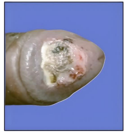
Figure 1: Primary tuberculosis of the glans penis showing ulcerated growth over dorsal part of the glans.
The patient was of average built and nourishment and worked at a steam press laundry facility at a garment export factory. He had type 2 diabetes mellitus controlled by regular medication and insulin and no history of tuberculosis. At the time of examination, the ulceration was about 2 c.m. X 2.5 c.m. on the dorsum of the glans which clinically looked like carcinoma of the penis. Inguinal lymph nodes were mildly enlarged and non-tender. Systemic examination did not reveal any abnormalities. The results of routine laboratory testing, such as CBC, urine analysis, liver and kidney function tests were all within normal ranges. Chest X-ray was also normal. Serology showed that human immunodeficiency virus, hepatitis B surface antigen and syphilis were nonreactive. Quantiferron TB Assay was found to be negative. Contrast-enhanced Magnetic Resonance Imaging (CE-MRI) of the pelvis was done which showed evidence of a poorly defined lesion ∼20X26.7X9.9 mm in dorsum of the distal penile shaft reaching up to the glans, seen to lie in the subcutaneous plane, showing heterogenous enhancement and having a non-enhancing necrotic centre with possibility of mitotic aetiology.
A biopsy and a following wedge biopsy of the ulcer was done (Figure 2).
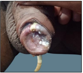
Figure 2: Image taken after wedge biopsy/mass excision was performed.
which were both sequentially inconclusive. ZN stain for AFB, tissue culture and GeneXpert for MtB/RIF were all negative. The patient was counselled about the need for further intervention in the form of partial
penectomy and histopathological examinations (HPE). The patient was admitted at the tertiary care hospital and operated under spinal anaesthesia (SA). Partial penectomy was done and corpora closed in continuous fashion. Urethra opened ventrally and was sutured with skin (Figure 3).

Figure 4: Image from post ATT of 6 months showing healing of the site with no signs of recurrence.
Despite genitourinary TB being a common form of extrapulmonary TB, penile presentation is one of its least common forms (<1>
When there is non-healing ulceration in the glans, tuberculosis should be taken into consideration, especially in nations where the disease is prevalent. Biopsies are necessary to rule out a malignant penile tumour and a chest X-ray and physical examination are required to ascertain whether a TB of the glans penis is a primary or secondary disease. Antitubercular medications are the cornerstone of therapy, although in questionable circumstances, surgery might be necessary. Treatment should aim for organ sharing surgery with fashioning of the penis and antitubercular therapy. Penile excisions are linked to a significant psychological impact, hence patient counselling is crucial.
The authors are highly grateful to the Department of Tuberculosis and Respiratory Diseases, National Institute of Tuberculosis and Respiratory Diseases, New Delhi for providing necessary logistics and support for the case study.
Ethical approval is not required for this study following local or national guidelines.
Both written and verbal consent were taken from the patient before communicating this case report.
The authors have no conflicts of interest to declare.
Dear Editorial Team, Clinical Medical Reviews and Reports. My experience with the journal was highly positive. The peer-review process was rigorous, constructive, and completed in a timely manner. The reviewers provided valuable comments that helped improve the quality and clarity of our manuscript. The editorial office was professional, responsive, and supportive throughout all stages of the publication process. Communication was clear and efficient, and any questions were addressed promptly. Overall, I found the journal to maintain high scientific standards and an excellent publication workflow. I would be pleased to consider submitting future work to this journal. Best wishes from, Elena Popa.

It was my pleasure to submit my testimonial concerning the Reviewer Board of our Scientific Journal “Brain and Neurological Disorders”. The Reviewers focused on some modifications and their contribution was helpful. The ladies of our Editorial Office were also supported my efforts. It was my honor to have such a co-operation and I am looking forward for more collaboration.

Dear Grace Pierce, Editorial Coordinator of Journal of Clinical Research and Reports, Thank you for the speedy and efficient peer review process. I appreciate the fact that your peer reviewers do not take months to respond like with some other journals. I would also like to thank the editorial office for responding quickly to my questions. It is an excellent journal. I plan to submit more manuscripts in the future. Best wishes from, Robert W. McGee

Dear Grace Pierce, Editorial Coordinator of Journal of Clinical Research and Reports, Working with you and your team on our recent publication in JCRR has been a truly wonderful and enjoyable experience. The responses were prompt, and the reviewers were patient, constructive, and highly professional. One reviewer in particular gave me the feeling that a professor was carefully reading and commenting on my coursework, which was deeply touching. The entire process was straightforward and hassle‑free, with no tedious online forms to complete. I highly recommend this journal. Best wishes from, DR Aibing Rao, Head of R&D

I Appreciate the Opportunity to Share my Experience with the Journal of Clinical Research and Reports. The peer review process was timely and constructive, and the feedback provided helped improve the quality of our manuscript. The editorial office was professional, responsive, and supportive throughout the process, ensuring smooth communication and efficient handling of the submission. Overall, it was a positive experience collaborating with your team.

Dear Mercy Grace, Editorial Coordinator of Obstetrics Gynecology and Reproductive Sciences, We would like to express our gratitude for your help at all stages of publishing and editing the article. The editors of the magazine answer all the necessary questions and help at every stage. We will definitely continue to cooperate and publish other works in the Obstetrics Gynecology and Reproductive Sciences! Best wishes from, Alla Konstantinovna Politova,
