
Research Article | DOI: https://doi.org/10.31579/2766-2314/110
1 Kerala Veterinary and Animal Sciences University,
2 CRP-6, Rajiv Gandhi Centre for Biotechnology
*Corresponding Author: Priya Srinivas, CRP-6, Rajiv Gandhi Centre for Biotechnology.
Citation: Krithiga Kuppusamy, Arathi Rajan, Geetu R. Varghese, Neetha R. Latha, Priya Srinivas, et all, (2023), Pathological and Molecular Analysis of Spontaneous Canine Mammary Carcinomas and its Prognostic implications, J, Biotechnology and Bioprocessing, 4(5); DOI:10.31579/2766-2314/110
Copyright: © 2023, Priya Srinivas. This is an open access article distributed under the Creative Commons Attribution License, which permits unrestricted use, distribution, and reproduction in any medium, provided the original work is properly cited.
Received: 27 June 2023 | Accepted: 06 July 2023 | Published: 17 July 2023
Keywords: canine mammary carcinomas; cytological grading; cancer stem cells; cancer model
Purpose: The canine mammary tumours (CMT) and human breast cancers (HBC) are postulated to resemble each other in genesis, progression, presentation and prognostication. Thus, studies involving naturally occurring CMT may aid in better understanding of HBC. The study also aims at replicating the techniques used to study the HBC in CMT and to find whether the canine model can be utilized for HBC research and also provide diagnostic methods for patients with CMT.
Methods: Samples from spontaneous CMT cases were collected and a cohort of canine mammary carcinomas (CMC) was utilised for this study after histopathological examination and grading. Immunophenotyping and identifying the cancer stem cells (CSC) which are the most acclaimed cause of recurrence, metastasis, and treatment failures in CMC was performed by using suitable markers.
Results: Expression of CD44+/24-/low CSC phenotype, CD24 overexpression, ALDH1 in higher grades, decreased E cadherin and increased N cadherin in recurrence/ metastasis were observed by immunohistochemistry. The qRTPCR results showed increased Oct-4, Sox-2, Nanog expression in higher grades of tumours, while the E and N cadherin switch was observed in recurrent/ metastatic cases. A survival analysis of a 36 months follow-up study revealed that prognosis was poor in patients with higher grades and in CMC with CD44+/24-/low or CD24 overexpression.
Conclusion: It could be deciphered from the study that the human and canine breast cancers share common diagnostic and prognostic signatures and can serve as better model to study the human disease.
Canine mammary neoplasms are the second most common cancer reported in intact aged female dogs next to skin cancer. The domesticated canines are exposed to similar external predisposing factors of carcinogenesis as that of humans as they share similar environment (Reif., 2011). Thus, dogs are supposed to be suitable breast cancer model to study mechanism of tumourigenesis. The naturally occurring CMT are a heterogeneous group of cancers which have almost similar characteristics of HBC (Gray et al., 2020).
The clinico-pathological similarities between CMT and HBC characteristics such as spontaneous occurrence, onset of the disease, hormonal aetiology and course of disease is discernible. The molecular characteristics like expression of steroid receptor, epidermal growth factor expression, proliferation markers, metalloprotienases and cyclo-oxygenase overexpression and mutation rate of p53 mutation is similar in CMT and HBC (Quierago et al., 2011).
The histological evidence proves that there is remarkable resemblance in the origin, distribution, and behaviour of mammary neoplasia in humans and canines. the neoplastic epithelial cells proliferate and invade through the walls of the glandular ductules into the surrounding stroma in simple tubular carcinoma and invading ductal carcinoma in CMT and HBC, respectively. It is noteworthy that HBC and CMT exhibit features like individual and clustered neoplastic cells exhibiting desmoplasia and invading the lymphatics; necrosis and dissolution of the tubules in the center of the lesion as in comedeo-carcinoma and mucin secreting neoplastic cells in mucinous carcinoma (Al-Mansour et al., 2018).
The tumour behaviour such as the risk of developing new primary tumors in canines as stated by Sorenmo et al., 2009, was significantly greater in cases with previous malignancy and the same picture is depicted in women with earlier malignancy, pre-malignant and benign lesions which were observed by Berstein et al. (2003), while studying the epidemiology of HBC in women.
Identification of breast cancer stem cells (BCSCs) has redefined the concept of cellular origin, of tumours and its maintenance and progression. They are responsible for tumour relapse by their relative resistance to drugs (Liu and Wicha, 2010) and hence, are identified as better targets for therapeutic intervention. Similarly, studies linking CSCs in CMT utilizing canine mammary cell lines have proved the presence of such cells resulting in chemo and radio-resistance (Pang et al., 2011).
There is paucity of literature in the area of linking CSC and prognosis in spontaneous cases of canine mammary carcinomas (CMC), which is the commonly occurring cancer phenotype. The current study aimed at studying the spontaneous cases and compared with HBC to identify the analogy between them. This would help in the treatment of canine cancer patients and also to identify the possibility of naturally occurring CMC to act as better models to study HBC.
Sample collection and follow-up study
The samples from fifty-five cases of spontaneous CMC were collected during the study. Samples were immediately collected in 10% Neutral buffered formalin (NBF) after surgical removal with owner’s consent from Veterinary Institutions across Kerala. The tumour samples were collected immediately after surgical excision with the owner’s consent. Preserved and paraffin embedded tissues (PET), and FNA smears were shared by Veterinary Institutions in Tamil Nadu and Kerala. The samples were examined and thirty carcinoma cases were included in the cohort under study. The cases were followed-up for a period of 36 months between November 2018 to April 2022. The animals which died due to causes other than recurrence/ metastasis was censored from the study. The study subjects did not receive any chemo or radio-therapy except for surgical resection of the tumour mass. The cases were categorized based on grades, histopathological appearance and hormonal receptor status.
Histopathology and grading
The tissue samples collected in 10% NBF were further processed and 4–5µm sections were taken for histopathology studies. The tissue sections were stained with Haematoxylin and Eosin(Suvarna et al., 2018). Histopathological examination was done to identify and grade the carcinomas as Grade I (GI), Grade II (GII) and Grade III (GIII) based on Goldschmidt et al., 2011.
Immunohistochemical analysis of the tissue samples
Tissue sections of 4 µm thickness were deparaffinized and antigen-retrieval was done using citrate buffer containing 0.147g of Trisodium citrate in 50 ml of distilled water (pH 6) and Tri EDTA buffer containing 1.21g of Tris and 0.15g of ETA in 100 ml of distilled water (pH 9). Heat based antigen retrieval was performed for 15–20 minutes using standard protocol. The PET sections were subjected to IHC using various primary antibodies (Table 1),
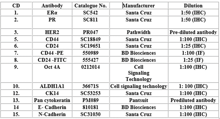
Table 1: Details of antibodies used in this study
Table 2: Primers used for qRT-PCR
polyexcel HRP/DAB detection system-one step from Pathnsitu biotechnologies for signal detection and Meyer’s haematoxylin as counterstain along with corresponding controls. The stained sections were scored using standard procedures. The scoring was done as follows 0 = No positive staining, < 10%- score 1, 10–50% of positive cells-score 2, 51–75% of positive cells-score 3, > 75% of positive cells-score 4. Score of 1 is considered as low staining. (Figueroa et al., 2015). The scoring of the hormonal receptors was based on Allred scoring system (Allred et al., 1998), Her2 (Koeppen et al., 2001). The ERp method of calculating the immunoreactivity in canine mammary tumours by Nieto et al. (2000) was used for ascertaining the ERα positivity.
qRT-PCR for CSC markers
The mRNA expression level of the CSC markers was evaluated using suitable qRT-PCR primers (Table.2) designed using Gene Walker software and Pang et al. (2011). The extraction of mRNA was done by Trizol method and cDNA was synthesized using the Applied 106 Biosystem cDNA synthesis kit. The reaction was carried out using 1µl (50 ng) of cDNA per 10 µl reaction.
Statistical analysis was carried out using Graph pad prism version 8 and Microsoft excel software. Log rank (Mantel-cox) test was used to find the significance in the survival analysis. Mann-Whitney U test and Wilcoxon test were performed to find significance of expression of CSC markers. Statistical significance represented as (*) if p ≤ 0.05, (**) ≤ 0.01, (***) ≤ 0.001. The quantitative data are taken from experiments conducted in triplicates and expressed as mean ± standard deviation (S.D). Error bars were given based on calculated S.D values.
Patient particulars
Animals in the age group of 6–11 years had higher chances for predisposition to develop mammary tumours. This study had more of larger breeds of animals like Labrador and German- Shepherd (14 nos.) with CMCs while the smaller breeds like Spitz and Dachshund (10 nos.) and non-descript (mongrels) animals (6 nos.) were less in number. Nearly 90% of the animals were not spayed (ovariohysterectomy) indicating an active reproductive cycle under the influence of hormones. The average size of the tumours used in the study was between 5–10 cm in diameter as measured using vernier calipers.
Histopathological presentation and grouping of the CMCs
The plethora of presentations in CMCs makes the process of analysis a difficult task unless otherwise, they are grouped into defined categories. This classification and grouping were done as per Goldschmidt et al., 2011 and Rogez et al., 2019. The CMC taken fell into three groups (Supplement. 1) with 11 cases of tubular simple, 4 cases of tubular complex and 15 cases of solid carcinomas (Figure. 1a-c).
Figure 1: This grouping facilitates the study of prognosis based on the different histological phenotypes which are also considered important in cases of HBC patients.
Hormonal receptor and triple negative status in CMC
The hormonal receptor status of ERα, PR along with the HER2 analysis are important factors that help to decide the treatment regimen for HBCs. Most of the CMC are triple negative 73.3Percentage (ER- /PR-/ HER2-); 10Percentage belong to luminal A (ER+/PR+/-/HER2-); 13.3Percentage luminal B (ER+/PR+/-/HER2+) and 3.33% of HER2 alone (ER-/PR-/-/HER2+). The IHC analysis data is provided in Supplement.2. The triple negative cases were subjected to Pancytokeratin and CK14 immunostaining which indicated that 64% of the CMCs had basal like properties (Fig. 1d-h). ERα expression was noticed in the nuclei of neoplastic cells in 23.3Percentage of CMCs under study and cytoplasmic staining could also be observed in some cases. PR expression was noticed only in 23.3Percentage in the cohort under study. The positive immunostaining was confined to lesser number of cells for the hormonal receptors. Her-2 immunoreaction was observed as a membranous and cytoplasmic reaction. The association between the increased tumour size and hormonal receptors is analysed in the CMC between the masses which are less than 5cm in diameter and greater than 5cm in diameter. A highly significant association between the tumour size and the ERα and PR expression is appreciated (p = 0.0078**) (Supplement 3). The results suggest that as the tumour size increases the dependency of the tumours on the hormonal stimulation decreases and they become independent.
Expression of CSC in CMC
IHC was performed on serial sections of the PET tissue samples for the cohort of CMC and evaluated. It is found that 27Percentage of the samples expressed the CD44+/24−/low phenotype, 7% has CD44+/24+ phenotype and 30% has the CD24 overexpressing (CD44−/low/ CD24++/+++) phenotype (Fig. 2a-f)

Figure 2
irrespective of the hormonal receptor/ HER2 based immunophenotype. The IHC analysis data is provided in Supplement.4. It is also note-worthy that though all the above expression of the surface markers is encountered in the GI or GII/III tumours; most of them are triple negative. However, all the triple negative CMCs (TNCMCs) do not express the markers. The widely studied CSC phenotype of CD44+/24−/low is observed in TNCMCs and luminal B CMCs with a predominance of TNCMCs. The transcription factors Oct-4, Sox-2 and Nanog are said to send continuous signals of self-renewal that help the CSC maintenance and proliferation. The qRT-PCR results indicate that the expression of Oct-4, Sox-2 and Nanog are up regulated in the GII/III tumours than GI (Figure. 2g-i). Immunohistochemical analysis of the GII/III carcinomas show higher expression of Oct-4A than GI (Figure. 2j, k). It indicates that the chances for presence of the CSC in CMC are more in higher grades of tumour.
Characterisation of the E and N Cadherin switch in CMC
The cadherin adhesion system which plays a major role in cell adhesion and migration indicate that the carcinomas that can undergo metastasis have a higher expression of N-cadherins than the E-cadherins (E and N Cadherin switch). The mRNA expression levels of the above cadherins did not suggest a switch when compared against the grades but showed such a phenomenon when metastasis/ recurrence potential of the carcinomas is considered (Figure. 3a, b).

Figure 3
The E and N cadherin switch was better observed in the cases with recurrence where a decrease in the protein expression of E cadherin and increase in protein expression of N cadherin could be noticed (3c, d). Hence, the E and N cadherin switch is a key factor in predicting the recurrence / metastatic potential of CMC. The data pertaining to E-Cadherin: NM/NR (Non-Metastatic/Non-Recurrent vs R/M (Recurrent/ Metastatic), (2) N-Cadherin: NM/NR vs R/M and (3) N/E ratio for each sample for NM/NR and R/M is provided in Supplement.4. It was found that there is significant difference (p = 0.0020**) between the E-cadherin expression in NM/NR vs R/M, while the N-cadherin expression though higher in R/M than in NR/NM did not show any statistical significance (p = 0.2717ns).
Significance of ALDH1A1 in CMCs as a prognostic marker
The ALDH1A1 expression level is one of the widely studied CSC marker in HBC. The IHC analysis carried out with the ALDH1A1 which is said to be a specific CSC marker, revealed that the GII/III recurrent carcinomas express higher levels of the marker when compared against the GI, indicating the excess presence of CSC population in higher grades of the tumours with recurrence (Fig. 3e, f). Most of the tumours analysed for this marker are triple-negative and basal like. The IHC quantification data is given in Supplement.5.
Survival analysis of CMCs based on grades and CSC markers
Kaplan-Meier survival analysis was carried out after a follow-up period of 36 months. Animals which died due to other causes rather than the recurrence were censored. During the follow-up period 20% of the animals died due to the recurrence/ metastasis of CMC within a period of 24 months and 30% of the total animals died during the follow-up period of 36 months. The survival analysis when carried out based on the expression of the surface receptors, histological grouping and grades. Survival analysis based on the surface markers CD44/CD24, showed that both the CSC CD44+/24−/low (n = 9) and the CD44−/low/ 24+/++ (n = 12) phenotype had a mean survival time of 28 months (Fig. 3g). This indicates that the lone expression of CD24 or its overexpression cannot be neglected while dictating the prognosis of CMCs much against the most proclaimed CD44+/24−/low CSC phenotype.
The Kaplan-meier survival analysis was carried out based on the histological phenotypes tubular simple (n = 12), tubular complex (n = 6) and solid carcinomas (n = 12). Statistical significance could be observed while performing the Log-rank (Mantel-Cox) test between the tubular simple and the tubular complex (p = 0.00688**); solid type (p = 0.0274*) CMC during a period of 24 months. However, such a phenomenon could be observed over a period of 36 months only between the tubular simple and the tubular complex (p = 0.00688**) phenotypes (Fig. 3h-j), with tubular complex CMC having a mean survival time of 22 months.
It is found that GI (n = 12) have a better survival time over the GII/III (n = 18; mean survival time = 28 months) carcinomas when the grades are compared against each other (Fig. 3k); log-rank (Mantel-Cox) test indicated that this is a statistically significant (p = 0.0065 **). This has also been proved by the fact that the CSC with CD44+/24−/low and the CD44−/low/24+/++ expressions were recorded substantially in GII/III carcinomas and most of them are triple negative.
It is interesting to observe that the event of death occurring within the follow up time of 24 months recorded in the present study occurred predominantly in canines having the solid and tubular complex histological type of carcinomas when compared to the tubular simple type of CMC. However, the event of death in the solid carcinoma phenotype occurred within the period of 24 months.
Reduction of CSC population after treatment with chemotherapeutic agents
The CSC are said to be chemo and radio-resistant and hence capable of aiding recurrence of cancers. Hence an attempt to evaluate the effect of cisplatin on CSCs in the REM134 cell line was performed in this study. Cisplatin is used in human triple-negative breast cancers (TNBCs) with proven effect (CD44+/CD24−/low phenotype) on CSCs. The natural plant product plumbagin has been proved to be effective in BRCA1 defective HBCs in reducing CSC, has shown reduction in REM134 CMC cell line also. REM134 CMC cell line was used to study the effect of the CSC population exhibiting the phenotype CD44+/CD24−/low. The cells after treatment with cisplatin 36µM, plumbagin 0.8 µM and a combination of cisplatin 9µM with plumbagin 0.2 µM showed a decreased population of the CD44+/24−/low. The control used for analysis showed a population of 26.8% exhibiting the CD44+/CD24−/lowphenotype. Cisplatin 36µM treatment resulted in the decrease of this population to 5.47%. Plumbagin 0.8 µM treatment reduced the population to 5.87%. The combination of both the drugs with cisplatin 9µM and plumbagin 0.2 µM (one-fourth of IC50 of both drugs) showed that there was a population of 15.77% of cells exhibiting the CD44+/24−/low phenotype (Fig. 4a-d).

Figure 4
It is noteworthy that the IC50 concentration of both anti-neoplastic agents decreased the CSC (CD44+/CD24−/low phenotype) population in the cell line. Interestingly, the one-fourth of the IC50 also has noticeable effect on reducing the CSC population (CD44+/CD24−/low phenotype). Hence, both the chemotherapeutic agents have a potential to act on the CSC population which will give a favourable prognosis when used for treatment.
Mammary Tumours are the most common neoplasia in female dogs as noticed in women and are said to share clinical, pathological, immunological, and molecular similarities with humans (Abdelmegeed and Mohammed., 2018). Hence, the study of the frequently occurring CMCs will not only improve the diagnostic and therapeutic aspects for the canines, but also open new areas of research using them as models to study HBCs.
The analysis of the epidemiological aspects recorded in this study revealed the occurrence of CMCs in the age group of 6–11 years (which corresponds to 40–65 years in women as noted by Metzger et al., 2005) as observed by earlier reports (MacEwen and Winthrow, 1996; Perez Alenza et al. 2000 and Sorenmo et al., 2009).
The CMC grading and grouping with the clinic-pathological features will help the field veterinarian to assess prognosis. Survival analysis carried out on the cohort based on grades revealed that the GI cases have better survival time when compared with the GII/III cases which is statistically significant as noticed by previous workers (Karayannopoulou et al., 2005 and Canadas et al., 2019). When the survival analysis carried out with the histological type, the tubular simple type trailed by the tubular complex indicate a better survival time over the solid type as also observed by Rasotto et al., 2017 and Rogez et al., 2019. The variation in the survival analysis between the follow-up period of 24 and 36 months can be attributed to the fact that the CMC death cases with the solid phenotype took place during the 24 months follow-up period itself. The aggressive behaviour, rapid proliferation of the solid carcinomas, which also includes the vicious adenosquamous and comedocarcinomas may be a cause of worse outcome in cases with solid type of CMC.
The hormonal receptor status has always decided the treatment regimen and prognosis in HBC (Howalder et al., 2018 and Amirkhani Namagerdi et al., 2020). It has been postulated that CMC with reduced expression of ERα has resulted in poor prognosis. The heterogeneity and sparse immunostaining expressed of ERα in the CMC has also been reported by de Andres et al., 2022, when compared against menopausal HBC patients. The expression of ER and PR was less in case of carcinomas; most of the patients in this study also exhibited triple negative tumours (Milanta et al., 2005; Port Louis et al., 2012). The absence of the steroid receptors could also be attributed to the fact that the animals included in the study had advanced or larger tumours (> 5–10 cm in diameter) and as the disease progress the neoplastic cells lose their dependency on the hormones for further proliferation. The cytoplasmic immunoreactivity for Her-2 has also been mentioned by Saefei et al., 2019 in breast cancer and has also stated that such reactions should also be considered for immunotyping. The triple negative CMC (TNCMC) are said to be more aggressive (Varallo et al., 2019) as also reported in the HBC (Howlader et al., 2018 and Amirkhani Namagerdi et al., 2020). Thus, immunophenotyping of the CMC is very much essential, as studies pertaining to drugs against hormonal receptors will not serve the purpose in the TNCMC, when used as models. The presence of CK14 expression in TNCMCs indicate the basal-like nature as the basal-like tumours of CMTs and HBCs express CK14 (Amirkhani Namgerdi et al., 2020).
It has been identified that a sub-population of cells with CD44+/CD24−/low could represent CSC in animal models the tumours in animal models (Al-Hajj et al., 2003). In the present study, the cohort was evaluated for the presence of CD44 and CD24 markers where membranous as well as membrano-cytoplasmic expression of the markers could be appreciated in the CMC as reported in human cancers (Moon et al., 2018; El-Din Ayoub et al., 2018).The CSC phenotype (CD44+/ CD24−/low) was mostly observed in the TNCMC belonging to GII/ III indicating that the aggressive and advanced cases are enriched with such a population of cells which was also observed in earlier work in CMT (Figueroa-Magalhães et al., 2014; Magalhães et al., 2013) and in HBC (Elbaiomy et al., 2020 and Fultang et al., 2021). The stemness of the CSC is said to be predominant in TNBC when compared to the non-TNBC, probably that could also be the reason for increased recurrence seen in TNCMC cases.
It is postulated that CD44 can act as a double-edged sword with both poor and favourable prognosis. This property has been attributed to the splice variants of CD44 namely CD44v and CD44s, CD44s is associated with stem cell signature in breast tissue while the CD44v does not have CSC property (Prochazka et al., 2014 and Zhang et al., 2019). In this study, the CD44 is studied as such and not the splice variants. This could also be a reason for the non-recurrence in some cases of CMC with the CD44+/CD24−/low.
Rogez et al., 2019, have claimed that CD24 is a better marker for poor prognosis in CMT rather than the CD44+/CD24−/low CSC phenotype. Jing et al., 2018 have also suggested that the overexpression of CD24 in HBC is correlated with worse prognosis and associated with higher Bloom-Richardson grade and triple-negative, basal like cancers. Survival analysis of the animals exhibiting the CD44+/CD24−/low and CD44−/low/ CD24+/++ phenotypes indicate that the CSC phenotype alone should not be considered while determining the prognosis in CMC as recurrence/ metastasis culminating in death was observed in both phenotypes. Extensive research employing more subjects over extended period and meta-analysis can be carried out to further confirm the above statements. The CD44+/24+, CD44−/24− phenotypes of the CMC in this study could not be associated with grades or recurrence as observed by Rogez et al., 2019.
The transcription factors namely Oct-4, Sox-2 and Nanog are very much essential for self-renewal and maintenance of the stem cell population and their upregulation in the GII/III CMC was noticed You et al., 2018., where it has been observed a positive correlation between higher grades and Oct-4 expression. Gwak et al., 2017 studied Oct-4 expression in HBC and stated that its expression is related to poor clinical outcome and tamoxifen resistance. These studies indicate the potential role of these factors and their involvement during the tumourigenesis with Sox-2 playing a major role in the early stages and along with Oct-4 and Nanog playing an important role in maintaining the self-renewal and chemo-resistant characters of the CSC population.
The decreased E cadherin and increased N cadherin expression could be seen in cases with metastasis/ recurrence but not appreciated across the grades as there was no consistent expression of the aforesaid markers in the GI or GII/III CMC. Ilano Kaszak et al., 2020, have reviewed the role of the Cadherin system in human and animal cancers and opined that due to some similarities exhibited by the E-cadherin in human and animal systems, a comparative study could unravel the role of the molecule in tumourigenesis. It could be deciphered that lower E-cadherin levels are linked with poorer prognosis and can be therapeutic targets in CMC.
A widely studied CSC marker in all types of cancers is ALDH1, an aldehyde dehydrogenase involved in the energy metabolism of the CSCs. In this study, it is observed that there is higher expression of ALDH1 in GII/III CMCs and vice-versa could be appreciated in the GI. The association between higher grades and expression of ALDH1 in cases of CMTs were recorded in previous works (Yoshioka et al., 2011; Rabinovich et al., 2018 and Marzban and Sasani, 2020). Most of the CMC expressing ALDH1were triple negative and basal like. Ricardo et al., 2011, observed the expression of ALDH1 mostly confined to the triple-negative basal-like HBC. The isoform ALDH1A1 is involved in conferring drug resistance and its presence has been recorded in CSCs of different cancers (Tomita et al., 2016). IHC analysis of the ALDH1A1 of this cohort, helped to appreciate higher level of expression of this marker in GII/III CMCs which were mostly TNCMC. ALDH1A1 was associated with higher grades of HBC and was related to poor prognosis (Liu et al., 2014 and Althobiti et al., 2020).
After analysing the expression of various CSC markers, the effect of a naturally occurring anti-neoplastic agent, plumbagin, extracted from Plumbago zeylanica and cisplatin – a platinum based anti-neoplastic agent used in TNBC were studied. Both the agents were used either singly or in combination on the REM134 cell line. Plumbagin generates reactive oxygen species (Srinivas et al., 2004). From our own laboratory the therapeutic efficacy of plumbagin on BRCA-1 defective HBC and ovarian cancers has been demonstrated (Reshma et al., 2016 and Somasundaram et al., 2016).
Since drug resistance is a hallmark of the CSCs, the cisplatin and plumbagin treated REM134 cells when analysed, showed significantly reduced CSC (CD44+/ CD24−/low) population and even one-fourth of the IC50 (IC50/4) of both drugs when combined gave significant reduction in CSC population.
Cisplatin has been used successfully used against TNBC (Huang et al., 2017) along with other anti-neoplastic drugs. As more TNCMT were encountered in this study, the above observation suggests that it can be a better therapeutic agent for TNCMC also. The REM134 cell line exhibit mainly CSC phenotype CD44+/CD24−/low and hence the efficacy of both the agents can be evaluated for such a presentation only. Therefore, using CMC cell lines / primary cultures more research studies must be done on the reduction of CD24+/++ cells upon treatment with plumbagin and cisplatin to evaluate their exact effect on the CMCs, that can be translated to the study on HBCs. Further, clinical trials using these anticancer agents will help to decide the performance of the drugs in a clinical set-up.
The study began with the utilization of basic diagnostic techniques used in the diagnosis of HBC and moved further to the analysis of markers relating to CSC in the CMC and compare them against the HBC. The results obtained from the CMC are mostly comparable with that of the HBC in the diagnostic criteria and clinico-pathological features except that the CMC are having high grade as they are diagnosed mostly in late stages. Hence, the CMC can be treated akin to HBC, and they can serve as animal models to study the human disease. However, further studies on a much more sizeable cohort and experiments focussing on the molecular mechanisms and signalling pathways associated with the expression of the markers in the CMC will be necessary.
Data Availability Statement
The data sets generated for this study are available on request to the corresponding author.
KK and PS conceived and designed the study and drafted the manuscript. AR, GRV, NRL, NK, DP, AW and SB helped during experimentation and data collection. NDN acted as Pathologist to study histopathology. PS coordinated the study and approved the manuscript.
The authors have no relevant financial or non-financial conflict of interests to disclose.
We thank the Rajiv Gandhi Centre for Biotechnology (intramural grant), University of Kerala, Department of Science and Technology (Inspire Fellowship to AR), Indian Council of Medical Research (NRL), University Grants Commission (DP and NK) and Science and Engineering Research Board (No. EMR/2017/002222) for funding.
The authors also thank Dr. N. Divakaran Nair, Professor (Retd), Department of Veterinary Pathology, College of Veterinary and Animal Sciences, Mannuthy, Kerala, for constant support. We also thank Dr. A. Sabareeswaran, SCIMST, Bio-medical wing, Thiruvananthapuram; Dr. Devi, S.S., Assistant Professor, CVAS, Mannuthy, Kerala; Dr. S. Vairamuthu, Dr. R. Madheswaran, Dr. M. Thangapandian, Tamil Nadu Veterinary and Animal Sciences University for the technical help rendered. The authors thank Dr. Ajith, Dr. Anoop Rajamony, Dr. Suman, Dr. Geethika, Dr. Saira Govinda Kurup, Veterinary Surgeons, who helped during the sample collection. We also thank the kind gesture of Dr. David Argyle, Dr. Lisa Pang, Ms. Rhona Muirhead, The University of Edinburgh, for providing the REM134 cell line. We also acknowledge Dr. Palaniappan Ramanathan, University of Texas Medical Branch at Galveston, for his constant support and encouragement.
Dear Editorial Team, Clinical Medical Reviews and Reports. My experience with the journal was highly positive. The peer-review process was rigorous, constructive, and completed in a timely manner. The reviewers provided valuable comments that helped improve the quality and clarity of our manuscript. The editorial office was professional, responsive, and supportive throughout all stages of the publication process. Communication was clear and efficient, and any questions were addressed promptly. Overall, I found the journal to maintain high scientific standards and an excellent publication workflow. I would be pleased to consider submitting future work to this journal. Best wishes from, Elena Popa.

It was my pleasure to submit my testimonial concerning the Reviewer Board of our Scientific Journal “Brain and Neurological Disorders”. The Reviewers focused on some modifications and their contribution was helpful. The ladies of our Editorial Office were also supported my efforts. It was my honor to have such a co-operation and I am looking forward for more collaboration.

Dear Grace Pierce, Editorial Coordinator of Journal of Clinical Research and Reports, Thank you for the speedy and efficient peer review process. I appreciate the fact that your peer reviewers do not take months to respond like with some other journals. I would also like to thank the editorial office for responding quickly to my questions. It is an excellent journal. I plan to submit more manuscripts in the future. Best wishes from, Robert W. McGee

Dear Grace Pierce, Editorial Coordinator of Journal of Clinical Research and Reports, Working with you and your team on our recent publication in JCRR has been a truly wonderful and enjoyable experience. The responses were prompt, and the reviewers were patient, constructive, and highly professional. One reviewer in particular gave me the feeling that a professor was carefully reading and commenting on my coursework, which was deeply touching. The entire process was straightforward and hassle‑free, with no tedious online forms to complete. I highly recommend this journal. Best wishes from, DR Aibing Rao, Head of R&D

I Appreciate the Opportunity to Share my Experience with the Journal of Clinical Research and Reports. The peer review process was timely and constructive, and the feedback provided helped improve the quality of our manuscript. The editorial office was professional, responsive, and supportive throughout the process, ensuring smooth communication and efficient handling of the submission. Overall, it was a positive experience collaborating with your team.

Dear Mercy Grace, Editorial Coordinator of Obstetrics Gynecology and Reproductive Sciences, We would like to express our gratitude for your help at all stages of publishing and editing the article. The editors of the magazine answer all the necessary questions and help at every stage. We will definitely continue to cooperate and publish other works in the Obstetrics Gynecology and Reproductive Sciences! Best wishes from, Alla Konstantinovna Politova,
