
Short communication | DOI: https://doi.org/ 10.31579/2768-0487/007
1Advisor in Pediatrics and Pediatric Psychiatry, Children Teaching Hospital of Baghdad Medical City.
2Head, Iraq Headquarter of Copernicus Scientists International Panel, Baghdad, Iraq.
*Corresponding Author: Aamir Jalal Al Mosawi, 1Advisor in Pediatrics and Pediatric Psychiatry, Children Teaching Hospital of Baghdad Medical City, Head, Iraq Headquarter of Copernicus Scientists International Panel, Baghdad, Iraq.
Citation: Aamir Jalal Al Mosawi. (2021) Partial atrioventricular canal defect: An educational ultrasound image. Journal of Clinical and Laboratory Research. 2(1) DOI: 10.31579/2768-0487/007
Copyright: ©2021 Aamir Jalal Al Mosawi. This is an open-access article distributed under the terms of the Creative Commons Attribution License, which permits unrestricted use, distribution, and reproduction in any medium, provided the original author and source are credited.
Received: 08 February 2021 | Accepted: 19 February 2021 | Published: 24 February 2021
Keywords: partial atrioventricular canal defect; educational ultrasound image
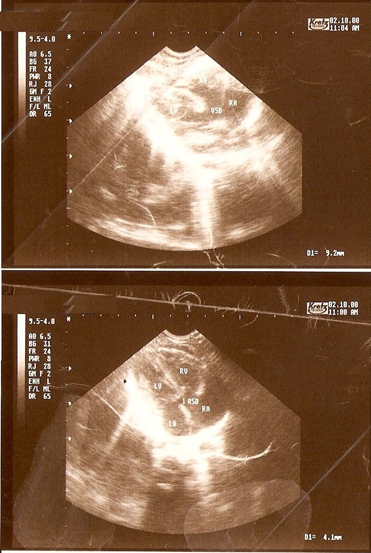
Atrioventricular canal defect results from an abnormal or inadequate fusion of the superior and inferior endocardial cushions. Both the complete and partial types of the defect are associated with the ostium primum defect in the lowermost portion of the atrial septum, left ventricular outflow narrowing and the atrioventricular valve abnormalities. The clinical diagnosis of partial atrioventricular canal defect can be confirmed by cardiac ultrasound. The aim of this paper is to preset an educational ultrasound image of partial atrioventricular canal defect.
Atrioventricular septal defect which is also atrioventricular canal defect, common atrioventricular canal, and endocardial cushion defect. It results from an abnormal or inadequate fusion of the superior and inferior endocardial cushions. Both the complete and partial types of the defect are associated with the ostium primum defect in the lowermost portion of the atrial septum, left ventricular outflow narrowing and the atrioventricular valve abnormalities. Cardiac ultrasound helps in establishing the diagnosis of various types of endocardial cushion defects. Figure-1 shows an ultrasound image of an infant with partial atrioventricular.
The atrioventricular canal is the "classic" congenital heart defect seen in patients with Down syndrome. In Down syndrome, complete atrioventricular canal is the prevalent defect, which is commonly associated with tetralogy of Fallot.
Partial atrioventricular canal and left-sided anomalies are more common in patients without Down syndrome.
Dear Editorial Team, Clinical Medical Reviews and Reports. My experience with the journal was highly positive. The peer-review process was rigorous, constructive, and completed in a timely manner. The reviewers provided valuable comments that helped improve the quality and clarity of our manuscript. The editorial office was professional, responsive, and supportive throughout all stages of the publication process. Communication was clear and efficient, and any questions were addressed promptly. Overall, I found the journal to maintain high scientific standards and an excellent publication workflow. I would be pleased to consider submitting future work to this journal. Best wishes from, Elena Popa.

It was my pleasure to submit my testimonial concerning the Reviewer Board of our Scientific Journal “Brain and Neurological Disorders”. The Reviewers focused on some modifications and their contribution was helpful. The ladies of our Editorial Office were also supported my efforts. It was my honor to have such a co-operation and I am looking forward for more collaboration.

Dear Grace Pierce, Editorial Coordinator of Journal of Clinical Research and Reports, Thank you for the speedy and efficient peer review process. I appreciate the fact that your peer reviewers do not take months to respond like with some other journals. I would also like to thank the editorial office for responding quickly to my questions. It is an excellent journal. I plan to submit more manuscripts in the future. Best wishes from, Robert W. McGee

Dear Grace Pierce, Editorial Coordinator of Journal of Clinical Research and Reports, Working with you and your team on our recent publication in JCRR has been a truly wonderful and enjoyable experience. The responses were prompt, and the reviewers were patient, constructive, and highly professional. One reviewer in particular gave me the feeling that a professor was carefully reading and commenting on my coursework, which was deeply touching. The entire process was straightforward and hassle‑free, with no tedious online forms to complete. I highly recommend this journal. Best wishes from, DR Aibing Rao, Head of R&D

I Appreciate the Opportunity to Share my Experience with the Journal of Clinical Research and Reports. The peer review process was timely and constructive, and the feedback provided helped improve the quality of our manuscript. The editorial office was professional, responsive, and supportive throughout the process, ensuring smooth communication and efficient handling of the submission. Overall, it was a positive experience collaborating with your team.

Dear Mercy Grace, Editorial Coordinator of Obstetrics Gynecology and Reproductive Sciences, We would like to express our gratitude for your help at all stages of publishing and editing the article. The editors of the magazine answer all the necessary questions and help at every stage. We will definitely continue to cooperate and publish other works in the Obstetrics Gynecology and Reproductive Sciences! Best wishes from, Alla Konstantinovna Politova,
