
Review Article | DOI: https://doi.org/10.31579/2690-1897/163
*Corresponding Author: Bhattacharyya S. Associate Professor Microbiology, AIIH&PH, Kolkata, India.
Citation: Roy B, Das T, Bhattacharyya S. (2023), Overview on Old and New Biochemical Test for Bacterial Identification, J, Surgical Case Reports and Images 6(5); DOI:10.31579/2690-1897/163
Copyright: © 2023, Bhattacharyya S. This is an open access article distributed under the Creative Commons Attribution License, which permits unrestricted use, distribution, and reproduction in any medium, provided the original work is properly cited.
Received: 26 July 2023 | Accepted: 05 August 2023 | Published: 15 August 2023
Keywords: bacteria; isolates; biochemical
Despite the advent of many newer tests, biochemical tests are still pivotal for bacterial identification. Many biochemical tests are there for bacterial identification and they have to be inoculated depending on which bacteria is suspected. Flow charts have to be prepared sequentially for deducing bacterial identification by biochemical tests. These aspects have been elaborated in this chapter.
Surgical and other infections can be caused by bacteria which need to be identified by phenotypic tests like staining and biochemical tests. Proper identification is the prerequisite for empirical therapy. Newer methods like MALDI-TOF and whole genome sequencing are available now in many places for accurate identification of bacteria from colonies, but biochemical tests are still one of the preferred identification methods for decades, not only just because they are reliable and inexpensive but also because of their quick and accurate results [1]. Identification of unknown microbial culture is a key step in medical, industrial and research institutes, and there are more than thousands of methods of identification of a particular species of microbes. However biochemical tests are still more preferable for many reasons from past till now [2]. Microbial identification at first glance is challenging for experienced research scientists also, and different screening steps are there which make classification schemes smaller and smaller and more specific. Firstly one should go for phenotypic and morphological studies; second, nutrient requirements in different nutrient media; third, Gram staining and acid fact staining ; fourth, biochemical properties of particular strain, fifth is PCR and sequencing [1]. After performing following screening tests, a researcher should have all details about that particular species. Although PCR and sequencing is more specific and expensive among all, mostly microbial origin source is identified in fourth step as well but to confirm the results, PCR and sequencing are done [3].
Biochemical tests are the oldest methods to identify microorganisms, by phenotypic traits. The cornerstone of most biochemical tests is the ability of bacteria to use specific biomolecules, producing valuable organic chemicals for themselves. There are several types of biochemical tests where different bacteria are identified or distinguished on the basis of different criteria. Simple visual confirmation of the organism's growth in the presence of essential nutrients by increasing turbidity in the liquid medium is one of the old approaches that is frequently used. However, in other experiments, the results are dependent on how the medium's colour changes as a result of the medium's pH changes. The way that microorganisms respond to these tests can be used to categorize them into distinct categories. Even down to the species level, several tests allow for the differentiation of microorganisms. . Nevertheless, there are several drawbacks to biochemical tests. Although affordable and providing both quantitative and qualitative data regarding the variety of microorganisms present in a sample, these procedures are time- and labour-intensive, and results take several days to appear. False positive results can occasionally be achieved, especially when similar microbial species are taken into consideration [4].
Different biochemical test and their discovery during time for identification of microbial culture are listed below:
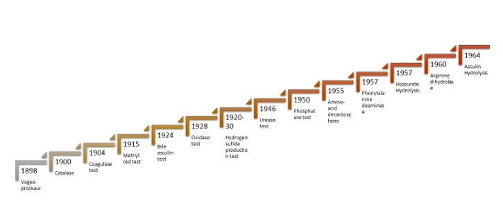
Figure : Evolution of biochemical test for microbial identification ( source:- author)
Oxidase test – This test was first postulated by Gordon and McLeod in 1928. The cytochrome oxidase test is useful for identifying bacteria that can manufacture the enzyme. The test aids in identifying the families of Pseudomonaceae that produce oxidase and order Enterobacterales that do not. The mechanism of cytochrome oxidase is the transmission of electrons from the donor (the electron transport chain) to the final acceptor (oxygen), and reduction results in the formation of water. The electron donor will be oxidized by cytochrome oxidase, changing the color to dark purple due to formation of indophenol. This test is carried out by impregnating filter paper with 1 % tetra-methyl-p-phenylenediamine dihydrochloride(TMPPD) which serves as an artificial electron donor, and drying it. The bacterial colonies are applied to a paper strip, and changes in color to blue-purple are detected within ten seconds [5]. Alternatively, the liquid reagent can be poured over the colonies directly and seen for colour change of the liquid. In modified oxidase, 6% TMPPD in DMSO is used for better cell wall penetration, in case of Micrococcus spp. Sometimes Bacillus spp. and many yeasts also give Oxidase positivity. Oxidase should never be done from colonies on blood agar because hemoglobin of blood has positive oxidase activity. Also, Oxidase should be done by picking the colony by edge of alme sterilized cover slip or glass slide and not nichrome loop, because Nichrome has Iron which has Positive oxidase activity itself. However,. Platinum loop may be used.
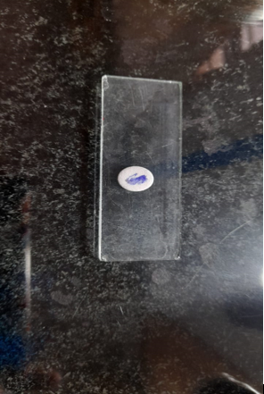
Figure. 1. Positive Oxidase disk test in P. aeruginosa
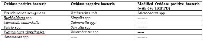
Table 1. Oxidase positive and negative bacteria of medical importance
Catalase test - Catalase test is used to identify microorganisms that possess the catalase enzymes. These bacteria create the catalase enzymes, which will neutralize the hydrogen peroxide and cause bubbles by producing nascent Oxygen, indicating a positive test result. O. Loew in 1900 discovered Catalase test. Most commonly, facultative anaerobic bacteria and obligate aerobes produce the catalase enzyme. Bacterial colony is mixed with a few drops of 3% H2O2 on a slide or in a test tube, and observed for bubble formation within 10 seconds (6). Most bacteria are catalase positive because this enzyme helps in neutralization of oxidizing free radicals. Catalase should never be done from colonies on blood agar due to positive Oxidase activity of hemoglobin . Catalase should be done by picking the colony by edge of sterilized cover slip or glass slide and not nichrome loop, because Nichrome has Iron which has Positive catalase activity itself. However, Platinum loop may be used Also, for Mycobacterium tuberculosis, 30% H2O2 is used for semiquantitative catalase test (tube catalase).
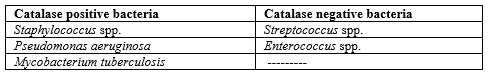
Table 2: Catalase positive and negative bacteria
Coagulase test - The subsequent test is performed first by Loeb in 1904 to identify microorganisms that can produce the coagulase enzyme. It generally aids in detecting Staphylococcus aureus, which is a coagulase- and catalase-positive bacterium. Coagulase is one of S. aureus's virulence-inducing components. During the reaction process, the coagulase enzyme will cause the blood plasma to coagulate by converting fibrinogen into fibrin. In order to perform this test, rabbit plasma and bacterial colonies in saline suspension are mixed. Bacteria will produce the coagulase enzyme, which will cause the plasma to coagulate as an indication of a positive reaction [7]. There are 2 types of coagulase tests :- free ( tube) and bound( slide ). Tube coagulase positivity is indicated by gellification of the plasma within 4-6 hours when incubated with liquid bacterial culture. It is to be note here that the gel again liquifies after 18-24 hours if kept at 37 degree C. Rabbit plasma is best for coagulase test but pooled human plasma can also be used. Staphylococcus aureus is positive for both slide and tube coagulase tests.
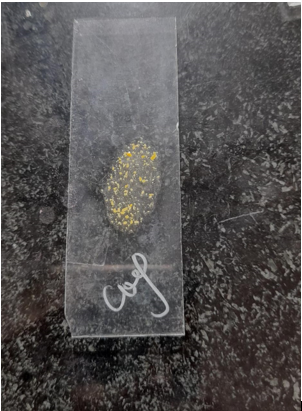
Figure 3. Picture of positive slide coagulase test (image: author)
Indole production test - The test described below is useful in identifying microorganisms with the capacity to produce the tryptophanase enzyme. Tryptophan is an amino acid that gets transformed into indole and Indole acetic acid by this enzyme. As a result, it can be tested by introducing various reagents like Ehrlich's reagent or Kovac's reagent (which gives red colour). Kovac's reagent has paradimethyl amino benzaldehyde( pDMAB) in isoamyl alcohol and concentrated HCl, whereas Ehrlich's have ethanol instead of isoamyl alcohol. As a result of the reagent and indole reaction, red rosindole dye is produced, a sign of positive test.
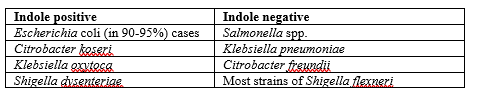
Table 2: Table showing Indole test results
< src>
Figure 4: Positive Indole test (image: authors)
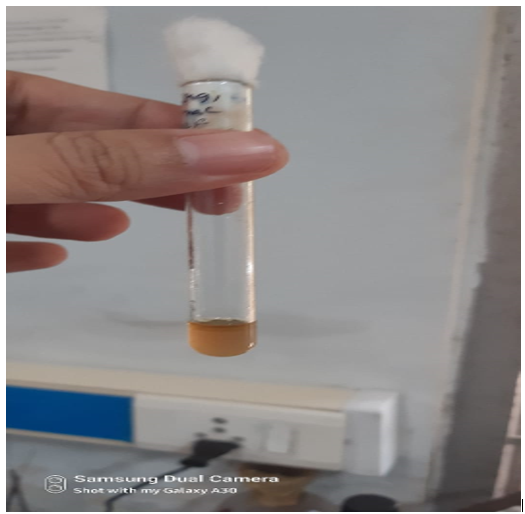
Figure 5: Negative Indole test (image: authors)
Spot indole test:- Here cinnamaldehyde is used instead of pDMAB. It is used mostly for anaerobic bacteria, and the isolate is rubbed on a filter paper containing cinnamaldehyde. Development of a dark brown to reddish colour within seconds indicates positive result.
Urease test - A urease test aids in identifying microorganisms that can produce the urease enzyme. Christensen in 1946 threw light on urease rest. Urease is a member of the superfamilies of amidohydrolases and phosphodoesterases. Urea is hydrolysed by urease enzyme into NH3 and Carbon dioxide. The ammonia production will cause the medium's pH to shift to an alkaline level and its colour to pink at pH 8.1 provided phenol red is used as pH indicator, which indicates a positive result. Helicobacter pylori , which is urease positive can be detected rapidly with this test. The bacterium uses the urease enzyme to produce an alkaline environment to tackle gastric acid. Urease test is carried out by inoculating bacterial colonies in urea broth or urea agar. This test needs 18 to 22 hours of incubation for results but for H. pylori and Proteus spp., positive results may appear as rapidly as 4-6 hours after inoculating [9].
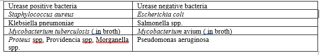
Table 3 : List of urease positive and negative bacteria
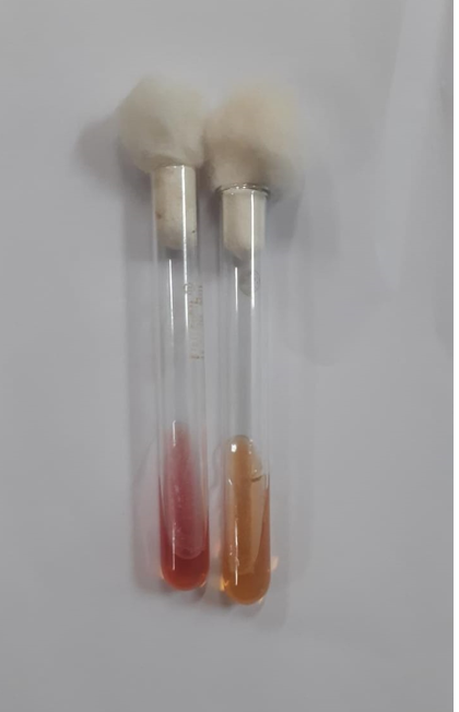
Figure 6: Urease positive (left) and negative tubes(right). (image: authors)
Citrate utilization test (CUT) - The identification of microorganisms with the capacity to use citrate as a single source of Carbon, is made easier with the use of this test. For this test, Simmons’ citrate agar is employed, which contains inorganic ammonium and citrate as sources of Nitrogen and Carbon, respectiveily. The CUT test is useful in identifying microorganisms that make the enzyme citrate permease, which transforms citrate into pyruvate and subsequently enters the metabolic cycle of organisms to generate energy and growth on culture media. The ammonium ions are converted into NH3 when microorganisms use citrate, raising the pH of the medium. When the pH rises above 7.6, bromothymol blue's colour will change from green to blue due to the pH change. Bromothymol blue is used as an indicator. As a result it should form colonies on the top of the slant and change colour into royal blue. If colour has been changer but colonies does not appear it also contemplated as negative. Colonies must be there to deduce a positive result [10]. Also inoculum on Citrate agar should be light and colonies should first be suspended in normal saline before inoculation. Colonies should never be directly inoculated form solid media on Citrate agar, because colonies may also contain dead bacterial cells and living bacterial cells may utilise Carbon from dead cells to give false positive Citrate utilization result.
Table 4:- examples of Citrate utilization positive and negative important bacteria.
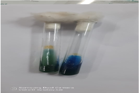
Figure 7: Citrate utilization positive (right) and negative (left) tubes
Triple sugar iron test
This test is useful in identifying microorganisms that are members of the Enterbacteriacea family. The test medium contains three sugars: lactose, sucrose, and glucose, each at a concentration of 1% (no, glucose is as 0.1%). As indicators, phenol red and ferric ammonium citrate are used. Sodium thiosulphate is also there as source of Sulphur. The medium is butted and slanted and both are inoculated. Since almost all inoculated bacteria use glucose, the concentration of glucose is kept low in comparison to other sugars. Within 16-18 hours of inoculation, the colour of the slant and butt will change to yellow as a result of acid production if the bacteria can use glucose in both aerobic and anaerobic circumstances. The media will continue to be yellow and acid production will continue if the bacteria can use the sucrose and lactose. If the bacteria are unable to use lactose or sucrose, they begin to use amino acids, which turns the medium alkaline and causes it to turn red from phenol red. If the bacteria is a strict aerobe, the colour of the butt remains the same, and the reaction will only happen in slant. Both will react if the bacteria is a facultative anaerobe. Agar media may rise or crack as a result of the production of hydrogen peroxide gas by reduction of thiosulfate by certain bacterial species [11]. TSI is interpreted as a/a and k/k , depending on acid formation in slant and butt. Slant reaction is used as numerator and butt reaction in denominator in a/a and k/k. H2S is detected by formation of black colour due to formation of Iron-sulfur complex.
Mannitol motility test
Mannitol motility test is required for confirmation of weather the bacteria are motile or not. Mannitol is a sugar which are fermented by some bacteria and it turns into pink in colour, if Andrade’s indicator is used in this medium. For identification of Bacillus cereus, mannitol should not be fermented and motility should be positive. It takes 18 to 22 hours of incubation for showing result. It is also useful for detecting Enterococcus casseliflavus.
Voges-Proskaur (VP) Test
This is an expansion of the methyl red test that locates organisms with the capacity to create the product butylene. The intermediate of this reaction, discovered by the use of alpha-naphthol and 40% KOH, is acetoin. Medium used is Glucose phosphate broth and the isolate needs to be grown for 48 hours at least. If KOH is present, acetoin will oxidize to diacetyl. Diacetyl will react with the guanidine component of peptone in the presence of alpha-naphthol, producing a deep red colour that indicates a positive result. The Methyl Red (MR) test is used for this. First identified during 1898. [12]. Write in table examples of VP positive and negative important bacteria. Examples of VP positive bacteria: Enterobacter spp., Klebsiella spp. and Staphylococcus aureus. Staphylococcus aureus is positive for both MR and VP. However, a bacterium that is positive for MR test is not usually positive for VP test.
Bile asculin agar test
This test was first described by Rochaix in 1924 and Swan first introduce the use of bile Esculin agar in 1954, is used to determine which bacteria hydrolyze esculin when bile is present. This test is a selective and differentiating medium for enterococcus identification. Bile and sodium azide are the selective media, while esculin is the differential medium. In contrast to sodium azide, bile will prevent the growth of Gram-ve bacteria, with the exception of enterococci and a few species of streptococci. When bile is present, some bacteria can hydrolyze esculin to produce esculetin, which reacts with ferric citrate in the medium to form phenolic iron complex, changing the color of agar from dark brown to black to indicate a positive test, such as for Enterococcus and Streptococcus bovis. The color will not alter in negative case [13]. Write in table examples of Bile aesculin hydrolysis positive and negative important bacteria.
Methyl Red-
Discovered by Clark and Lubs in 1915. This test used to identify coliform bacteria and their potential to produce acid from the glucose. And ultimately lowered the pH at about 4. Medium used is Glucose phosphate broth. A few drops of methyl red as an indicator when added to the 48-hour old microbial culture, a bright red colour is contemplated as positive. Shades fall intermediate between yellow and red result as doubtful positive. Methyl red indicator is prepared by dissolving 0.1 g methyl red in 300 ml 95% ethyl alcohol, which is then diluted to 500 ml with distilled water [14].
β-Galactosidase (ONPG)- This test is to identify late-lactose-fermenting paracolon organisms (like Enterobacter spp., Citrobacter spp., Shigella sonnei) known by their unique β-galactosidase activity which make them different from non lactose- fermenting bacteria like Salmonella and Proteus spp. From o-nitrophenyl-/3-D-galactopyranoside (ONPG), β – Galactosidase releases o-nitrophenol. Test organism should be grown in ONPG broth made by adding 250ml of ONPG solution to 750 ml of peptone water and stored at 4°C before use. The ONPG solution contains 0.6% ONPG in 0.01M Na2HPO4 buffer at pH 7.5. Alternatively, it can be done with ONPG disk. Normal saline is taken in 2 ml amounts and inoculated with loopful of the test bacterial culture previously grown on agar and ONPG disk. If there is yellow colour within 4-5 hours, it means that o-nitrophenol is formed, which indicates β -galactosidase activity. The colour may be change to bright yellow within 3 hours of incubation at 37°C. Tubes showing no colour change between 24 h considered as negative [14]. It is useful for detecting Late lactose fermenters like Shigella sonnei, Citrobacter spp. and some Enterobacter species([4].
Aesculin Hydrolysis
Discovered by Gemmell and Hodgkiss in 1964 to detect lactobacilli by incorporating 1
Surgical site infections and many other infections can be caused by medically important bacteria. Despite new identification methods like PCR and sequencing, phenotypic and biochemical tests still hold the key for laboratory identification due to their lower cost and ease in carrying out and interpretation. Newer modifications of biochemical tests have come like Rapid carbohydrate utilization test, API -20E and others, but they have not been able to replace old conventional biochemical tests. Hence they are still very important to know and discuss. Tests like Catalase, coagulase and Indole production are still the mainstay for proper laboratory diagnosis of many bacterial infections. Also, one needs to form flow charts for proper and accurate identification, like if one gets Gram positive cocci in clusters and golden-yellow pigmented colonies on Nutrient agar or Salt-milk agar, and if the isolate is catalase and coagulase positive, it is most likely to be Staphylococcus aureus. Likewise if one gets Lactose fermenting Gram negative bacilli and the isolate is catalase positive and Indole positive with no Citrate utilization and Urease activities, and in TSI one gets a/a with gas but no H2S, it may be Escherichia coli. Such flow charts help in more precise and accurate identification. Also sometimes 2 or 3 biochemical tests can be combined in 1 medium or tube to reduce workload, like SIM medium (Sulfide-Indole-motility), MIU (Motility-Indole-Urease) medium and others. They also yield satisfactory results.
Among the biochemical tests, assays such as catalase test, amylase and nitrate test are used to identify the gram positive bacteria while oxidase test, urease test, indole test, arginine dihydrolase test, hydrogen sulfide test, methyl red test, and voges-proskauer test are used to encounter gram negative bacteria. Some special test like heamolysis test, phosphatase test, amino acid decarboxylase test, phenylalanine deaminase test and aromatic ring cleavage test is used to illuminate some special groups of bacteria carrying unique properties. Through this paper, one can get a clear overview of biochemical test and their advancement with time can be understandable easily. Biochemical test is the key for easily microbial identification for researchers in past, present and also in future, from various sites of infections.
Dear Editorial Team, Clinical Medical Reviews and Reports. My experience with the journal was highly positive. The peer-review process was rigorous, constructive, and completed in a timely manner. The reviewers provided valuable comments that helped improve the quality and clarity of our manuscript. The editorial office was professional, responsive, and supportive throughout all stages of the publication process. Communication was clear and efficient, and any questions were addressed promptly. Overall, I found the journal to maintain high scientific standards and an excellent publication workflow. I would be pleased to consider submitting future work to this journal. Best wishes from, Elena Popa.

It was my pleasure to submit my testimonial concerning the Reviewer Board of our Scientific Journal “Brain and Neurological Disorders”. The Reviewers focused on some modifications and their contribution was helpful. The ladies of our Editorial Office were also supported my efforts. It was my honor to have such a co-operation and I am looking forward for more collaboration.

Dear Grace Pierce, Editorial Coordinator of Journal of Clinical Research and Reports, Thank you for the speedy and efficient peer review process. I appreciate the fact that your peer reviewers do not take months to respond like with some other journals. I would also like to thank the editorial office for responding quickly to my questions. It is an excellent journal. I plan to submit more manuscripts in the future. Best wishes from, Robert W. McGee

Dear Grace Pierce, Editorial Coordinator of Journal of Clinical Research and Reports, Working with you and your team on our recent publication in JCRR has been a truly wonderful and enjoyable experience. The responses were prompt, and the reviewers were patient, constructive, and highly professional. One reviewer in particular gave me the feeling that a professor was carefully reading and commenting on my coursework, which was deeply touching. The entire process was straightforward and hassle‑free, with no tedious online forms to complete. I highly recommend this journal. Best wishes from, DR Aibing Rao, Head of R&D

I Appreciate the Opportunity to Share my Experience with the Journal of Clinical Research and Reports. The peer review process was timely and constructive, and the feedback provided helped improve the quality of our manuscript. The editorial office was professional, responsive, and supportive throughout the process, ensuring smooth communication and efficient handling of the submission. Overall, it was a positive experience collaborating with your team.

Dear Mercy Grace, Editorial Coordinator of Obstetrics Gynecology and Reproductive Sciences, We would like to express our gratitude for your help at all stages of publishing and editing the article. The editors of the magazine answer all the necessary questions and help at every stage. We will definitely continue to cooperate and publish other works in the Obstetrics Gynecology and Reproductive Sciences! Best wishes from, Alla Konstantinovna Politova,
