AUCTORES
Globalize your Research

Research Article | DOI: https://doi.org/10.31579/2766-2314/044
*Corresponding Author: Siavash Hosseinpour Chermahini, Department of Bioprocess Engineering, Faculty of Chemical Engineering, Universiti Teknologi, Malaysia, 81310 Johor Bahru, Johor, Malaysia.
Citation: Siavash H. Chermahini, Fadzilah A. A. Majid, Azila A. Aziz and Anvari R., (2021) The tower of Babylon – bioreactor. J, Biotechnology and Bioprocessing 2(7); DOI: 10.31579/2766-2314/044
Copyright: © 2021, Siavash Hosseinpour Chermahini, This is an open access article distributed under the Creative Commons Attribution License, which permits unrestricted use, distribution, and reproduction in any medium, provided the original work is properly cited.
Received: 16 May 2021 | Accepted: 13 July 2021 | Published: 20 August 2021
Keywords: niosome, human skin fibroblast, inflammation, IL-6, TNF-α, bromelain
The topical delivery of bromelain as an anti-inflammatory solution for skin inflammation has attracted the attention of researchers. Due to the skin barrier issue, a new method was designed for the effective delivery of specific doses of bromelain to the desired action sites. A niosome was selected as a novel and practical transdermal vehicle for the delivery of bromelain to inflamed sites. In this regard, a lipopolysaccharide (LPS)-induced human skin fibroblast (HSF1184) cell line was assembled in-vitro as a simulated model. The levels of interleukin-6 (IL-6) and tumour necrosis factor alpha (TNF-α), the two immune-modulatory regulators of cell responses to inflammation, were measured to determine the response towards the niosome-encapsulated bromelain treatment. The results showed that the niosome-encapsulated bromelain significantly reduced the levels of IL-6 and TNF-α compared to the non-encapsulated bromelain, the vehicle (niosome) and the control.
Bromelain has demonstrated many beneficial properties in-vitro and in-vivo such as anti-oedematous, anti-thrombotic, and fibrinolytic properties. Most importantly, clinical trials of bromelain have confirmed its anti-inflammatory properties, which include, but are not limited to, breast engorgement during lactation [1]; osteoarthritis of the knee and hip [2, 3]; rhinosinusitis [4]; sepsis in children [5]; and urogenital inflammation [6]. Experimental evidence also exists on its effects on blood coagulation, where increases in the serum fibrinolytic activity and prostaglandin levels have been recorded due to a decrease in PGE2 and thromboxane A2. Essentially, this phenomenon is important for reducing inflammation [7]. However, there are many internal and external factors that trigger the transcription of pro-inflammatory cytokines in the human body, for instance, viral and bacterial infections, cuts, wounds, and obesity. Bromelain inhibits bacterial endotoxin LPS-induced NF-kB activity as well as the expression of PGE2 and Cox-2 [8]. To explain this mechanism, it was hypothesized that bromelain induces the cleavage of cell surface markers such as CD14 [9]. Among the secreted regulators of inflammation that are connected to the NF-kB pathways and that respond to bromelain are IFN-γ, TNF-α, IL-1β, and IL-6. Depending on the context and micro-environment, these regulators can either stimulate tumour growth and invasion or activate immune responses and cause tumour regression [10–13]. Experimental evidence derived from an analysis of peripheral blood mononuclear cells (PBMC) from healthy volunteers as well as mouse macrophages suggest that bromelain can activate TNF-α, IL-1β and IL-6 secretions in an IFN-γ-dependent mechanism [14,15]. However, the IFN-γ production is also encouraged by the presence of bromelain [16]. These data led to the hypothesis that bromelain has the potential to activate a healthy immune system to ensure a rapid response to pathogens and cellular stress. Nevertheless, in situations when immune cells have already been stimulated, bromelain reduces the secretion of TNF-α and IL-6 [17]. According to the summary by Chobotova et al. (2010) [18] on the influence of bromelain on inflammation regulation in relation to the cancer network, an over-production of cytokines is expected in inflammation-induced cases. A study has also found that in the presence of LPS, which can stimulate an acute inflammatory reaction, bromelain reduces elevated TNF-α, IL-1β, and IL-6 expressions in human PBMC [19]. The reduction of TNF-α and IFN-γ expressions has also been observed in inflamed tissues of patients with inflammatory bowel disease (IBD) that have been treated with bromelain [20]. The described data demonstrate that the effects of bromelain on cytokine expressions depend on the presence of inflammation-inducing conditions. This underlines the potential of bromelain for the treatment of inflammation-based pathologies. However, the challenge of the transdermal therapeutic system (TTS) is the stratum corneum (SC). The stratum corneum (SC) is the main barrier to the transportation of nutritional compounds via the skin [21]. In this regard, the delivery and release of bioactive materials like bromelain to different parts of the body are practically controlled by nanocarriers [22]. Several types of nanocarrier systems are available for TTSs such as vesicular phospholipid gels (VPG) [23], microspheres [24], nanospheres [25], nanoliposomes [26] liposomes [27] archaeosomes [28], complexes [29], ethosomes [30], dendrimers [31] nanoemulsions [32], and niosomes [33]. The effective delivery of bromelain to the affected sites depends on the delivery system used in the topical formulation [34]. Niosomes have specific characteristics for topical delivery [35] as theypossess high chemical long-term stability; high solubility for lipophilic and hydrophilic components; complete drug delivery; and large membrane flexibility to be effective against the SC barrier [36]. Niosomes are thermodynamically stable because they contain two volumes of liquids, namely water and oil, which are combined into a single phase by means of a surfactant to produce a non-ionic surfactant. Moreover, niosomes are highly compatible with biological systems and have no toxicity because of their non-ionic nature [37]. Due to the technological and physicochemical characteristics of niosomes, researchers have been motivated to consider them as ideal for carrying drugs for topical administration [38, 39]. For instance, Bouwstra and Hofland (1996) [40] loaded ellagic acid (EA) onto a niosome as an antioxidant drug application by transdermal administration. LPS is a suitable agent in stimulating inflammation since it can be used to induce inflammation both in-vitro and in-vivo. [41]. In order to detect LPS-induced inflammation, 2 cytokines, namely IL-6 and TNF-α, were selected in this study. IL-6 and TNF-αare immuno-modulating agents that act as regulators of host responses to infection, immune responses, inflammation, and trauma. They include various groups of soluble proteins, peptides, or glycoproteins which act as hormonal regulators or signalling molecules from nanomolar to picomolar concentrations [42]. Some of them are pro-inflammatory, being necessary for initiating the inflammatory response that is needed for the recruitment of granulocytes and, later on, lymphocytes, to fight disease. However, excessive inflammation is sometimes the pathogenicity of certain diseases. Other cytokines are anti-inflammatory and serve to reduce inflammation and promote healing [43]. Essentially, the definition of inflammation is that the cells are alive up to a certain point but will die subsequently. In this study, niosome-encapsulated bromelain was shown to be an effective compound that reduces the levels of IL-6 and TNF-α in LPS-induced inflammation of the human skin fibroblast (HSF1184) cell line. This is the first study to use niosome-encapsulated bromelain as an anti-inflammatory application. In order to determine the effectiveness of the treatment in HSF1184, the LPS-induced inflammation was treated with niosome-encapsulated bromelain after four hours of induction. The inflammation responses were measured after four hours and 24hours.
2.1 Chemicals and Cell Culture
All the chemicals used in this study were purchased from Sigma Aldrich and Merck, unless noted otherwise. These included Span (40, 60, 80), Labrasol, dicetyl phosphate, chloroform, DMEM, trypsin, penicillin, streptomycin, MTT solution, and DMSO (sterile and non-sterile). The bromelain was purchased from Merck, while the LPS from Escherichia coli (0111:B4) was purchased from Sigma Aldrich. The TNF-a kit (Catalogue Number RAB0476) and IL-6 kit (Catalogue Number RAB0307) were purchased from Sigma Aldrich (USA). Acetaminophen tablets (80 mg) were purchased from Guardian Pharmacy. HSF1184 was purchased from ATCC (USA) (Catalogue No. 107-75a). The cell culture grade chemicals and analytical grade chemicals used in this study were from Sigma Aldrich.
2.2 Preparation of Niosome-Encapsulated Bromelain
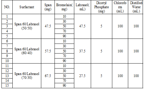
The niosome was created based on the protocol of Arora and Sharma (2010) [44] with minor modifications. First of all, a mixture of the vesicle-forming agents, i.e., the surfactant (40, 60, 80) and Labrasol, was dissolved in chloroform (a volatile organic solvent) in a round-bottom flask at a mole ratio of 1:1. The organic solvent was removed at a temperature of 45°C using a rotary evaporator (40 rpm), leaving a thin film of solid mixture deposited on the wall of the flask. This dried surfactant film was then rehydrated with 100 mL of aqueous-phase distilled water and was then agitated gently for an hour to yield multi-lamellar niosomes. The mixture was subjected to probe sonication using a Fisher Scientific sonicator at an amplitude of 40 m, energy of 2000 J, and power of 30 W for two minutes to yield uniform nanoparticles of niosomes. After selecting a span of 60 (40, 60, 80), different concentrations of bromelain (10, 30, 50, 70, 90) with different ratios of span/labrasol (50/50, 60/40, 70/30) were tested in this study.
2.3 Cell Line Culture and Maintenance
The cell culture and maintenance followed the protocol of Freshney (2008) [45]. The cells were cultured between passages 7 and 9. A water bath at 37ºC was used to pre-warm all the media and solutions before use. The cells were maintained routinely in a 75-cm2 flask. Moreover, the cells that had been cultured in Dulbecco’s modified Eagle’s medium (DMEM) containing penicillin (100 units/ml), streptomycin (100 lg/ml), and foetal bovine serum (10% v/v) were also incubated in a humidified incubator with 5% CO2/ 95% air at 37°C. The cells took between 24-48 hours to reach a confluent state. Cells from the exponential growth phase were used for the experimental work, where 2 x 10⁵cells/mL were loaded into each well and incubated for 24 hours for cell attachment before the start of the experiment.
2.4 Inflammation Induction of HSF1184 with Serial Dilution of LPS
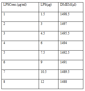
The serial dilution of LPS was modified from the protocol of Freshney (2008) [45]. After mixing 1mg of LPS with 1 mL of deionized water (DIW) according to the manufacturer’s instructions (stock LPS solution), serial dilutions of LPS were performed from 12 μg/mL to 1.5μg/mL (1.5 μg/mL, 3 μg/mL, 4.5 μg/mL, 6 μg/mL, 7.5 μg/mL, 9 μg/mL, 10.5 μg/mL, and 12 μg/mL) by diluting the solution in an appropriate volume of DMEM.
200μl of cells at a concentration of 2 x10⁵ cells/well were seeded in a 96-well plate and incubated (5% CO2 and 37°C) for two days to reach a confluent state. Then, the cells were exposed to different concentrations of LPS (1.5, 3, 4.5, 6, 7.5, 9, 10.5, 12 μg/mL) for four hours or 24 hours using separate plates. The arrangement of the different concentrations of LPS on the plate is presented in Figure 3. DMEM was used as the blank, while cells without the LPS induction were used as the control. The cell induction was terminated accordingly after four hours and 24 hours.
2.5 Quantification of Cell Viability Using MTT Assay
The cell viability after the LPS induction was quantified using an MTT assay, as recommended by Freshney (2008) [45]. The assay was used to investigate the effects of niosome-encapsulated bromelain, niosome without bromelain (vehicle), and bromelain alone on the viability of HSF1184 cells treated with 1.5μg/mL of LPS-induced inflammation. The HSF1184 cells were seeded at 2-4 x 10⁴ cells per/mL on two 96-well plates (4hrs and 24hrs) and were left to grow for two days to a confluent state, with media changes being made once per day. On completion of the confluence cells, inflammation was triggered in the plates treated with 1.5 μl/mL of LPS and the cells were incubated for four hours at 37°C in a humidified atmosphere with 5% CO2. Then, they were treated with acetaminophen (2μg/mL), bromelain (25μg/mL), niosome with 10% bromelain (20μl/mL), and niosome as a vehicle (20μl/mL) before being incubated for either four or 24 hours. The induced cells were washed twice with PBS, and 20μl of MTT solution (5mg/1ml PBS) was added to each experimental well. The plate was wrapped with aluminium foil and incubated for four hours. In continue, the MTT solution was carefully removed from all the wells using fine tips before being replaced with 200μl of non-sterile DMSO and mixed well with MTT formazan. A model ELX 808 BIO-TEK plate reader was used to measure the optical density of the colour formations of each sample at a wavelength of 570 nm and reference wavelength of 630nm. The procedure for 24 hours was similar to that for four hours. The average of six repetitions taken thrice from each sample was taken as the result. The OD was changed to a percentage of cell viability. Since the control (cells and DMEM) had 100% viability, the other treatments were compared to the control.
2.6 Quantification of IL-6 and TNF-α Concentrations
In accordance with Freshney (2008) [45] protocol, after each culture medium had been collected and centrifuged for three minutes at 3300 rpm and the supernatant had been separated, the resulting cells in the well plates (supernatant) were processed for IL-6 and TNF-α according to the manufacturer’s instructions for the ELISA protocol. The standards and samples were pipette into the wells of the kits. The IL-6 and TNF-α presented in the sample were bound to the wells by the immobilized antibody. The wells were then washed and biotinylated with the added anti-human IL-6 and TNF-α antibodies. After washing away the unbound biotinylated antibodies, HRP-conjugated streptavidin was pipette into the wells. The wells were again washed before a TMB substrate solution was added to the wells. The colour that developed was in proportion to the amount of IL-6 and TNF-α that had been bound. The colour of the stop solution changed from blue to yellow. The intensity of the colour was measured at 450 nm, and the result was based on pg/mL. From the standard curve equation (y = a * ln(x) +b) of both cytokines (IL-6 and TNF-α), since y (OD) = 450 nm and x is in pg/mL, then with various amounts of y in the above stated equation, various amounts of x in pg/mL were obtained.
2.7 Treatment of LPS-Induced Inflammation using Niosome-Encapsulated Bromelain
The LPS induction was carried out according to the protocol by Zdarilova et al. (2009) [46] with minor modifications, as described above. 2 – 4 x10⁴ cells/mL were prepared at the exponential growth phase, and 200 μL of cell suspension was transferred to each well on two 96-well plates according to the plate design. The plates were then incubated in 5% CO2 at 37°C for two days with a daily change of medium until the cells reached a confluent state. Then, 1.5 μg/mL of LPS was added to each well and the plates were incubated for another four hours. After four hours of post induction incubation, the designated wells in both the four-hour and 24-hour plates were treated with acetaminophen (2μg/mL), bromelain (25μg/mL), niosome with bromelain 10% (20μl/mL), and niosome as a vehicle (20μl/mL), and then incubated again for another four hours or 24 hours.
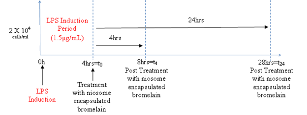
The summary of the LPS induction and treatment with the niosome-encapsulated bromelain for both plates is presented in Figure 1. The cell viability was determined using the MTT assay as described above. The levels of both the IL-6 and TNF-α cytokines present in the wells were measured accordingly based on the above protocol.
In this experiment, two pairs of samples were compared using the t-Test because these two groups contributed a pair of scores. This statistical technique is often called a paired sample t-test or correlated t-test that tells whether there is a statistically significant difference in the mean scores for niosome-encapsulated bromelain and the other treatments. When P<0> it means that there is a significant difference.
4.1 Determination of suitable LPS Concentration to Induce Inflammation in HSF1184
A suitable concentration of LPS to induce inflammation in HSF1184 was determined based on the cell viability and IL-6 response. The purpose was to select the lowest concentration of LPS that would cause inflammation on the cells without causing cell death.
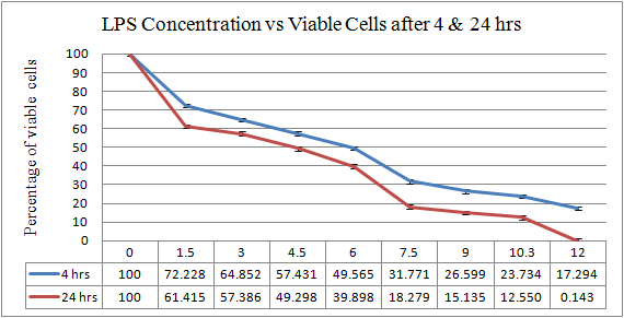
As shown in Figure 2, the HSF1184 cell line was treated with different concentrations of LPS to induce inflammation after 4 hours and 24 hours. Then, the percentage of viable cells was calculated. By using the paired sample t-Test in the analysis and comparing the LPS concentration of 1.5μg/mL with other concentrations, the difference was most significant for the 1.5μg/mL LPS. The interleukin-6 (IL-6) and tumour necrosis factor alpha (TNF-α) were used as indicators for the host’s responses to the inflammation. The suitable concentration of LPS-induced inflammation in the HSF1184 cell line was determined based on the response of the cytokine. The determination of the inflammation-inducing concentration of LPS was based on the production of TNF-α and IL-6 by the cells.

In Figure 3, the HSF1184 cell line was treated with different concentrations of LPS-induced inflammation for four hours and 24 hours. The supernatant of the HSF1184 cell line that was inflamed by the LPS was measured by the response of the cytokine (IL-6). By using the paired sample t-Test to compare the concentration of 1.5μg/mL LPS with other concentrations, the 1.5μg/mL LPS showed a more significant difference than the other concentrations (p<0>in-vitro simulated model. This LPS concentration was determined using quantitative experiments such as quantitative cell viability using an MTT assay and quantitative cytokines concentration. Thus, 1.5μg/mL of LPS was selected to induce inflammation in the HSF1184 cell line in the following experiments.
4.2 Cell Viability of LPS-Induced HSF1184 Treated with Niosome-Encapsulated Bromelain
In the MTT assay, inflammation was induced in the HSF1184by the 1.5 μg/mL LPS. It was treated with the niosome-encapsulated bromelain, non-encapsulated bromelain, and the vehicle (niosome). In this experiment, acetaminophen at a concentration of 2 μg/mL was considered as the positive control, LPS at a concentration of 1.5 μg/mL was the negative control, and DMEM-plus cells with a volume of 1 mL was the control specimen. The concentrations for the other treatments involving niosome-encapsulated bromelain, bromelain alone, and niosome (vehicle) were 20 μg/mL, 25 μg/mL, and 20 μg/mL, respectively, as determined from previous experiments.
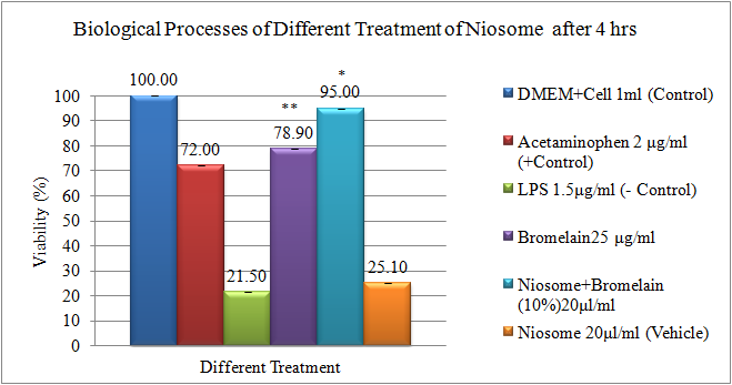
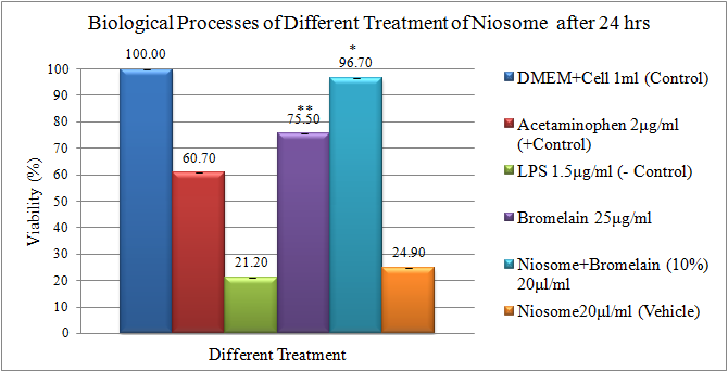
Figure 4 and Figure 5 show the differences in the results for various treatments of niosome after four hours and 24 hours. Based on the presented figures, the biggest difference in the percentage of viability was related to the niosome-encapsulated 10% bromelain, which had a viability of about 96.70 percent in Figure 5 and a viability of 95 percent in Figure 4. On the contrary, the vehicle had the lowest viability at 24.90 percent in Figure 5 and 25.10 percent in Figure 4. However, the viability of bromelain alone was 78.90 percent in Figure 4 and 75.50 percent in Figure 5; this was the second highest after the niosome-encapsulated10% bromelain (Figure 4 and Figure 5). As a positive control, acetaminophen ranked third with 72 percent viability in Figure 4 and 60.70 percent in Figure 5. The free and encapsulated bromelain were evaluated by MTT assays using four different cell lines, which were HeLa, HEK293, MCF-7 and A549, for 24, 48 and 72 hours, respectively [48]. It was found that the encapsulated bromelain needed significantly (p<0>P<0>
4.3 IL-6 and TNFα Response in LPS Induction of HSF1184 Treated with Niosome-Encapsulated Bromelain
Interleukin-6 (IL-6) and tumour necrosis factor alpha (TNF-α), the two immune-modulatory regulators of cell responses to inflammation, were measured in response to 1.5 μg/mL LPS-induced inflammation and were then treated with niosome-encapsulated bromelain, non-encapsulated bromelain, and the vehicle. The cytokines, IL-6 and TNF-α, were used because these pro-inflammatory cytokines play a key role in the inflammatory response and can be easily quantified in the supernatant.
4.3.1 Determination of Inflammation Stage Using the IL-6 Response to Different Niosome Treatments:
Interleukin-6 (IL-6) was measured in response to 1.5 μg/mL LPS and was then treated with niosome-encapsulated bromelain at a concentration of 20μg/mL, bromelain alone with a concentration of 25 μg/mL, and niosome (vehicle) with a concentration of20 μg/mL. 1 mL DMEM was used as the blank sample and 1 mL of DMEM-plus cells was used as the control. All the concentrations resulted from previous experiments in this study.
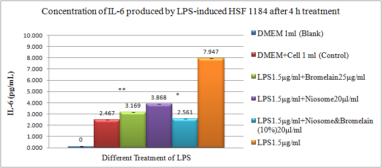

Figure 6 and Figure 7 show that the lowest amounts of 2544 pg/mL in Figure 7 and 2561 pg/mL in Figure 6 belonged to the niosome-encapsulated bromelain. The highest amounts, marked at 3908 pg/mL in Figure 7 and 3868 pg/mL in Figure 6, were related to the vehicle. However, bromelain alone, reported at 3169 pg/mL in Figure 6 and 3489 pg/mL in Figure 7, was ranked second. Strong evidence on the anti-inflammatory effect of theniosome-encapsulated 10% bromelain was found when the result of the quantitative cytokines concentration showed the highest induction for LPS without any treatment and vehicle. The lowest induction of inflammatory cytokines was observed in the niosome-encapsulated bromelain and the bromelain-treated cells, as previously mentioned by Mosmann (1983) [51]. The statistical analysis of the niosome-encapsulated 10% bromelain showed a significant difference compared to other treatments of niosome, where P<0>
4.3.2 Determination of Inflammation Stage Using TNF-α Response to Different Niosome Treatments
The tumour necrosis factor alpha (TNF-α) was measured in response to 1.5 μg/mL LPS, and then to treatments with niosome-encapsulated bromelain at a concentration of 20 μg/mL, bromelain alone at a concentration of 25 μg/mL, and niosome (vehicle) at a concentration of 20 μg/mL. The setup for the control and blank samples was similar to that of the IL-6.
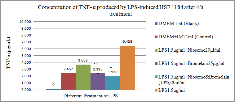

Figure 8 and Figure 9 show that the lowest amounts, marked at 1978 pg/mL in Figure 8 and 1991 pg/mL in Figure 9, were from the niosome-encapsulated bromelain. The highest amounts, reported at 3697 pg/mL in Figure 9 and 3688 pg/mL in Figure 8, were related to the vehicle. However, bromelain alone was ranked second since the reported readings were 2396 pg/mL in Figure 8 and 2410 pg/mL in Figure 9.
Unlike IL-6, strong evidence on the anti-inflammatory effect of niosome-encapsulated 10% bromelain was from the low induction of inflammatory cytokines with the niosome-encapsulated 10% bromelain and bromelain. The highest induction was observed in the LPS without any treatment and the vehicle-treated cells (Mosmann, 1983) [51]. Again, the niosome-encapsulated 10% bromelain was statistically significant. The efficacy of the bromelain-niosome system on skin inflammation around the knee was evaluated. The potential anti-inflammatory properties of the niosome-encapsulated bromelain were investigated in the HSF1184 cell line because this model produces high concentrations of IL-6 and TNF-α in cultures upon activation by LPS. Studies on the effects of bromelain on pro-inflammatory cytokines are limited. Moreover, no previous studies have evaluated the effects of niosome-encapsulated bromelain on IL-6 and TNF-α cytokines. Greenspan et al. (2005) [52] stimulated human endothelial cells with TNF-α and found that co-treatment with bromelain inhibited the production of TNF-α for LPS induction. However, in another study evaluating the inhibitory effects of bromelain on TNF-α production, Mastuda et al. (2012) [53] showed that bromelain inhibited the antigen IgE-mediated TNF-α secretion in RBL-2H3 mast cells. A potential reason for the differences in the results may be due to differences in the stimuli and cell types used. These findings are partly coherent with the results of this study, which show that niosome-encapsulated bromelain significantly impaired LPS-induced IL-6 and TNF-α production. Apart from this finding, in different comparisons of niosome encapsulation, the niosome-encapsulated bromelain could be affected in a higher range compared to other treatments like bromelain alone or a vehicle. Moreover, the results showed that niosome alone as a vehicle did not affect the IL-6 and TNF-α levels in the cultured supernatant and suggest that the immuno-stimulatory properties of the bromelain were not due to the presence of the vehicle [54]. On the other hand, during the post treatment, a pattern was observed where the anti-inflammatory cytokines peaked at four hours and 24 hours. A similar time period has been reported in numerous studies on humans [55]. Other key factors are the biological conditions of the HSF1184 cell line, for instance, inflammation induced by other components. Future perspectives should include an understanding on the molecular basis for the inhibitory effects of niosome-encapsulated bromelain on the HSF1184 cell line. The importance of niosome-encapsulated bromelain on the elimination of inflammation in the HSF1184 cell line infected by LPS-induced inflammation was evaluated. The result showed that niosome-encapsulated 10% bromelain significantly reduced the production of IL-6 and TNF-α in the LPS-induced human skin fibroblast cell line (HSF1184) after four hours post treatment as compared to the non-encapsulated bromelain and vehicle.
This study set out with the aim of assessing the importance of niosome-encapsulated bromelain in the elimination of inflammation in the HSF1184 cell line infected by LPS-induced inflammation. The results showed that niosome-encapsulated 10% bromelain significantly reduced the production of IL-6 and TNF-α in the LPS-induced human skin fibroblast cell line (HSF1184) after 4hours of post treatment as compared to the non-encapsulated bromelain and vehicle.
Clearly Auctoresonline and particularly Psychology and Mental Health Care Journal is dedicated to improving health care services for individuals and populations. The editorial boards' ability to efficiently recognize and share the global importance of health literacy with a variety of stakeholders. Auctoresonline publishing platform can be used to facilitate of optimal client-based services and should be added to health care professionals' repertoire of evidence-based health care resources.

Journal of Clinical Cardiology and Cardiovascular Intervention The submission and review process was adequate. However I think that the publication total value should have been enlightened in early fases. Thank you for all.

Journal of Women Health Care and Issues By the present mail, I want to say thank to you and tour colleagues for facilitating my published article. Specially thank you for the peer review process, support from the editorial office. I appreciate positively the quality of your journal.

Journal of Clinical Research and Reports I would be very delighted to submit my testimonial regarding the reviewer board and the editorial office. The reviewer board were accurate and helpful regarding any modifications for my manuscript. And the editorial office were very helpful and supportive in contacting and monitoring with any update and offering help. It was my pleasure to contribute with your promising Journal and I am looking forward for more collaboration.

We would like to thank the Journal of Thoracic Disease and Cardiothoracic Surgery because of the services they provided us for our articles. The peer-review process was done in a very excellent time manner, and the opinions of the reviewers helped us to improve our manuscript further. The editorial office had an outstanding correspondence with us and guided us in many ways. During a hard time of the pandemic that is affecting every one of us tremendously, the editorial office helped us make everything easier for publishing scientific work. Hope for a more scientific relationship with your Journal.

The peer-review process which consisted high quality queries on the paper. I did answer six reviewers’ questions and comments before the paper was accepted. The support from the editorial office is excellent.

Journal of Neuroscience and Neurological Surgery. I had the experience of publishing a research article recently. The whole process was simple from submission to publication. The reviewers made specific and valuable recommendations and corrections that improved the quality of my publication. I strongly recommend this Journal.

Dr. Katarzyna Byczkowska My testimonial covering: "The peer review process is quick and effective. The support from the editorial office is very professional and friendly. Quality of the Clinical Cardiology and Cardiovascular Interventions is scientific and publishes ground-breaking research on cardiology that is useful for other professionals in the field.

Thank you most sincerely, with regard to the support you have given in relation to the reviewing process and the processing of my article entitled "Large Cell Neuroendocrine Carcinoma of The Prostate Gland: A Review and Update" for publication in your esteemed Journal, Journal of Cancer Research and Cellular Therapeutics". The editorial team has been very supportive.

Testimony of Journal of Clinical Otorhinolaryngology: work with your Reviews has been a educational and constructive experience. The editorial office were very helpful and supportive. It was a pleasure to contribute to your Journal.

Dr. Bernard Terkimbi Utoo, I am happy to publish my scientific work in Journal of Women Health Care and Issues (JWHCI). The manuscript submission was seamless and peer review process was top notch. I was amazed that 4 reviewers worked on the manuscript which made it a highly technical, standard and excellent quality paper. I appreciate the format and consideration for the APC as well as the speed of publication. It is my pleasure to continue with this scientific relationship with the esteem JWHCI.

This is an acknowledgment for peer reviewers, editorial board of Journal of Clinical Research and Reports. They show a lot of consideration for us as publishers for our research article “Evaluation of the different factors associated with side effects of COVID-19 vaccination on medical students, Mutah university, Al-Karak, Jordan”, in a very professional and easy way. This journal is one of outstanding medical journal.

Dear Hao Jiang, to Journal of Nutrition and Food Processing We greatly appreciate the efficient, professional and rapid processing of our paper by your team. If there is anything else we should do, please do not hesitate to let us know. On behalf of my co-authors, we would like to express our great appreciation to editor and reviewers.

As an author who has recently published in the journal "Brain and Neurological Disorders". I am delighted to provide a testimonial on the peer review process, editorial office support, and the overall quality of the journal. The peer review process at Brain and Neurological Disorders is rigorous and meticulous, ensuring that only high-quality, evidence-based research is published. The reviewers are experts in their fields, and their comments and suggestions were constructive and helped improve the quality of my manuscript. The review process was timely and efficient, with clear communication from the editorial office at each stage. The support from the editorial office was exceptional throughout the entire process. The editorial staff was responsive, professional, and always willing to help. They provided valuable guidance on formatting, structure, and ethical considerations, making the submission process seamless. Moreover, they kept me informed about the status of my manuscript and provided timely updates, which made the process less stressful. The journal Brain and Neurological Disorders is of the highest quality, with a strong focus on publishing cutting-edge research in the field of neurology. The articles published in this journal are well-researched, rigorously peer-reviewed, and written by experts in the field. The journal maintains high standards, ensuring that readers are provided with the most up-to-date and reliable information on brain and neurological disorders. In conclusion, I had a wonderful experience publishing in Brain and Neurological Disorders. The peer review process was thorough, the editorial office provided exceptional support, and the journal's quality is second to none. I would highly recommend this journal to any researcher working in the field of neurology and brain disorders.
Dear Agrippa Hilda, Journal of Neuroscience and Neurological Surgery, Editorial Coordinator, I trust this message finds you well. I want to extend my appreciation for considering my article for publication in your esteemed journal. I am pleased to provide a testimonial regarding the peer review process and the support received from your editorial office. The peer review process for my paper was carried out in a highly professional and thorough manner. The feedback and comments provided by the authors were constructive and very useful in improving the quality of the manuscript. This rigorous assessment process undoubtedly contributes to the high standards maintained by your journal.

International Journal of Clinical Case Reports and Reviews. I strongly recommend to consider submitting your work to this high-quality journal. The support and availability of the Editorial staff is outstanding and the review process was both efficient and rigorous.

Thank you very much for publishing my Research Article titled “Comparing Treatment Outcome Of Allergic Rhinitis Patients After Using Fluticasone Nasal Spray And Nasal Douching" in the Journal of Clinical Otorhinolaryngology. As Medical Professionals we are immensely benefited from study of various informative Articles and Papers published in this high quality Journal. I look forward to enriching my knowledge by regular study of the Journal and contribute my future work in the field of ENT through the Journal for use by the medical fraternity. The support from the Editorial office was excellent and very prompt. I also welcome the comments received from the readers of my Research Article.

Dear Erica Kelsey, Editorial Coordinator of Cancer Research and Cellular Therapeutics Our team is very satisfied with the processing of our paper by your journal. That was fast, efficient, rigorous, but without unnecessary complications. We appreciated the very short time between the submission of the paper and its publication on line on your site.

I am very glad to say that the peer review process is very successful and fast and support from the Editorial Office. Therefore, I would like to continue our scientific relationship for a long time. And I especially thank you for your kindly attention towards my article. Have a good day!

"We recently published an article entitled “Influence of beta-Cyclodextrins upon the Degradation of Carbofuran Derivatives under Alkaline Conditions" in the Journal of “Pesticides and Biofertilizers” to show that the cyclodextrins protect the carbamates increasing their half-life time in the presence of basic conditions This will be very helpful to understand carbofuran behaviour in the analytical, agro-environmental and food areas. We greatly appreciated the interaction with the editor and the editorial team; we were particularly well accompanied during the course of the revision process, since all various steps towards publication were short and without delay".

I would like to express my gratitude towards you process of article review and submission. I found this to be very fair and expedient. Your follow up has been excellent. I have many publications in national and international journal and your process has been one of the best so far. Keep up the great work.

We are grateful for this opportunity to provide a glowing recommendation to the Journal of Psychiatry and Psychotherapy. We found that the editorial team were very supportive, helpful, kept us abreast of timelines and over all very professional in nature. The peer review process was rigorous, efficient and constructive that really enhanced our article submission. The experience with this journal remains one of our best ever and we look forward to providing future submissions in the near future.

I am very pleased to serve as EBM of the journal, I hope many years of my experience in stem cells can help the journal from one way or another. As we know, stem cells hold great potential for regenerative medicine, which are mostly used to promote the repair response of diseased, dysfunctional or injured tissue using stem cells or their derivatives. I think Stem Cell Research and Therapeutics International is a great platform to publish and share the understanding towards the biology and translational or clinical application of stem cells.

I would like to give my testimony in the support I have got by the peer review process and to support the editorial office where they were of asset to support young author like me to be encouraged to publish their work in your respected journal and globalize and share knowledge across the globe. I really give my great gratitude to your journal and the peer review including the editorial office.

I am delighted to publish our manuscript entitled "A Perspective on Cocaine Induced Stroke - Its Mechanisms and Management" in the Journal of Neuroscience and Neurological Surgery. The peer review process, support from the editorial office, and quality of the journal are excellent. The manuscripts published are of high quality and of excellent scientific value. I recommend this journal very much to colleagues.

Dr.Tania Muñoz, My experience as researcher and author of a review article in The Journal Clinical Cardiology and Interventions has been very enriching and stimulating. The editorial team is excellent, performs its work with absolute responsibility and delivery. They are proactive, dynamic and receptive to all proposals. Supporting at all times the vast universe of authors who choose them as an option for publication. The team of review specialists, members of the editorial board, are brilliant professionals, with remarkable performance in medical research and scientific methodology. Together they form a frontline team that consolidates the JCCI as a magnificent option for the publication and review of high-level medical articles and broad collective interest. I am honored to be able to share my review article and open to receive all your comments.

“The peer review process of JPMHC is quick and effective. Authors are benefited by good and professional reviewers with huge experience in the field of psychology and mental health. The support from the editorial office is very professional. People to contact to are friendly and happy to help and assist any query authors might have. Quality of the Journal is scientific and publishes ground-breaking research on mental health that is useful for other professionals in the field”.

Dear editorial department: On behalf of our team, I hereby certify the reliability and superiority of the International Journal of Clinical Case Reports and Reviews in the peer review process, editorial support, and journal quality. Firstly, the peer review process of the International Journal of Clinical Case Reports and Reviews is rigorous, fair, transparent, fast, and of high quality. The editorial department invites experts from relevant fields as anonymous reviewers to review all submitted manuscripts. These experts have rich academic backgrounds and experience, and can accurately evaluate the academic quality, originality, and suitability of manuscripts. The editorial department is committed to ensuring the rigor of the peer review process, while also making every effort to ensure a fast review cycle to meet the needs of authors and the academic community. Secondly, the editorial team of the International Journal of Clinical Case Reports and Reviews is composed of a group of senior scholars and professionals with rich experience and professional knowledge in related fields. The editorial department is committed to assisting authors in improving their manuscripts, ensuring their academic accuracy, clarity, and completeness. Editors actively collaborate with authors, providing useful suggestions and feedback to promote the improvement and development of the manuscript. We believe that the support of the editorial department is one of the key factors in ensuring the quality of the journal. Finally, the International Journal of Clinical Case Reports and Reviews is renowned for its high- quality articles and strict academic standards. The editorial department is committed to publishing innovative and academically valuable research results to promote the development and progress of related fields. The International Journal of Clinical Case Reports and Reviews is reasonably priced and ensures excellent service and quality ratio, allowing authors to obtain high-level academic publishing opportunities in an affordable manner. I hereby solemnly declare that the International Journal of Clinical Case Reports and Reviews has a high level of credibility and superiority in terms of peer review process, editorial support, reasonable fees, and journal quality. Sincerely, Rui Tao.

Clinical Cardiology and Cardiovascular Interventions I testity the covering of the peer review process, support from the editorial office, and quality of the journal.

Clinical Cardiology and Cardiovascular Interventions, we deeply appreciate the interest shown in our work and its publication. It has been a true pleasure to collaborate with you. The peer review process, as well as the support provided by the editorial office, have been exceptional, and the quality of the journal is very high, which was a determining factor in our decision to publish with you.

The peer reviewers process is quick and effective, the supports from editorial office is excellent, the quality of journal is high. I would like to collabroate with Internatioanl journal of Clinical Case Reports and Reviews journal clinically in the future time.

Clinical Cardiology and Cardiovascular Interventions, I would like to express my sincerest gratitude for the trust placed in our team for the publication in your journal. It has been a true pleasure to collaborate with you on this project. I am pleased to inform you that both the peer review process and the attention from the editorial coordination have been excellent. Your team has worked with dedication and professionalism to ensure that your publication meets the highest standards of quality. We are confident that this collaboration will result in mutual success, and we are eager to see the fruits of this shared effort.

Dear Dr. Jessica Magne, Editorial Coordinator 0f Clinical Cardiology and Cardiovascular Interventions, I hope this message finds you well. I want to express my utmost gratitude for your excellent work and for the dedication and speed in the publication process of my article titled "Navigating Innovation: Qualitative Insights on Using Technology for Health Education in Acute Coronary Syndrome Patients." I am very satisfied with the peer review process, the support from the editorial office, and the quality of the journal. I hope we can maintain our scientific relationship in the long term.

Dear Monica Gissare, - Editorial Coordinator of Nutrition and Food Processing. ¨My testimony with you is truly professional, with a positive response regarding the follow-up of the article and its review, you took into account my qualities and the importance of the topic¨.

Dear Dr. Jessica Magne, Editorial Coordinator 0f Clinical Cardiology and Cardiovascular Interventions, The review process for the article “The Handling of Anti-aggregants and Anticoagulants in the Oncologic Heart Patient Submitted to Surgery” was extremely rigorous and detailed. From the initial submission to the final acceptance, the editorial team at the “Journal of Clinical Cardiology and Cardiovascular Interventions” demonstrated a high level of professionalism and dedication. The reviewers provided constructive and detailed feedback, which was essential for improving the quality of our work. Communication was always clear and efficient, ensuring that all our questions were promptly addressed. The quality of the “Journal of Clinical Cardiology and Cardiovascular Interventions” is undeniable. It is a peer-reviewed, open-access publication dedicated exclusively to disseminating high-quality research in the field of clinical cardiology and cardiovascular interventions. The journal's impact factor is currently under evaluation, and it is indexed in reputable databases, which further reinforces its credibility and relevance in the scientific field. I highly recommend this journal to researchers looking for a reputable platform to publish their studies.

Dear Editorial Coordinator of the Journal of Nutrition and Food Processing! "I would like to thank the Journal of Nutrition and Food Processing for including and publishing my article. The peer review process was very quick, movement and precise. The Editorial Board has done an extremely conscientious job with much help, valuable comments and advices. I find the journal very valuable from a professional point of view, thank you very much for allowing me to be part of it and I would like to participate in the future!”

Dealing with The Journal of Neurology and Neurological Surgery was very smooth and comprehensive. The office staff took time to address my needs and the response from editors and the office was prompt and fair. I certainly hope to publish with this journal again.Their professionalism is apparent and more than satisfactory. Susan Weiner

My Testimonial Covering as fellowing: Lin-Show Chin. The peer reviewers process is quick and effective, the supports from editorial office is excellent, the quality of journal is high. I would like to collabroate with Internatioanl journal of Clinical Case Reports and Reviews.

My experience publishing in Psychology and Mental Health Care was exceptional. The peer review process was rigorous and constructive, with reviewers providing valuable insights that helped enhance the quality of our work. The editorial team was highly supportive and responsive, making the submission process smooth and efficient. The journal's commitment to high standards and academic rigor makes it a respected platform for quality research. I am grateful for the opportunity to publish in such a reputable journal.

My experience publishing in International Journal of Clinical Case Reports and Reviews was exceptional. I Come forth to Provide a Testimonial Covering the Peer Review Process and the editorial office for the Professional and Impartial Evaluation of the Manuscript.

I would like to offer my testimony in the support. I have received through the peer review process and support the editorial office where they are to support young authors like me, encourage them to publish their work in your esteemed journals, and globalize and share knowledge globally. I really appreciate your journal, peer review, and editorial office.

Dear Agrippa Hilda- Editorial Coordinator of Journal of Neuroscience and Neurological Surgery, "The peer review process was very quick and of high quality, which can also be seen in the articles in the journal. The collaboration with the editorial office was very good."

I would like to express my sincere gratitude for the support and efficiency provided by the editorial office throughout the publication process of my article, “Delayed Vulvar Metastases from Rectal Carcinoma: A Case Report.” I greatly appreciate the assistance and guidance I received from your team, which made the entire process smooth and efficient. The peer review process was thorough and constructive, contributing to the overall quality of the final article. I am very grateful for the high level of professionalism and commitment shown by the editorial staff, and I look forward to maintaining a long-term collaboration with the International Journal of Clinical Case Reports and Reviews.

To Dear Erin Aust, I would like to express my heartfelt appreciation for the opportunity to have my work published in this esteemed journal. The entire publication process was smooth and well-organized, and I am extremely satisfied with the final result. The Editorial Team demonstrated the utmost professionalism, providing prompt and insightful feedback throughout the review process. Their clear communication and constructive suggestions were invaluable in enhancing my manuscript, and their meticulous attention to detail and dedication to quality are truly commendable. Additionally, the support from the Editorial Office was exceptional. From the initial submission to the final publication, I was guided through every step of the process with great care and professionalism. The team's responsiveness and assistance made the entire experience both easy and stress-free. I am also deeply impressed by the quality and reputation of the journal. It is an honor to have my research featured in such a respected publication, and I am confident that it will make a meaningful contribution to the field.

"I am grateful for the opportunity of contributing to [International Journal of Clinical Case Reports and Reviews] and for the rigorous review process that enhances the quality of research published in your esteemed journal. I sincerely appreciate the time and effort of your team who have dedicatedly helped me in improvising changes and modifying my manuscript. The insightful comments and constructive feedback provided have been invaluable in refining and strengthening my work".

I thank the ‘Journal of Clinical Research and Reports’ for accepting this article for publication. This is a rigorously peer reviewed journal which is on all major global scientific data bases. I note the review process was prompt, thorough and professionally critical. It gave us an insight into a number of important scientific/statistical issues. The review prompted us to review the relevant literature again and look at the limitations of the study. The peer reviewers were open, clear in the instructions and the editorial team was very prompt in their communication. This journal certainly publishes quality research articles. I would recommend the journal for any future publications.

Dear Jessica Magne, with gratitude for the joint work. Fast process of receiving and processing the submitted scientific materials in “Clinical Cardiology and Cardiovascular Interventions”. High level of competence of the editors with clear and correct recommendations and ideas for enriching the article.

We found the peer review process quick and positive in its input. The support from the editorial officer has been very agile, always with the intention of improving the article and taking into account our subsequent corrections.

My article, titled 'No Way Out of the Smartphone Epidemic Without Considering the Insights of Brain Research,' has been republished in the International Journal of Clinical Case Reports and Reviews. The review process was seamless and professional, with the editors being both friendly and supportive. I am deeply grateful for their efforts.

To Dear Erin Aust – Editorial Coordinator of Journal of General Medicine and Clinical Practice! I declare that I am absolutely satisfied with your work carried out with great competence in following the manuscript during the various stages from its receipt, during the revision process to the final acceptance for publication. Thank Prof. Elvira Farina

Dear Jessica, and the super professional team of the ‘Clinical Cardiology and Cardiovascular Interventions’ I am sincerely grateful to the coordinated work of the journal team for the no problem with the submission of my manuscript: “Cardiometabolic Disorders in A Pregnant Woman with Severe Preeclampsia on the Background of Morbid Obesity (Case Report).” The review process by 5 experts was fast, and the comments were professional, which made it more specific and academic, and the process of publication and presentation of the article was excellent. I recommend that my colleagues publish articles in this journal, and I am interested in further scientific cooperation. Sincerely and best wishes, Dr. Oleg Golyanovskiy.

Dear Ashley Rosa, Editorial Coordinator of the journal - Psychology and Mental Health Care. " The process of obtaining publication of my article in the Psychology and Mental Health Journal was positive in all areas. The peer review process resulted in a number of valuable comments, the editorial process was collaborative and timely, and the quality of this journal has been quickly noticed, resulting in alternative journals contacting me to publish with them." Warm regards, Susan Anne Smith, PhD. Australian Breastfeeding Association.

Dear Jessica Magne, Editorial Coordinator, Clinical Cardiology and Cardiovascular Interventions, Auctores Publishing LLC. I appreciate the journal (JCCI) editorial office support, the entire team leads were always ready to help, not only on technical front but also on thorough process. Also, I should thank dear reviewers’ attention to detail and creative approach to teach me and bring new insights by their comments. Surely, more discussions and introduction of other hemodynamic devices would provide better prevention and management of shock states. Your efforts and dedication in presenting educational materials in this journal are commendable. Best wishes from, Farahnaz Fallahian.

Dear Maria Emerson, Editorial Coordinator, International Journal of Clinical Case Reports and Reviews, Auctores Publishing LLC. I am delighted to have published our manuscript, "Acute Colonic Pseudo-Obstruction (ACPO): A rare but serious complication following caesarean section." I want to thank the editorial team, especially Maria Emerson, for their prompt review of the manuscript, quick responses to queries, and overall support. Yours sincerely Dr. Victor Olagundoye.

Dear Ashley Rosa, Editorial Coordinator, International Journal of Clinical Case Reports and Reviews. Many thanks for publishing this manuscript after I lost confidence the editors were most helpful, more than other journals Best wishes from, Susan Anne Smith, PhD. Australian Breastfeeding Association.

Dear Agrippa Hilda, Editorial Coordinator, Journal of Neuroscience and Neurological Surgery. The entire process including article submission, review, revision, and publication was extremely easy. The journal editor was prompt and helpful, and the reviewers contributed to the quality of the paper. Thank you so much! Eric Nussbaum, MD

Dr Hala Al Shaikh This is to acknowledge that the peer review process for the article ’ A Novel Gnrh1 Gene Mutation in Four Omani Male Siblings, Presentation and Management ’ sent to the International Journal of Clinical Case Reports and Reviews was quick and smooth. The editorial office was prompt with easy communication.

Dear Erin Aust, Editorial Coordinator, Journal of General Medicine and Clinical Practice. We are pleased to share our experience with the “Journal of General Medicine and Clinical Practice”, following the successful publication of our article. The peer review process was thorough and constructive, helping to improve the clarity and quality of the manuscript. We are especially thankful to Ms. Erin Aust, the Editorial Coordinator, for her prompt communication and continuous support throughout the process. Her professionalism ensured a smooth and efficient publication experience. The journal upholds high editorial standards, and we highly recommend it to fellow researchers seeking a credible platform for their work. Best wishes By, Dr. Rakhi Mishra.

Dear Jessica Magne, Editorial Coordinator, Clinical Cardiology and Cardiovascular Interventions, Auctores Publishing LLC. The peer review process of the journal of Clinical Cardiology and Cardiovascular Interventions was excellent and fast, as was the support of the editorial office and the quality of the journal. Kind regards Walter F. Riesen Prof. Dr. Dr. h.c. Walter F. Riesen.

Dear Ashley Rosa, Editorial Coordinator, International Journal of Clinical Case Reports and Reviews, Auctores Publishing LLC. Thank you for publishing our article, Exploring Clozapine's Efficacy in Managing Aggression: A Multiple Single-Case Study in Forensic Psychiatry in the international journal of clinical case reports and reviews. We found the peer review process very professional and efficient. The comments were constructive, and the whole process was efficient. On behalf of the co-authors, I would like to thank you for publishing this article. With regards, Dr. Jelle R. Lettinga.

Dear Clarissa Eric, Editorial Coordinator, Journal of Clinical Case Reports and Studies, I would like to express my deep admiration for the exceptional professionalism demonstrated by your journal. I am thoroughly impressed by the speed of the editorial process, the substantive and insightful reviews, and the meticulous preparation of the manuscript for publication. Additionally, I greatly appreciate the courteous and immediate responses from your editorial office to all my inquiries. Best Regards, Dariusz Ziora

Dear Chrystine Mejia, Editorial Coordinator, Journal of Neurodegeneration and Neurorehabilitation, Auctores Publishing LLC, We would like to thank the editorial team for the smooth and high-quality communication leading up to the publication of our article in the Journal of Neurodegeneration and Neurorehabilitation. The reviewers have extensive knowledge in the field, and their relevant questions helped to add value to our publication. Kind regards, Dr. Ravi Shrivastava.

Dear Clarissa Eric, Editorial Coordinator, Journal of Clinical Case Reports and Studies, Auctores Publishing LLC, USA Office: +1-(302)-520-2644. I would like to express my sincere appreciation for the efficient and professional handling of my case report by the ‘Journal of Clinical Case Reports and Studies’. The peer review process was not only fast but also highly constructive—the reviewers’ comments were clear, relevant, and greatly helped me improve the quality and clarity of my manuscript. I also received excellent support from the editorial office throughout the process. Communication was smooth and timely, and I felt well guided at every stage, from submission to publication. The overall quality and rigor of the journal are truly commendable. I am pleased to have published my work with Journal of Clinical Case Reports and Studies, and I look forward to future opportunities for collaboration. Sincerely, Aline Tollet, UCLouvain.

Dear Ms. Mayra Duenas, Editorial Coordinator, International Journal of Clinical Case Reports and Reviews. “The International Journal of Clinical Case Reports and Reviews represented the “ideal house” to share with the research community a first experience with the use of the Simeox device for speech rehabilitation. High scientific reputation and attractive website communication were first determinants for the selection of this Journal, and the following submission process exceeded expectations: fast but highly professional peer review, great support by the editorial office, elegant graphic layout. Exactly what a dynamic research team - also composed by allied professionals - needs!" From, Chiara Beccaluva, PT - Italy.

Dear Maria Emerson, Editorial Coordinator, we have deeply appreciated the professionalism demonstrated by the International Journal of Clinical Case Reports and Reviews. The reviewers have extensive knowledge of our field and have been very efficient and fast in supporting the process. I am really looking forward to further collaboration. Thanks. Best regards, Dr. Claudio Ligresti

Dear Chrystine Mejia, Editorial Coordinator, Journal of Neurodegeneration and Neurorehabilitation. “The peer review process was efficient and constructive, and the editorial office provided excellent communication and support throughout. The journal ensures scientific rigor and high editorial standards, while also offering a smooth and timely publication process. We sincerely appreciate the work of the editorial team in facilitating the dissemination of innovative approaches such as the Bonori Method.” Best regards, Dr. Matteo Bonori.

I recommend without hesitation submitting relevant papers on medical decision making to the International Journal of Clinical Case Reports and Reviews. I am very grateful to the editorial staff. Maria Emerson was a pleasure to communicate with. The time from submission to publication was an extremely short 3 weeks. The editorial staff submitted the paper to three reviewers. Two of the reviewers commented positively on the value of publishing the paper. The editorial staff quickly recognized the third reviewer’s comments as an unjust attempt to reject the paper. I revised the paper as recommended by the first two reviewers.

Dear Maria Emerson, Editorial Coordinator, Journal of Clinical Research and Reports. Thank you for publishing our case report: "Clinical Case of Effective Fetal Stem Cells Treatment in a Patient with Autism Spectrum Disorder" within the "Journal of Clinical Research and Reports" being submitted by the team of EmCell doctors from Kyiv, Ukraine. We much appreciate a professional and transparent peer-review process from Auctores. All research Doctors are so grateful to your Editorial Office and Auctores Publishing support! I amiably wish our article publication maintained a top quality of your International Scientific Journal. My best wishes for a prosperity of the Journal of Clinical Research and Reports. Hope our scientific relationship and cooperation will remain long lasting. Thank you very much indeed. Kind regards, Dr. Andriy Sinelnyk Cell Therapy Center EmCell

Dear Editorial Team, Clinical Cardiology and Cardiovascular Interventions. It was truly a rewarding experience to work with the journal “Clinical Cardiology and Cardiovascular Interventions”. The peer review process was insightful and encouraging, helping us refine our work to a higher standard. The editorial office offered exceptional support with prompt and thoughtful communication. I highly value the journal’s role in promoting scientific advancement and am honored to be part of it. Best regards, Meng-Jou Lee, MD, Department of Anesthesiology, National Taiwan University Hospital.

Dear Editorial Team, Journal-Clinical Cardiology and Cardiovascular Interventions, “Publishing my article with Clinical Cardiology and Cardiovascular Interventions has been a highly positive experience. The peer-review process was rigorous yet supportive, offering valuable feedback that strengthened my work. The editorial team demonstrated exceptional professionalism, prompt communication, and a genuine commitment to maintaining the highest scientific standards. I am very pleased with the publication quality and proud to be associated with such a reputable journal.” Warm regards, Dr. Mahmoud Kamal Moustafa Ahmed

Dear Maria Emerson, Editorial Coordinator of ‘International Journal of Clinical Case Reports and Reviews’, I appreciate the opportunity to publish my article with your journal. The editorial office provided clear communication during the submission and review process, and I found the overall experience professional and constructive. Best regards, Elena Salvatore.

Dear Mayra Duenas, Editorial Coordinator of ‘International Journal of Clinical Case Reports and Reviews Herewith I confirm an optimal peer review process and a great support of the editorial office of the present journal

Dear Editorial Team, Clinical Cardiology and Cardiovascular Interventions. I am really grateful for the peers review; their feedback gave me the opportunity to reflect on the message and impact of my work and to ameliorate the article. The editors did a great job in addition by encouraging me to continue with the process of publishing.

Dear Cecilia Lilly, Editorial Coordinator, Endocrinology and Disorders, Thank you so much for your quick response regarding reviewing and all process till publishing our manuscript entitled: Prevalence of Pre-Diabetes and its Associated Risk Factors Among Nile College Students, Sudan. Best regards, Dr Mamoun Magzoub.

International Journal of Clinical Case Reports and Reviews is a high quality journal that has a clear and concise submission process. The peer review process was comprehensive and constructive. Support from the editorial office was excellent, since the administrative staff were responsive. The journal provides a fast and timely publication timeline.

Dear Maria Emerson, Editorial Coordinator of International Journal of Clinical Case Reports and Reviews, What distinguishes International Journal of Clinical Case Report and Review is not only the scientific rigor of its publications, but the intellectual climate in which research is evaluated. The submission process is refreshingly free of unnecessary formal barriers and bureaucratic rituals that often complicate academic publishing without adding real value. The peer-review system is demanding yet constructive, guided by genuine scientific dialogue rather than hierarchical or authoritarian attitudes. Reviewers act as collaborators in improving the manuscript, not as gatekeepers imposing arbitrary standards. This journal offers a rare balance: high methodological standards combined with a respectful, transparent, and supportive editorial approach. In an era where publishing can feel more burdensome than research itself, this platform restores the original purpose of peer review — to refine ideas, not to obstruct them Prof. Perlat Kapisyzi, FCCP PULMONOLOGIST AND THORACIC IMAGING.

Dear Mayra Duenas, Editorial Coordinator of the journal IJCCR, I write here a little on my experience as an author submitting to the International Journal of Clinical Case Reports and Reviews (IJCCR). This was my first submission to IJCCR and my manuscript was inherently an outsider’s effort. It attempted to broadly identify and then make some sense of life’s under-appreciated mysteries. I initially had responded to a request for possible submissions. I then contacted IJCCR with a tentative topic for a manuscript. They quickly got back with an approval for the submission, but with a particular requirement that it be medically relevant. I then put together a manuscript and submitted it. After the usual back-and-forth over forms and formality, the manuscript was sent off for reviews. Within 2 weeks I got back 4 reviews which were both helpful and also surprising. Surprising in that the topic was somewhat foreign to medical literature. My subsequent updates in response to the reviewer comments went smoothly and in short order I had a series of proofs to evaluate. All in all, the whole publication process seemed outstanding. It was both helpful in terms of the paper’s content and also in terms of its efficient and friendly communications. Thank you all very much. Sincerely, Ted Christopher, Rochester, NY.

Dear Grace Pierce, Editorial Coordinator of the journal IJCCR, I had a very positive experience with Auctores - Journal throughout the publication process. The Editorial Team was highly responsive, professional, and supportive at every stage. I would like to extend my sincere thanks to the Editor: Grace Pierce, for her guidance and assistance. The peer-review process was smooth and constructive, helping improve the quality of my work. I would gladly recommend Auctores Journal to fellow researchers and authors. Dr. SABITA SINHA, Medical Oncologist, MD (Electro Homeopathy).

Dear Maria Emerson, Editorial Coordinator of - Journal of Clinical Research and Reports. ''I am pleased to provide this testimonial following the publication of our recent case report in this journal. The peer review process was rigorous, constructive, thorough, and conducted in a timely manner. The reviewers’ comments were thoughtful, detailed, and highly constructive, contributing substantially to the refinement, clarity, and scientific robustness of our manuscript. The process was conducted with professionalism and academic integrity throughout. The support provided by the editorial office was exemplary. Communication was consistently prompt, clear, and courteous at all stages of the submission and publication process. The editorial team demonstrated a high level of organization and responsiveness, ensuring that all queries were addressed efficiently and that the process remained transparent and well-coordinated. The overall quality of the journal is reflected in its strong editorial standards, commitment to scientific excellence, and dedication to publishing clinically meaningful research. It has been a privilege to publish our work in this journal, and we would welcome the opportunity to contribute further in the future.'' Best wishes from, Dr. Efstratios Trogkanis, Cardiologist.

Dear Reader: We have published several articles in the Auctores Publishing, LLC, journal, Clinical Medical Reviews and Reports in recent years (CMRR). This is an ‘open access’ journal and the following are our observations. From the initial invitation to submit an article, to the final edits of galley proofs, we have found CMRR personnel to be professional, responsive, rapid and thorough. This entire process begins with Catherine Mitchell, Editorial Coordinator. She is simply outstanding, and, I believe, unparalleled in her capacity. I cannot imagine a more responsive and dedicated Editorial Coordinator. As I read the dates and timing of her correspondence with us, it seems that she never sleeps. I hope Auctores Publishing, LLC, appreciates her efforts as much as these authors do. Thank you to Auctores Publishing, LLC, to the Editorial Staff/Board, and to Catherine Mitchell from a grateful author(s).
