
case report | DOI: https://doi.org/10.31579/2642-9756/113
1The Sastry Foundation Advanced Imaging Laboratory, Wayne State School of Medicine, 4201 St Antoine, Detroit, MI 48201, USA.
*Corresponding Author: Samuel Lichtman-Mikol, The Sastry Foundation Advanced Imaging Laboratory, Wayne State School of Medicine, 4201 St Antoine, Detroit, MI 48201, USA.
Citation: Oludamilola Olufosoye, Samuel L-Mikol, Biren A. Shah (2022). Neurofibromatosis 1 Presence in Breast Tissue: Case Report. J. Women Health Care and Issues. 5(3); DOI:10.31579/2642-9756/113
Copyright: © 2022 Samuel Lichtman-Mikol, This is an open access article distributed under the Creative Commons Attribution License, which permits unrestricted use, distribution, and reproduction in any medium, provided the original work is properly cited.
Received: 03 April 2022 | Accepted: 27 April 2022 | Published: 04 May 2022
Keywords: neurofibromatosis 1; neurofibromin; breast imaging; mammogram
Neurofibromatosis 1, also known as Von Recklinghausen disease, is the most common of the three neurofibromatoses. It is a multi-organ disease that is characterized by the development of cutaneous neurofibromas, plexiform neurofibromas, optic nerve gliomas, astrocytomas, Lisch nodules, and pheochromocytomas. We present a case of 66-year-old woman with NF-1. The disease presents with multiple cutaneous neurofibromas in both breasts. People living with NF-1 disease might have a different course of malignancy or other associated symptoms than the average individual without NF-1. It is essential that patients presenting with NF-1 symptoms are followed longitudinally to ensure that the progression of their symptoms are appropriately treated.
Neurofibromatosis type 1 is the most common of the neurocutaneous syndromes (phakomatosis). Neurofibromatoses are nerve sheath tumors that include neurofibromatosis 1 (NF 1), neurofibromatosis 2 (NF 2), and schwannomatosis of which all are autosomal-dominant inherited genetic disorders [1]. Neurofibromatosis 1, also known as Von Recklinghausen disease, is the most common of the three neurofibromatoses. It is a multi-organ disease that is characterized by the development of cutaneous neurofibromas, plexiform neurofibromas, optic nerve gliomas, astrocytomas, Lisch nodules, and pheochromocytomas.
Neurofibromas are a major tumor associated with NF1. A neurofibroma is a benign nerve sheath tumor that consists of fibroblasts, mast cells, perineural like cells and Schwann cells. The NF1 gene is found on chromosome 17q11.2. [3] The NF1 gene produces neurofibromin which functions in the downregulation of the RAS, a proto-oncogene involved in cell growth and differentiation. [2] NF1 tumors can present with varying symptoms due to its expression in most tissues and different systems. [3] Patients presenting with this mutation are at an increased risk of cancer such as gliomas, malignant peripheral nerve sheath tumors, juvenile chronic myelomonocytic leukemia, rhabdomyosarcoma and pheochromocytoma. [1, 2] In addition, both NF1 and BRCA1 are found on chromosome 17q, which could indicate a possible interaction, and women with NF1 have a 3.5-fold increase overall risk of developing breast cancer, and a 4.9-fold increased risk of developing breast cancer prior to age 50. [4] Patients with NF1, who present with any of the associated tumor manifestations, require a unique approach to patient care, as these findings may have a different clinical course as compared to sporadic occurring tumors. [2]
The purpose of this case study is to show a case of multiple cutaneous neurofibromas seen on mammography and how cutaneous neurofibromas can be distinguished from masses within the breast parenchyma.
History:
A 66-year-old woman presents to a breast imaging center for routine screening mammography. The patient has a known history of neurofibromatosis type 1, hypertension, and dyslipidemia.
Imaging studies:
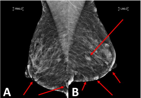
Mediolateral oblique and craniocaudal images of both breasts demonstrate multiple round and oval circumscribed cutaneous masses in both breasts of varying sizes. (Figure 1, 2).

Diagnosis:
The multiple round and oval cutaneous masses in both breasts represented multiple neurofibromas in a patient with known history of NF1 rather than masses within the breast parenchyma.
NF-1 is one of the most common genetic disorders, it occurs in 90% of neurofibromatosis cases and has a prevalence of one in 3000 births [5].The main clinical manifestations of NF-1 is café au lait which occurs within the first year of life and in most patients with the NF-1 mutation [6]. Lisch nodules, optic gliomas, neurological impairment, scoliosis, oral and maxillofacial abnormalities, malignant tumors of the nerve sheath, pheochromocytoma, and bone deformities being additional common clinical findings. [5,6]
Mutations to the gene occur during the embryonic period before neural crest differentiates. [3,5] Due to the large size of the NF1 gene, a high degree of sporadic mutations are observed along with an approximation of 50
In cases of spontaneous NF1 mutations that present with neurofibromin tumors, it is crucial that physicians are aware of how the tumors might progress to prevent further complications, as they are at increased risk for developing cancers. Patients presenting with NF1 tumors should be monitored with systemic therapies and follow-up screening for development of new lesions and to ensure current tumors are not evolving. This can be achieved by annual screening mammography, with the radiologist paying close attention to detail to differentiate between cutaneous and intraparenchymal masses. Understanding the classic appearance of NF1 on mammography can help prevent unnecessary workup while also appropriately identifying suspicious findings requiring further evaluation.
Dear Editorial Team, Clinical Medical Reviews and Reports. My experience with the journal was highly positive. The peer-review process was rigorous, constructive, and completed in a timely manner. The reviewers provided valuable comments that helped improve the quality and clarity of our manuscript. The editorial office was professional, responsive, and supportive throughout all stages of the publication process. Communication was clear and efficient, and any questions were addressed promptly. Overall, I found the journal to maintain high scientific standards and an excellent publication workflow. I would be pleased to consider submitting future work to this journal. Best wishes from, Elena Popa.

It was my pleasure to submit my testimonial concerning the Reviewer Board of our Scientific Journal “Brain and Neurological Disorders”. The Reviewers focused on some modifications and their contribution was helpful. The ladies of our Editorial Office were also supported my efforts. It was my honor to have such a co-operation and I am looking forward for more collaboration.

Dear Grace Pierce, Editorial Coordinator of Journal of Clinical Research and Reports, Thank you for the speedy and efficient peer review process. I appreciate the fact that your peer reviewers do not take months to respond like with some other journals. I would also like to thank the editorial office for responding quickly to my questions. It is an excellent journal. I plan to submit more manuscripts in the future. Best wishes from, Robert W. McGee

Dear Grace Pierce, Editorial Coordinator of Journal of Clinical Research and Reports, Working with you and your team on our recent publication in JCRR has been a truly wonderful and enjoyable experience. The responses were prompt, and the reviewers were patient, constructive, and highly professional. One reviewer in particular gave me the feeling that a professor was carefully reading and commenting on my coursework, which was deeply touching. The entire process was straightforward and hassle‑free, with no tedious online forms to complete. I highly recommend this journal. Best wishes from, DR Aibing Rao, Head of R&D

I Appreciate the Opportunity to Share my Experience with the Journal of Clinical Research and Reports. The peer review process was timely and constructive, and the feedback provided helped improve the quality of our manuscript. The editorial office was professional, responsive, and supportive throughout the process, ensuring smooth communication and efficient handling of the submission. Overall, it was a positive experience collaborating with your team.

Dear Mercy Grace, Editorial Coordinator of Obstetrics Gynecology and Reproductive Sciences, We would like to express our gratitude for your help at all stages of publishing and editing the article. The editors of the magazine answer all the necessary questions and help at every stage. We will definitely continue to cooperate and publish other works in the Obstetrics Gynecology and Reproductive Sciences! Best wishes from, Alla Konstantinovna Politova,
