
Case report | DOI: https://doi.org/10.31579/2578-8868/361
*Corresponding Author: Sérend Hipe, IFPEA, Rue des Saint-Pères, 75 006 Paris, France.
Citation: Sérend Hipe (2025), Neuro-Anatomical Approach to the Humano-Murian Brain based on Cerebral Imaging Studies, J. Neuroscience and Neurological Surgery, 17(4); DOI:10.31579/2578-8868/361
Copyright: ©, 2025, Sérend Hipe. This is an open-access article distributed under the terms of The Creative Commons Attribution License, which permits unrestricted use, distribution, and reproduction in any medium, provided the original author and source are credited
Received: 31 January 2025 | Accepted: 12 March 2025 | Published: 02 April 2025
Keywords: human brain ; Scan studies ; humano-murian ; motor skills ; corpus callosum
We present the case of a humano-murian victim of a left stroke who presented with impaired motor skills on the left side of his body. Brain imaging studies (Scan, Pet Scan) have shown that the brain is functionally separated into two parts. The white corners and the corpus callosum are calcified and leave almost no possibility for inter-hemispheric communication as is the case for us. Basically, the entire anatomical substrate that allows our human brain to communicate between the two hemispheres is completely inactivated in humano-murians. It is among the latter that the term di-encephalon takes on its full meaning.
We present a case study of anatomical pathology by medical imaging after having been able to study a case of humano-murian hospitalized after a stroke with loss of sensitization of the right hemi-body, associated motor disorders (hemiplegia) and anosognosia that will give way after a few days. The patient was 78 years old (Zia, 2021), male and in possession of all means prior to the stroke.
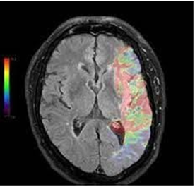
Our recent research has shown that humano-murians have two brains. Understand that they do not have two brains in the skull, but that their brains are functionally separated into two parts. The white corners and the corpus callosum are calcified (Figure 1 and 2) and leave almost no possibility for interhemispheric communication as is the case for us. Basically, the entire anatomical substrate that allows our human brain to communicate between the two hemispheres is totally inactivated in humano-murians. It is in the latter that the term diencephalon takes on its full meaning
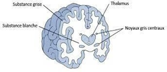
Figure 1: The white matter allows the communication of the two cerebral hemispheres. It is totally calcified in Humano-Murians
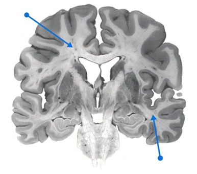
Figure 2: White matter makes up a large proportion of the brain. Its difference in mineral composition between that of humans and human-murians makes it a unique cerebral constitution (Martin and al., 2024).
This has considerable consequences on their brain and sensory capacities (Liew and al., 2023). Indeed, on the motor level, there is no longer any lateral predominance, so all humano-murians are completely ambidextrous. Their movements sometimes seem wider to us while, paradoxically, they are permanently parasitized by a slight tremor that we had mistakenly taken for one of the effects of the polar cold that reigns on the Wall. These tremors are similar to Parkinson's movements. As a result, there is no pyramidal decussation (Figure 3 and 4), so motor skills are controlled by the homolateral cerebral hemisphere, which is not the case in humans (in whom the right hemibody is controlled by the motor cortex of the left cerebral hemisphere). The human-murian has developed frightening motor qualities by being able to dissociate the left hemibody and the right hemibody, which can carry out two different motor activities.
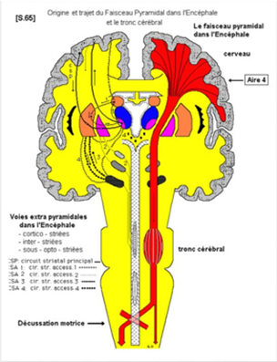
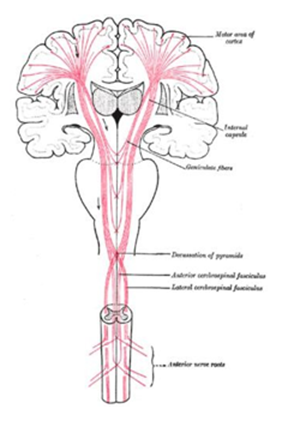
Figure 3 and 4: Pyramidal decussation (Riley and al., 2011).
According to Ramón y Cajal (Hope and al., 2013), pyramidal decussation was related to that of sensory pathways: in the optic chiasma we also observe the decussation of a large part of the optic nerve fibers, which adapts to perception by allowing both hemispheres to have complete
information about what both eyes perceive and can generate complete images that can be located in space. In this sense, the displacement necessary to react to a possible threat would be that of muscle groups, unlike the part of the brain that perceives them. If there is no pyramid discussion, the information would first go to the other hemisphere, then process and react, which would be slower. This explains the particularity of the motor reactions of humano- murians, which we had initially taken as an almost constitutional nonchalance.
Each left and right "brain" has thus developed its own potential independently. If humano- murians use a very particular type of communication that we have described previously (message 15), it is now certain that it stems from this anatomophysiological difference. Human- Murian communication is a mixture of a powerful process of empathy that transmits intentions, images, and emotions to the interlocutor, before verbal language expresses the thought formulated in words. A communication by double channel, a verbal channel coupled with a cognitive channel, knowing that on the latter we humans are very good receivers to our Murian friends but are unable to activate it ourselves. The impression we have is to hear in a way within ourselves what the lips of humano-murians will pronounce with a slight delay. This seems to us to function as a preform of telepathy and allows almost any human to understand very quickly the humano-murian language and its multiple dialects all around the Wall. This mode of communication uses both a very organic substrate and follows primitive phylogenetic pathways (use of pheromone cloud as we have previously demonstrated) and very elaborate thought processes, similar to the secondarized psychic processes of the human psyche.
The patient who allowed us to discover these anatomical and physiological elements is in a way our "patient 1"(as Sperber and al., 2023 calls this type of founder patient). This humano-murian had indeed been the victim of a left stroke and had a motor impairment on the left side of his body (left hemiplegia, facial paralysis and oculomotor muscles of the left orbit) which seemed impossible to us. His name, Solpinan, will forever be associated with this discovery.

Photos 5 and 6: MRI images after stroke: significant initial temporo-parieto-occipital involvement, the last images (top) show a very rapid resorption.
The reality of the two cerebral hemispheres (Alexander and al., 2010, Brodtmann and al., 2020, Chen and al., 2022) has been known for a long time, on the other hand our explorations in imaging have made it possible to introduce a discussion around the notion of presence or absence of interhemispheric white substance. The proposed clinical case made it possible to highlight such a neuro-anatomical reality with unsuspected neurophysiological consequences. until then, the absence of white substance was perceived as a deficit. Our study seems to have demonstrated that this is, in our patients, a developmental and innate functioning of a cerebral organization typical of this type of population.
This is a case study of a human-murian who suffered a left stroke and had a motor impairment on the left side of his body. Brain imaging examinations (Scan, Pet Scan) have shown that the brain is functionally separated 5 into two parts. The white matter and corpus callosum are calcified and leave almost no possibility of inter-hemispheric communication as is the case in the human brain. Basically, the entire anatomical substrate that allows our human brain to communicate between the two hemispheres is totally inactivated in humano-murians. It is among the latter that the term diencephalon takes on its full meaning.
The author declares absence of conflict of interest.
Dear Editorial Team, Clinical Medical Reviews and Reports. My experience with the journal was highly positive. The peer-review process was rigorous, constructive, and completed in a timely manner. The reviewers provided valuable comments that helped improve the quality and clarity of our manuscript. The editorial office was professional, responsive, and supportive throughout all stages of the publication process. Communication was clear and efficient, and any questions were addressed promptly. Overall, I found the journal to maintain high scientific standards and an excellent publication workflow. I would be pleased to consider submitting future work to this journal. Best wishes from, Elena Popa.

It was my pleasure to submit my testimonial concerning the Reviewer Board of our Scientific Journal “Brain and Neurological Disorders”. The Reviewers focused on some modifications and their contribution was helpful. The ladies of our Editorial Office were also supported my efforts. It was my honor to have such a co-operation and I am looking forward for more collaboration.

Dear Grace Pierce, Editorial Coordinator of Journal of Clinical Research and Reports, Thank you for the speedy and efficient peer review process. I appreciate the fact that your peer reviewers do not take months to respond like with some other journals. I would also like to thank the editorial office for responding quickly to my questions. It is an excellent journal. I plan to submit more manuscripts in the future. Best wishes from, Robert W. McGee

Dear Grace Pierce, Editorial Coordinator of Journal of Clinical Research and Reports, Working with you and your team on our recent publication in JCRR has been a truly wonderful and enjoyable experience. The responses were prompt, and the reviewers were patient, constructive, and highly professional. One reviewer in particular gave me the feeling that a professor was carefully reading and commenting on my coursework, which was deeply touching. The entire process was straightforward and hassle‑free, with no tedious online forms to complete. I highly recommend this journal. Best wishes from, DR Aibing Rao, Head of R&D

I Appreciate the Opportunity to Share my Experience with the Journal of Clinical Research and Reports. The peer review process was timely and constructive, and the feedback provided helped improve the quality of our manuscript. The editorial office was professional, responsive, and supportive throughout the process, ensuring smooth communication and efficient handling of the submission. Overall, it was a positive experience collaborating with your team.

Dear Mercy Grace, Editorial Coordinator of Obstetrics Gynecology and Reproductive Sciences, We would like to express our gratitude for your help at all stages of publishing and editing the article. The editors of the magazine answer all the necessary questions and help at every stage. We will definitely continue to cooperate and publish other works in the Obstetrics Gynecology and Reproductive Sciences! Best wishes from, Alla Konstantinovna Politova,
