
Case Report | DOI: https://doi.org/10.31579/2692-9406/096
1 I Degree Specialist in Surgery, Assistant Professor, Provincial General University Hospital “Mártires del 9 de Abril”, Sagua la Grande, Villa Clara, Cuba.
2 I Degree Specialist in Surgery, Instructor Professor, Provincial General University Hospital “Mártires del 9 de Abril”, Sagua la Grande, Villa Clara, Cuba.
3 I Degree Specialist in Surgery, Provincial General University Hospital “Mártires del 9 de Abril”, Sagua la Grande, Villa Clara, Cuba.
4 3rd year resident in Surgery, Provincial General University Hospital “Mártires del 9 de Abril”, Sagua la Grande, Villa Clara, Cuba.
*Corresponding Author: Eida Moya Hernández, I Degree Specialist in Surgery, Assistant Professor, Provincial General University Hospital “Mártires del 9 de Abril”, Sagua la Grande, Villa Clara, Cuba.
Citation: Eida M Hernández, René V Estévez, Andrés P Morales, Yanet F Mirabal, Hanuary L. S Estebanés. (2022). Necrobiosis of a Uterine Fibroid in the Course of a Pregnancy. Biomedical Research and Clinical Reviews. 6(2); DOI: 10.31579/2692-9406/096
Copyright: © 2022 Eida Moya Hernández, This is an open-access article distributed under the terms of the Creative Commons Attribution License, which permits unrestricted use, distribution, and reproduction in any medium, provided the original author and source are credited.
Received: 13 November 2021 | Accepted: 03 January 2022 | Published: 21 January 2022
Keywords: uterine myoma; leiomyoma; liomyoma; fibromyoma
Uterine myoma is a very common benign neoplasm. A 32-year-old patient with 8 weeks of gestation was presented, who came to the emergency room presenting abdominal pain of two days of evolution, vomiting and low-grade fever in the course of an already known and desired pregnancy, with a diagnosis of necrobiosis of a uterine fibroid. The fibroid was resected and the gravid uterus is preserved. The patient is progressing satisfactorily, she being discharged seven days later, she is followed by outpatient consultation and 20 weeks later she maintains a normal pregnancy. Key words: necrobiosis, uterine fibroma, pregnancy.
Uterine myoma is a benign neoplasm of the uterus that is popularly known by the name of fibroma and from the anatomopathological point of view as leiomyoma, liomyoma and fibromyoma, according to the predominance of smooth muscle fibers or the amount of fibrous tissues [1].
Its frequency makes it one of the most common diagnoses in patients undergoing surgery in gynecology and general surgery services. Predominantly in women aged 30-50 years, although nowadays they are diagnosed earlier, before they produce the symptoms, due to the widespread use of ultrasound [2].
On the other hand, it can be single or multiple and be located anywhere in the uterus; A higher frequency is also reported in black, mestizo and multiparous women. Some authors [3] consider that the cause is unknown, others value the sustained or increased estrogenic stimulus, which activates the genitoblasts (immature muscular elements), mesodermal and embryonic cells that respond to this stimulus.
Many women with uterine fibroids have no symptoms and never require treatment; however, one in four women of reproductive age suffers from significant symptoms, which can vary depending on the location, size, and number of fibroids.
LMN patient, female, black race, housewife, 32 years old with 8 weeks of gestation, who went to the emergency room of the General Hospital “Dr. Loery Comba ”in Malabo, in the Republic of Equatorial Guinea, with abdominal pain of two days of evolution, vomiting and low-grade fever. She has a history of three previous full-term pregnancies and eutocic deliveries, no Family History of Chronic Hereditary Diseases is collected. Current desired pregnancy.
Physical exploration
The physical examination revealed a general condition, temperature of 37.6 ° C, blood pressure of 100/60 mmHg, heart rate of 100x min. Flat abdomen that follows respiratory movements, normal abdominal air-fluid sounds, on soft and depressible palpation, diffusely painful with peritoneal reaction towards the hypogastrium, especially the right iliac fossa, no tumor is palpated, normal percussion, negative digital rectal examination. Vaginal examination with an enlarged uterus that impresses more than 10cm, the glove comes out stained with blood.
Diagnostic evaluation
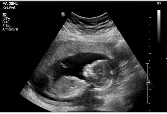
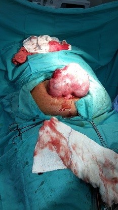
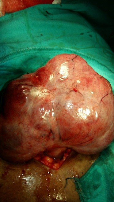
After the meticulous physical examination, laboratory tests were performed: HIV negative, Hepatitis B negative, Hepatitis C negative, Hct in 0.32, urine not useful, Eneb test positive, abdominal ultrasound (Fig. 1) that shows complex image in the body of the uterus Hypogenic predominance, with a single and living fetus inside, normal adnexa, a discrete amount of fluid in the Douglas sac. A diagnosis of necrobiosis of a uterine fibroid was raised during a pregnancy, the possibility of Acute Appendicitis was also assessed. Since it was a desired pregnancy, the patient and family members were explained the possibility of losing it and that action would be taken according to the intraoperative findings. In the operating room, after spinal anesthesia, an infraumbilical median laparotomy is performed and necrobiosis of a pedunculated fibroma of the fundus of the uterus is verified, we proceed to resection between forceps and continuous helical chrome-plated suture 0 in 2 planes, the hemostasis and it was verified that the pregnancy of the uterus did not present other manifestations and the abdomen is closed by planes. The patient recovers satisfactorily and is transferred to the conventional room with treatment: Rocephin (1g) 1bbo intravenous every 12 hours, hydration and Diclofenac Sodium (75mg) 1 amp intramuscular every 12 hours and also includes the proper care of the surgery.
At 18 hours after the operation, oral feeding is started and urinary catheter is removed. The patient was hospitalized for 7 days with the administration of the antibiotic and general measures. She discharged without difficulties and was followed up by outpatient consultation every 15 days, and once a month by ultrasonography, maintaining a normal pregnancy until 20 weeks, after which she stopped attending the consultation. About fourt weeks later, she was located through a native health worker who helped reincorporate her to follow-up. She remained the rest of the normal time, at 38 weeks she was admitted and an elective cesarean section was performed. The product was a 4200 gram newborn male with good vitality. He graduated on the 5th day, both with a satisfactory evolution. She was followed in the health area until 2 months without complications.
Uterine fibroids are a fairly common pathology in women. Normally they do not usually cause serious problems during pregnancy, although these pregnancies are normally controlled by the High Risk Obstetric Unit [4].
Cases similar to the one presented are described by other authors such as Moslemi et al [5] who describe a 36-year-old female, primigravida, who consulted at 11 weeks of gestation due to abdominal distension, edema in the lower extremities and scarce vaginal bleeding, and De Dios Perera et al [6] who present the clinical case of a 27-year-old patient with 17.2 weeks of pregnancy, who was under follow-up at the Gynecology consultation at the Mariana Grajales Coello Provincial Gynecological Hospital in Santiago de Cuba for presenting a symptomatic uterine myoma with Acute abdominal pain due to necrobiosis, which required urgent surgical resolution and the pregnant woman evolved satisfactorily.
Care for pregnant women with uterine myoma should preferably be conservative, as shown in the consulted literature; [7] therefore, perigravid myomectomies should be avoided. When there is an uncomplicated fibroid, it is not indicated to remove it by surgery during pregnancy or in the course of a cesarean section. In early pregnancy, leiomyomas smaller than 5 cm in diameter are often undetectable [8].
Myomectomy carries risks of haemorrhage and abortion, which is why it is only reserved for specific cases that do not respond to expectant management, as occurred in the case presented above, criteria coinciding with other authors [5,9].
The medical treatment of fibroids is a recent strategy and is attractive to many gynecologists because of its ease and fewer complications when compared to surgery, especially when the main intention is the preservation of fertility or the desire to preserve the uterus [10].
The current recommendation and experience indicate that it should be performed in the second trimester of pregnancy, although there are cases reported in the first trimester. Even these patients would have a better obstetric outcome than those treated expectantly.
Dear Editorial Team, Clinical Medical Reviews and Reports. My experience with the journal was highly positive. The peer-review process was rigorous, constructive, and completed in a timely manner. The reviewers provided valuable comments that helped improve the quality and clarity of our manuscript. The editorial office was professional, responsive, and supportive throughout all stages of the publication process. Communication was clear and efficient, and any questions were addressed promptly. Overall, I found the journal to maintain high scientific standards and an excellent publication workflow. I would be pleased to consider submitting future work to this journal. Best wishes from, Elena Popa.

It was my pleasure to submit my testimonial concerning the Reviewer Board of our Scientific Journal “Brain and Neurological Disorders”. The Reviewers focused on some modifications and their contribution was helpful. The ladies of our Editorial Office were also supported my efforts. It was my honor to have such a co-operation and I am looking forward for more collaboration.

Dear Grace Pierce, Editorial Coordinator of Journal of Clinical Research and Reports, Thank you for the speedy and efficient peer review process. I appreciate the fact that your peer reviewers do not take months to respond like with some other journals. I would also like to thank the editorial office for responding quickly to my questions. It is an excellent journal. I plan to submit more manuscripts in the future. Best wishes from, Robert W. McGee

Dear Grace Pierce, Editorial Coordinator of Journal of Clinical Research and Reports, Working with you and your team on our recent publication in JCRR has been a truly wonderful and enjoyable experience. The responses were prompt, and the reviewers were patient, constructive, and highly professional. One reviewer in particular gave me the feeling that a professor was carefully reading and commenting on my coursework, which was deeply touching. The entire process was straightforward and hassle‑free, with no tedious online forms to complete. I highly recommend this journal. Best wishes from, DR Aibing Rao, Head of R&D

I Appreciate the Opportunity to Share my Experience with the Journal of Clinical Research and Reports. The peer review process was timely and constructive, and the feedback provided helped improve the quality of our manuscript. The editorial office was professional, responsive, and supportive throughout the process, ensuring smooth communication and efficient handling of the submission. Overall, it was a positive experience collaborating with your team.

Dear Mercy Grace, Editorial Coordinator of Obstetrics Gynecology and Reproductive Sciences, We would like to express our gratitude for your help at all stages of publishing and editing the article. The editors of the magazine answer all the necessary questions and help at every stage. We will definitely continue to cooperate and publish other works in the Obstetrics Gynecology and Reproductive Sciences! Best wishes from, Alla Konstantinovna Politova,
