
Case Report | DOI: https://doi.org/10.31579/2690-8808/210
1Otorhinolaryngologist, PhD in Otorhinolaryngology, professor of de Medicine Course at Universidade do Sul de Santa Catarina, Brasil.
2Medical student at the Universidade do Sul de Santa Catarina, Brasil.
3Infectious Disease Specialist.
*Corresponding Author: Carlos Eduardo Monteiro Zappelini. Otorhinolaryngologist, PhD in Otorhinolaryngology, professor of de Medicine Course at Universidade do Sul de Santa Catarina, Brasil.
Citation: Carlos E. M. Zappelini, Rafaela Z. Machado, Isabela M. Destro, Giulia A. Mendes, Maria E. C. da Maia, et al, (2024), Nasal Histoplasmosis in an Immunocompetent Patient - A Case Report, J, Clinical Case Reports and Studies, 5(6); DOI:10.31579/2690-8808/210
Copyright: ©, 2024, Carlos Eduardo Monteiro Zappelini. This is an open access article distributed under the Creative Commons Attribution License, which permits unrestricted use, distribution, and reproduction in any medium, provided the original work is properly cited.
Received: 30 July 2024 | Accepted: 07 August 2024 | Published: 14 August 2024
Keywords: histoplasmosis; immunocompetence; fungal infection
The clinical case described below deals with the diagnosis and treatment of a fungal infection in the nasal region, rare in immunocompetent patients and with few reports in the literature. A 49-year-old woman from the south of Brazil, with no previous comorbidities, began to suffer from swelling in the right nasal and orbital region for around 4 weeks, accompanied by intense pain, fever and drainage of purulent secretion from the nostril. The first biopsy was inconclusive, but subsequent biopsies led to the identification of a fungal infection and finally the diagnosis of histoplasmosis. Treatment with antifungal drugs was then started, which consequently allowed the condition to improve after several hospitalizations.
Histoplasmosis is a systemic fungal disease caused primarily by the inhalation of spores of the fungus Histoplasma capsulatum which typically affects the lungs, but can affect other areas of the body such as the brain, spinal cord and head and neck [1]. Mucocutaneous involvement occurs in 10-25% of cases and is an important diagnostic sign of disseminated Histoplasmosis. In immunocompetent patients, the cure is usually spontaneous [2]. Its endemic regions are Central America, South America, Africa, Asia and Australia, but the emergence of HIV and the increasing use of immunosuppressants have led to the emergence of cases in non-endemic areas [3]. In Brazil, the disease is present in all regions.
A 49-year-old woman from the state of Santa Catarina, Brazil, with no previous comorbidities, presented with a 4-week history of edema in the nasal and orbital regions, along with drainage of purulent secretion from the nose, with intense pain in the right hemiface and eye, associated with fever. Physical examination revealed significant edema in the nasal region, bulging of the nasal wing internally and periorbital edema. A CT scan of the skull showed that the nasal cavity was occupied, especially on the right, by content with soft tissue attenuation and showing contrast enhancement, as well as occupying the right maxillary and sphenoid sinus. The initial diagnosis was periorbital and nasal cellulitis, and the patient was admitted for treatment with cefepime and vancomycin. However, the patient fled before the treatment was completed.
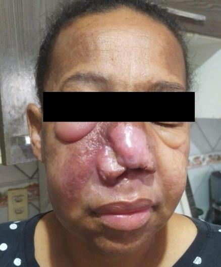
Figure 1: Edema in the nasal and orbital regions
After the patient's condition worsened, she returned to the hospital's emergency department with local redness and necrosis, as well as brownish discharge from her nostrils. She was taking Levofloxacin and Prednisone.
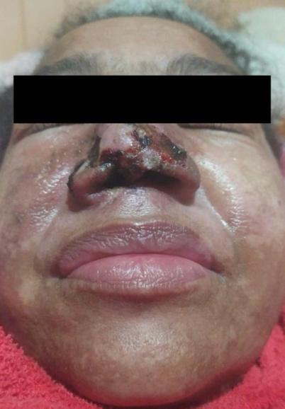
Figure 2: Local necrosis
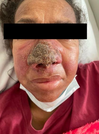
Figure 3: Progression of local necrosis.
Some biopsies were conducted during his hospitalization: the first showed only inflammation - where ceftazidime and clindamycin were started, and the culture collected was positive for Vancomycin-sensitive Staphylococcus CGN. Following this, a new biopsy was carried out showing a fungal infection. As a course of action, amphoterecin-B was started and a new biopsy was requested to elucidate the diagnosis. Debridement of the devitalized tissue was necessary and the lesion improved compared to the initial biopsy. During this hospitalization, the patient suffered from anemia and increase in nitrogenous compounds, requiring the prescription of concentrated red blood cells and correction of the Amphotericin-B dose according to the glomerular filtration rate.
A new skin biopsy showed Histoplasmosis and on the same day the patient once again expressed her wish to be discharged. The risks were explained and she was advised to continue her outpatient follow-up with an infectious disease specialist, as well as prescribed itraconazole on discharge.
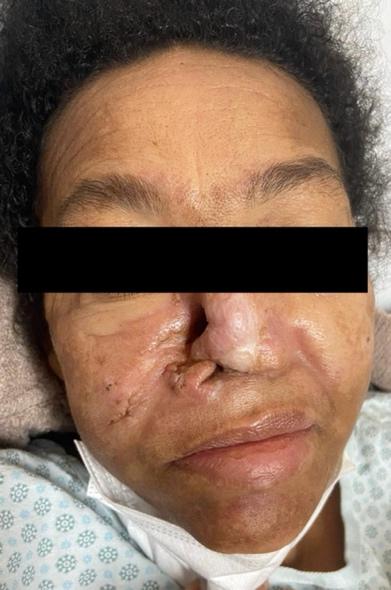
Figure 4: Devitalized tissue debridement
The patient once again returned to hospital with a lesion in the nasal region with purulent and spontaneous drainage, without airway obstruction and taking itraconazole correctly. The patient was admitted due to the findings of hyponatremia and respiratory alkalosis according to the laboratory tests carried out. Oxacillin and ceftriaxone were administered. According to the service's otorhinolaryngologist, there was no need for an endonasal approach at the moment, but nasal reconstruction would be necessary once the disease was under control. Patient discharged due to clinical improvement.
In July 2023, the patient returned to hospital complaining of dizziness and asthenia for 22 days, right-sided paresis for 20 days and epigastric pain for 2 weeks. At the moment the patient was being treated by an infectious disease specialist for a fungal lesion in the nose. The tests on arrival showed leukocytosis with a left shift, proteinuria and a hemoglobin of 9.9. Amphotericin-B and corticoids were started. The following day, the patient's general condition deteriorated, with drowsiness and a productive cough, no signs of respiratory distress and afebrile. 1 CHAD was administered. A CT scan of the skull identified a deep lesion from the temporal-insular to the nucleus-capsular on the left side, with vasogenic edema in between, and conservative neurosurgical treatment was adopted due to the impossibility of access. On the 15th, the patient's condition worsened, presenting with glasgow 7, respiratory discomfort, pulmonary auscultation with diffuse rhonchi and use of accessory muscles. IOT was performed and the patient was transferred to the ICU, amphotericin-B and decadron were maintained and Tazocin was introduced, and the patient was treated for bronchopneumonia. It was necessary to use noradrenaline. After a few days, vancomycin was started guided by cultures (Staphylococcus aureus) and Tazocin was suspended. Noradrenaline had to be increased, vasopressin associated and hemodialysis performed. On the 18th, the patient had fixed bilateral mydriasis without brainstem reflexes and a CT scan of the skull with diffuse cerebral ischemia affecting the brainstem. The patient presented with enterorrhagia and required a dose of platelets and a cryoprecipitate. On the 20th, a family conference was held and the Brain Death assessment protocol was started. The following day, the patient died at 9:52 a.m. with mydriatic pupils, no trunk reflexes, cold and cyanotic extremities, and no pulses.
Histoplasmosis is an endemic systemic mycosis in Brazil and is associated with seropositive patients in over 86% of cases. In this context, immunodepression seems to be a major risk factor for the development of more severe forms of the disease. [4] In general, the infection does not
usually cause major clinical repercussions and is asymptomatic in over 90% of cases. When present, the most common symptoms are fever, discomfort, headache, fatigue, dry cough and chest discomfort. The disease tends to resolve spontaneously in most cases within 2 to 4 weeks. [4] Histoplasma infection can progress to dissemination with or without symptoms, but this clinical form has been more associated with individuals with immunodepression due to AIDS, neoplasms (leukemias, lymphomas) and transplant patients. [4] The disseminated form of infection is rarely manifested by skin lesions, and is more common in reports from Latin America, affecting up to half of the cases when compared to reports from the United States, where 10% of infections are associated with histoplasma skin lesions. [4-6]. Evidence from studies attributes these differences to the skin tropism of the strains diagnosed in South America. The cutaneous manifestations of the disease are polymorphous and range from papules and nodules to ulcers and vegetating lesions. Therapy for disseminated Histoplasmosis is usually carried out using Amphotericin B and the second line of treatment is Itraconazole [6].
The case report shows an atypical presentation of Histoplasmosis in the nasal region. The differential diagnosis of these lesions should always be considered, especially in immunosuppressed patients, where delayed diagnosis and effective treatment can affect the patient's prognosis.
Dear Editorial Team, Clinical Medical Reviews and Reports. My experience with the journal was highly positive. The peer-review process was rigorous, constructive, and completed in a timely manner. The reviewers provided valuable comments that helped improve the quality and clarity of our manuscript. The editorial office was professional, responsive, and supportive throughout all stages of the publication process. Communication was clear and efficient, and any questions were addressed promptly. Overall, I found the journal to maintain high scientific standards and an excellent publication workflow. I would be pleased to consider submitting future work to this journal. Best wishes from, Elena Popa.

It was my pleasure to submit my testimonial concerning the Reviewer Board of our Scientific Journal “Brain and Neurological Disorders”. The Reviewers focused on some modifications and their contribution was helpful. The ladies of our Editorial Office were also supported my efforts. It was my honor to have such a co-operation and I am looking forward for more collaboration.

Dear Grace Pierce, Editorial Coordinator of Journal of Clinical Research and Reports, Thank you for the speedy and efficient peer review process. I appreciate the fact that your peer reviewers do not take months to respond like with some other journals. I would also like to thank the editorial office for responding quickly to my questions. It is an excellent journal. I plan to submit more manuscripts in the future. Best wishes from, Robert W. McGee

Dear Grace Pierce, Editorial Coordinator of Journal of Clinical Research and Reports, Working with you and your team on our recent publication in JCRR has been a truly wonderful and enjoyable experience. The responses were prompt, and the reviewers were patient, constructive, and highly professional. One reviewer in particular gave me the feeling that a professor was carefully reading and commenting on my coursework, which was deeply touching. The entire process was straightforward and hassle‑free, with no tedious online forms to complete. I highly recommend this journal. Best wishes from, DR Aibing Rao, Head of R&D

I Appreciate the Opportunity to Share my Experience with the Journal of Clinical Research and Reports. The peer review process was timely and constructive, and the feedback provided helped improve the quality of our manuscript. The editorial office was professional, responsive, and supportive throughout the process, ensuring smooth communication and efficient handling of the submission. Overall, it was a positive experience collaborating with your team.

Dear Mercy Grace, Editorial Coordinator of Obstetrics Gynecology and Reproductive Sciences, We would like to express our gratitude for your help at all stages of publishing and editing the article. The editors of the magazine answer all the necessary questions and help at every stage. We will definitely continue to cooperate and publish other works in the Obstetrics Gynecology and Reproductive Sciences! Best wishes from, Alla Konstantinovna Politova,
