
Review | DOI: https://doi.org/10.31579/2690-1919/205
1 Kujawy University, Mechanical Department, Hallera 32, 86-300 Grudziadz, Poland and CORSAR Engineering Industry, Glogowa 2, 86-031 Osielsko, Poland.
2 Tribochemistry Consulting, Salt Lake City, UT 84117, USA and University of Economy, Biotribology Lab, Garbary 2, 85-229, Bydgoszcz, Poland.
*Corresponding Author: Z. Pawlak, Tribochemistry Consulting, Salt Lake City, UT 84117, USA and University of Economy, Biotribology Lab, Garbary 2, 85-229, Bydgoszcz, Poland.
Citation: M.Sojka and Z. Pawlak. (2021). Nanostructures of phospholipids on articular cartilage surface and their functions. J Clinical Research and Reports, 9(3); DOI:10.31579/2690-1919/205
Copyright: © 2021, Z. Pawlak. This is an open access article distributed under the Creative Commons Attribution License, which permits unrestricted use, distribution, and reproduction in any medium, provided the original work is properly cited.
Received: 01 October 2021 | Accepted: 25 October 2021 | Published: 05 November 2021
Keywords: amphoteric cartilage; phospholipid bilayers; friction coefficient; wettability; hydrophilic and hydrophobic
Phospholipids bilayers fulfill an important role in natural joint lamellar-repulsive lubrication mechanism. Low friction between surfaces coated with negatively charged the phospholipid headgroup (–PO4-) as being due to a hydration layer. Wettability of the cartilage surface depends on the number of PLs that act as a lubricant. The cartilage can be classified as a group of intelligent material, which in the wet state has a contact angle of ~0º, and the air-dry state has a contact angle of ~104º.
1. Phospholipid bilayers adsorbed on the cartilage surface
Phospholipids bilayers fulfill an important role in natural joint lamellar-repulsive lubrication mechanism [1, 2]. Low friction between surfaces coated with negatively charged the phospholipid headgroups (–PO4-) as being due to a hydration layer. Articular cartilage (AC) is a specific type of connective tissue without blood vessels and innervation. There is hyaline cartilage 3-5mm thick in the joints, which feeds in motion based on diffusion. About 65-80% of cartilage is water [1, 2]. During the movements, it allows the bones to move in relation to each other in the joint.
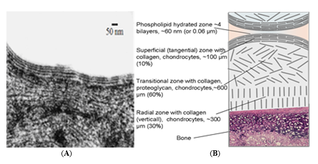
In Figure 1(B), four zones in the articular cartilage (~ 0.06)mm 3-5 phospholipid bilayers, 100 mm horizontally arranged collagen fibers with chondrocytes, ~ 600 mm middle zone with indirectly arranged collagen, proteoglycan and chondrocytes, ~ 300 mm collagen (vertical arrangement) with chondrocytes) were distinguished. The surface of the cartilage in the joint is hydrophilic, and the cartilage on the air loses its moisture and becomes hydrophobic.
2. The surface of AC and synovial fluid
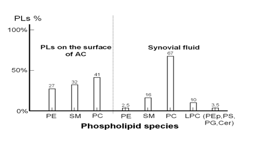
Figure 2 shows the range of concentrations of phospholipids present on the surface of AC [4] and the concentration of particular types of phospholipids in a healthy synovial fluid [5]. Concentrations (%) of phospholipids adsorbed on the AC surface are as follows: phosphatidylcholine (PC) 41%, sphingomyelin (SM) 32% and phosphatidylethanolamine (PE) 27% whereas the synovial fluid is dominated by PC (67%), sphingomyelin, lysophosphatidylcholine (16% and 10% respectively), PE 2.5% and 3.5% (PEp = phosphatidylethanolamine-based plasmalogens; PS = phosphatidylserine; PG = phosphatidylglycerol; Cer = ceramide).
Chondrocytes naturally produce phospholipids in the joints. Phospholipids support the majority of organ functions such as the cardiovascular system, nervous system, liver functions digestive system and, most importantly, selectively settle on the cartilage surface [4, 6] and create bilayers that are involved in border friction.
3. Hydrophilic and hydrophobic character cartilage surface
The uniqueness of natural cartilage tissue can be determined by its amphoteric, hydrophilic and hydrophobic character, wettability, and resistance to load. The cartilage can be classified as a group of intelligent material, which in the wet state has a contact angle of ~0º, and the air-dry state has a contact angle of ~104º.
One of the tribochemical parameters of the cartilage surface is its wettability. Wettability characterizes the surface of various materials, which are generally referred to as wettable (highly hydrophilic, ~ 0 to 45º) or non-wettable (highly hydrophobic, ~ 90º to 180º). Evaporation of surface water in the air leads to changes in surface energy and conformational changes of phospholipids on the surface, (a) the wet surface of the cartilage is hydrophilic (~ 0º contact angle), (b) after water evaporation, the bilayers self-reorganize into a monolayer (hydrophobic) 104º of the contact angle. Change in the contact angle depending on the time of drying of the cartilage surface in the air (see Figure 3).
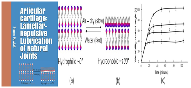
The contact angle of the cartilage surface will depend on the charge density of the amine (-NH2) and phosphate (–PO4-) functional groups, which is expressed in the number of PLs bilayers on the cartilage surface [2, 10]. During the evaporation of water molecules from the surface of the cartilage (dehydration), the process of transformation from the hydrophilic state to the hydrophobic state (HL → HB) takes place. The polar part of the phospholipid molecule (amino (-NH2) and phosphate (–PO4-), the outer bilayer is flipped (flip-flop) to have a contact with the moisture of the cartilage interior and the surface bilayer is reorganized into a monolayer [2, 8, 9]. The high value of the contact angle (dry surface measurement) corresponds to high hydrophilicity when the surface is wet (process reversibility) (see Fig. 4 a, b). A low contact angle (for dry cartilage) corresponds to low hydrophilicity when the surface is wet.
4. Friction vs wettability of the cartilage surface
Wettability of the cartilage surface depends on the number of PLs that act as a lubricant [2, 10]. The study aimed to confirm the hypothesis that healthy and degenerate cartilage samples can be easily identified by measuring wettability. Observations led to the conclusion that the coefficient of friction is significantly dependent on the wettability of the tissue surface. This part of the study analyzed the surfaces of healthy and degenerated samples of articular cartilage through atomic force microscopy (AFM) methods, by measurements of wettability of cartilage surfaces and friction of pairs (cartilage/cartilage) [2, 11].
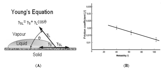
The most common characteristic of the degree of wettability of the joint cartilage surface is the wetting angle θ, which can be determined from the Young-Dupree relationship (Figure 5).

Figure 5A presents the energy distribution of the droplets on the surface of the cartilage and Figure 5B shows the dependence of the coefficient of friction (ƒ) vs. wettability for healthy and degenerate surfaces of bovine cartilage (result after 5 minutes of the test). It has been also shown that phospholipids organized in bilayers on the AC surface are a lubricant and reduce the friction of the surface.
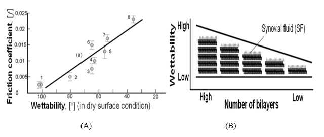
Figure 5A shows the dependence of the coefficient of friction vs. contact angle for pairs of healthy (cartilage/cartilage) and degenerate surfaces. The friction coefficient test confirms the hypothesis about the relationship between the number of phospholipid bilayers (wettability) and friction [12-14]. In the case of healthy cartilage, the coefficient of friction is f=0.004-0.012 and for unhealthy 0.015-0.024 cartilage. The coefficient of friction for sick cartilage increased 2 to 3 times. In Figure 6A, literature values of the coefficient of friction of pairs (cartilage/cartilage) of healthy samples and samples in various degeneration states were collected [6]. Own research supported by the observation of other authors has shown that wettability is an essential parameter in the evaluation of biological surfaces.
Coefficient of friction vs. wettability for natural joints with healthy and degenerated cartilage surfaces of bovine cartilage samples for partially and wholly degenerate samples is presented in Figure (5A and 5B) shows changes in wettability of the AC surface depending on the number of bilayers. Increased values of the coefficient of friction are interpreted by a decrease in the number of bilayers [2, 6]. The implication of this condition was observed in osteoarthritis, where the increase in the coefficient of friction was associated with the gradual loss of surface amorphous layer, SAL [16, 17]. Phospholipid lamellar phases and biomacromolecules in SF participate in the electrostatic repulsion of the surface during friction. Strongly hydrated lamellar PLs are expected to cover cartilage surfaces and participate in hydrophilic-lamellar lubrication [2, 16, 17].
The cartilage surface was characterized using a combination of the pH, wettability and friction coefficient testing methods to support lamellar-repulsive mechanism of hydration lubrication. Wettability of the cartilage surface depends on the number of PLs that act as a lubricant. Phospholipids bilayers fulfill an important role in natural joint lamellar-repulsive lubrication mechanism. The cartilage can be classified as a group of intelligent material, which in the wet state has a contact angle of ~0º, and the air-dry state has a contact angle of ~104º.
Dear Editorial Team, Clinical Medical Reviews and Reports. My experience with the journal was highly positive. The peer-review process was rigorous, constructive, and completed in a timely manner. The reviewers provided valuable comments that helped improve the quality and clarity of our manuscript. The editorial office was professional, responsive, and supportive throughout all stages of the publication process. Communication was clear and efficient, and any questions were addressed promptly. Overall, I found the journal to maintain high scientific standards and an excellent publication workflow. I would be pleased to consider submitting future work to this journal. Best wishes from, Elena Popa.

It was my pleasure to submit my testimonial concerning the Reviewer Board of our Scientific Journal “Brain and Neurological Disorders”. The Reviewers focused on some modifications and their contribution was helpful. The ladies of our Editorial Office were also supported my efforts. It was my honor to have such a co-operation and I am looking forward for more collaboration.

Dear Grace Pierce, Editorial Coordinator of Journal of Clinical Research and Reports, Thank you for the speedy and efficient peer review process. I appreciate the fact that your peer reviewers do not take months to respond like with some other journals. I would also like to thank the editorial office for responding quickly to my questions. It is an excellent journal. I plan to submit more manuscripts in the future. Best wishes from, Robert W. McGee

Dear Grace Pierce, Editorial Coordinator of Journal of Clinical Research and Reports, Working with you and your team on our recent publication in JCRR has been a truly wonderful and enjoyable experience. The responses were prompt, and the reviewers were patient, constructive, and highly professional. One reviewer in particular gave me the feeling that a professor was carefully reading and commenting on my coursework, which was deeply touching. The entire process was straightforward and hassle‑free, with no tedious online forms to complete. I highly recommend this journal. Best wishes from, DR Aibing Rao, Head of R&D

I Appreciate the Opportunity to Share my Experience with the Journal of Clinical Research and Reports. The peer review process was timely and constructive, and the feedback provided helped improve the quality of our manuscript. The editorial office was professional, responsive, and supportive throughout the process, ensuring smooth communication and efficient handling of the submission. Overall, it was a positive experience collaborating with your team.

Dear Mercy Grace, Editorial Coordinator of Obstetrics Gynecology and Reproductive Sciences, We would like to express our gratitude for your help at all stages of publishing and editing the article. The editors of the magazine answer all the necessary questions and help at every stage. We will definitely continue to cooperate and publish other works in the Obstetrics Gynecology and Reproductive Sciences! Best wishes from, Alla Konstantinovna Politova,
