Department of Urology, North Manchester General Hospital, United Kingdom.
*Corresponding Author:
Anthony Kodzo-grey Venyo, Department of Urology, North Manchester General Hospital, Retired Clinician, United Kingdom.
Citation: Anthony Kodzo-grey Venyo, (2024), Myointimoma Of The Penis: Review And Update,Clinical Medical Reviews and Reports; 6(7): DOI: 10.31579/2690-8794/231
Received:06 September 2024
| Accepted:13 September 2024
| Published:20 September 2024
Keywords: Myointimoma of the penis; angiocentric myofibroblastic tumour of the penis; penile mass; glans penis; corpus spongiosum; biopsy;
Abstract
Myointimoma, which is also referred to angiocentric myofibroblastic tumour, is an uncommon benign soft tissue neoplasm which is derived from intimal cells of the vascular spaces of the corpora cavernosa of the penis, histologically typified by multinodular/plexiform myofibroblastic proliferation within the vascular spaces of cavernous bodies of the penis. Myointimoma could afflict children as well as adult males and it does tend to present as a lump or mass upon the penis that had been noticed recently over a few weeks and which recently had been increasing in size or getting larger within the glans penis. Generally, patients who are afflicted by myointimoma tend to be asymptomatic apart from having noticed a lump in the penis. Diagnosis of myointimoma can be confirmed via pathology examination and immunohistochemistry staining studies and molecular and cytogenetics studies of biopsy specimens or excised specimens of the penile tumour. Total excision alone is stated to be sufficient for the management of myointimoma of the glans penis in view of the fact that the tumour tends to portend a benign indolent cause. Less than 30 cases of myointimoma of the penis had so far been reported in the global literature and for this reason, it would be envisaged that most clinicians in the world have never encountered a case of myointimoma of the penis before and they would tend not to be familiar with the presentation, diagnostic features, treatment, and outcome of the tumour ensuing its treatment. The important thing that needs to be appreciated is the fact that myointimoma of the penis which mimics more common lesions of the penis does require careful pathology examination and a high-index of suspicion by the clinician and the pathologist in order to differentiate myointimoma of the penis from its simulants including: plexiform fibrohistiocytic tumour of penis, epithelioid haemangioendothelioma of penis, myofibroma of the penis, intravascular fasciitis of penis, nerve sheath tumour of penis, leiomyoma of penis as well as various other types of lesions afflicting the penis.
Introduction
Myointimoma, which is also referred to as angiocentric myofibroblastic tumour, is an uncommon benign soft tissue tumour which is derived from intimal cells of the vascular spaces of the corpora cavernosa of the penis, histologically typified by multinodular / plexiform myofibroblastic proliferation within the vascular spaces of cavernous bodies. [1] The terminology of myointimoma was stated to be first introduced by Fetsch et al. [2]in 2000 and it was recognized as a distinctive histological entity in the World Health Organization Classification of the Tumours of the Urinary System and Male Genital Organs in 2016 [3]. It has been iterated that up to Nobember 2022, only 22 cases had been reported in the literature, of which only 10 had been reported in children and adolescents. Except of two small series [2] [4] of cases that had based upon a retrospective re-evaluation of few decades stored slides of tumours, these myointimoma tumours had been stated to be based upon isolated case reports. [1] It has been pointed out that in view of the rarity of myointimoma of the penis, the main importance is to be able to differentiate myointimoma from other tumours of variable biological behaviour. In view of the rarity of myointimoma of the penis, it would be envisaged that majority of clinicians all over the world would not have encountered a case of myointimoma of the penis before. The ensuing article on myointimoma of the penis is divided into two parts: (A) Overview which has discussed general aspects of myointimoma and (B) Miscellaneous narrations and discussions from some case reports, case series and studies related to myointimoma of the penis.
AIM
To review and update the literature on myointimoma of the penis.
METHOD
Internet data bases were searched including: Google; Google Scholar; Yahoo; and PUBMED. The search words that were used included: Myointimoma of the penis; Penile myointimoma; Myointimoma of glans penis. Twenty references were identified which were used to write the article which has been divided into two parts: (A) Overview which has discussed general aspects of myointimoma and (B) Miscellaneous narrations and discussions from some case reports, case series and studies related to myointimoma of the penis.
RESULTS
[A] OVERVIEW
Top of Form
Bottom of Form
Definition / general statement
It has been iterated that myointimoma is a terminology that is used for a benign myointimal proliferation with predilection to the corpus spongiosum of the glans penis. [5]
Essential features
The essential features of myointimoma had been summated as follows: [5]
It has been stated that myointimoma is a benign mesenchymal neoplasm which afflicts the corpus spongiosum of the glans penis. [5]
It has been iterated that myointimoma is an intravascular proliferation of the vascular intimal cells. [5]
It has been pointed out that myointimoma lesional cells upon immunohistochemistry staining studies stain with α smooth muscle actin but not desmin. [5]
Terminology
Some of the terminologies that tend to be used for myointimoma had been summated to include the ensuing: [5]
Penile myointimoma.
Myointimoma of the penis.
Epidemiology
The epidemiology of myointimoma had been summated as follows: [5]
It has been pointed out that myointimoma is a rare tumour with about 28 cases reported 28 cases reported in the literature. [2] [4] [6]
It has been iterated that myointimoma was in the past referred to as intravascular leiomyoma, leiomyomatosis, late-stage intravascular fasciitis, leiomyoma of the glans penis, solitary cutaneous myofibroma of the glans penis. [7-9]
Wide age range (2 - 74 years old). [2] [10]
Sites
With regard to the sites of the penis afflicted by myointimoma, it has been stated that myointimoma tends to afflict the corpus spongiosum of the glans penis. [5]
Pathophysiology
It has been iterated that the pathophysiology of myointimoma entails intravascular proliferation from the intimal cells of the inner layer of the corpus spongiosum vasculature in the glans penis. [2]
Aetiology
It has been pointed out that the myointimoma mesenchymal tumour is not related to the status of circumcision, history of trauma or the presence of disease. [2] [10]
Clinical features
The clinical features of myointimoma of the penis had been summated as follows: [5]
Myointimoma of the penis manifests as small, distinct, nonmobile, firm, painless nodule upon the glans penis. [11]
Myointimoma of the penis is a benign, rapidly growing lesion that remain stable f or years, even pursuant to incomplete excision of the lesion or penile lesions. [2]
Diagnosis
Laboratory tests, including urine analysis and ultrasonic evaluation of the abdomen and the scrotum to exclude other potential causes. [12-13]
It has been iterated that diagnosis of myointimoma of the penis is established based upon pathology examination of punch, incisional or excisional biopsy of specimens of the penile lesion. [2]
Prognostic factors
With regard to the prognosis of myointimoma, it has been stated that myointimoma of the penis is associated with a benign outcome with no recurrence; and also, spontaneous regression of myointimoma of the penis might occur. [2] [4] [6]
Treatment
It has been iterated that the treatment of myointimoma of the penis entails the undertaking of simple excision of the penile lesion. [5]
Gross description
It has been iterated that gross pathology of a specimen of myointimoma of the penis demonstrates a firm white mass of the penis which measures between 0.4 cm and 1.9 cm. [2] [4] [11]
Frozen section description
It has been pointed out that during frozen section examination of specimens of myointimoma of the penis tends to demonstrate that there may be evidence of presence of the lesion at the margin of excision. [4]
Microscopic (histologic) description
The microscopy pathology examination features of specimens of myointimoma of the penis had been summated as follows: [5]
The microscopy pathology examination of specimens of myointimoma of the penis tends to demonstrate multi-nodular or plexiform pattern that is composed of occlusive intravascular myointimal proliferation. [2]
The microscopy pathology examination of specimens of myointimoma of the penis tends to demonstrate nodules which contain spindle or stellate shaped cells embedded in abundant fibromyxoid matrix or sometimes chondroid matrix. [4]
The microscopy pathology examination of specimens of myointimoma of the penis tends to demonstrate cells that have long eosinophilic cytoplasmic processes, blunt ended nuclei, fine chromatin and juxtanuclear vacuoles. [5]
Microscopy pathology examination of specimens of myointimoma of the penis tends to demonstrate foci of degenerative changes which appear as ghost cells. [2] [5]
Microscopy pathology examination of specimens of myointimoma of the penis tends to demonstrate no cytologic atypia, nuclear pleomorphism, prominent nucleoli or mitoses. [5]
Microscopy pathology examination of specimens of myointimoma of the penis tends to demonstrate residual smooth muscle bundles surrounding the tumour. [5] [14]
Microscopy pathology examination of specimens of myointimoma of the penis tends to demonstrate overlying skin which may show slight hyperkeratosis. [5] [14]
Microscopy pathology examination of specimens of myointimoma of the penis tends to demonstrate no necrosis or significant inflammation. [5]
Positive stains
It has been iterated that immunohistochemistry staining studies tend to demonstrate positive staining for the ensuing tumour markers: [5]
Muscle specific actin [2].
Alpha smooth muscle actin.
Calponin.
Negative stains
It has been iterated that immunohistochemistry staining studies tend to demonstrate negative staining for the ensuing tumour markers: [5]
Desmin (which demonstrates the residual native smooth muscle of the vessel walls).
CD34 / CD31 / factor VIII related antigen (which highlight the penetrating capillaries and the residual endothelial cells).
S100 protein.
AE1 / AE3. [2]
Verhoeff-van Gieson elastic histochemical stain (which demonstrates the meshwork of the elastic fibres surrounding the nodules). [2]
actin positive and desmin negative.
Differential diagnoses
The differential diagnoses of myointimoma of the penis has been summated to include the ensuing: [5]
Plexiform fibrohistiocytic tumour: [5]
Not intravascular.
Common in extremities, trunk, head and neck.
Dimorphic population of cells:
Fascicles of spindle cells with no desmin reactive collarettes of residual native smooth muscles, which are seen in myointimoma
Nodules of histiocytes, including osteoclast-like giant cells (CD68+ and CD163+).
Epithelioid hemangioendothelioma: [5]
Neoplastic cells are more epithelioid with intraluminal vacuoles instead of intracytoplasmic juxtanuclear vacuoles.
CD34+, CD31+ and SMA-.
Myofibroma: [5]
Biphasic pattern of myoid nodules similar to myointimoma; however, there are hemangiopericytoma-like areas.
Can involve vessels but is not limited to intravascular lumina
Intravascular fasciitis: [5]
May overlap.
Intralesional inflammatory cells, mucoid pools.
Nerve sheath tumour: [5]
Diffuse nuclear S100.
Leiomyoma: [5]
Not intravascular.
Fascicular architecture not plexiform or multinodular pattern.
Fibromyxoid stroma is not a typical feature.
SMA+ and desmin+.
[B] MISCELLANEOUS NARRATIONS AND DISCUSSIONS FROM SOME CASE REPORTS, CASE SERIES, AND STUDIES RELATE
Myointimoma is a rare, benign soft-tissue tumour which is derived from the intimal cells of blood vessels.
Since little is known about this uncommon tumour entity, they had reported an additional case.
Cito et al. [15] reported a 49-year-old Caucasian man who had presented with a 12-month history of a palpable, firm, solitary lesion involving his glans penis. During his clinical examination, there was a 1 cm palpable, endophytic well-circumscribed nodule, that was located to the left side of his glans penis, close to his coronal sulcus, with disease-free external urethral orifice. The patient underwent complete excisional biopsy. A skin rhombus that measured 1.1 cm × 0.8 cm × 0.3 cm was removed and the biopsy sample, was fixed in 10% formaldehyde, sent to the pathology department. During his 18-month follow-up visit, he was clinically disease free. Histopathology examination of his excised penile lesion demonstrated a multi-nodular intra-vascular proliferation of the corpus spongiosum. The myointimal proliferation had comprised of bland predominantly spindle cells in an abundant fibromyxoid stroma. Immunohistochemistry staining for smooth muscle actin (1A4), cytokeratins (AE1/AE3, CAM5.2), and CD34 were undertaken utilising the avidin-biotin complex (ABC) immunoperoxidase method. The lesional cells had exhibited positive staining for smooth muscle actin and negative staining for cytokeratins and CD34. Cito et al. [15] made the ensuing concluding iterations:
Myointimoma has been confirmed to be a penile benign lesion that may be adequately treated with excisional biopsy.
Even pursuant to incomplete or marginal removal, the penile lesion had been shown to remain stable overtime or regress.
Differential diagnosis is essential to exclude similar histological entities which could be more aggressive or have possible systemic implications.
Casa et al. [6] made the ensuing iterations:
Penile myointimoma is an uncommon, benign tumour which afflicts the corpus spongiosum vasculature of the glans penis.
Up to the time of publication of their article in 2023, there had been twenty-three reported myointimoma tumours in the literature.
Casa et al. [6] reported four additional tumours of this unique myointimal proliferation. Casa et al. [6] summated the results as follows:
The ages of the patients had ranged from 20 years to 68 years and the patients had manifested with a firm mass upon the glans penis.
All four tumours had displayed distinctive morphological features which had consisted of a myointimal proliferation with plexiform architecture of bland myofibroblastic cells within a myxoid background within the corpus spongiosum vasculature.
Typifying cytoplasmic immunoreactivity of the lesional cells with smooth muscle actin in addition to a desmin positive staining had collated with those of native vessel smooth muscle was visualised within all four tumours.
No disease was reported in any of the patients during their last clinical follow-up assessment, which had ranged between 9 years and 15 years, pursuant to their biopsy or excision.
Casa et al. [6] made the ensuing concluding iterations:
Myointimoma is stated to be part of a rare group of mesenchymal tumours which had been recently classified by its distinctive location, morphology, and immunohistochemical reactivity.
For any nodular, spindle cell lesion of the corpus spongiosum, myointimoma should be included in the differential diagnosis given its unique characteristics and favorable clinical outcome.Vadar et al. [12] stated that Myointimoma was a recently described benign tumour, which is regarded to be a rare type of mesenchymal tumour of the penis. Vadar et al. [12] reported a patient, who was a 50-year-old man and who had manifested with a nodule that was located within his glans penis. He had a 2-month history of a mass. An excisional biopsy of the mass was undertaken. The histopathology examination findings revealed a multinodular tumour which was typified by spindle-shaped cells that were located within his intravascular area. Vadar et al. [12] made the ensuing additional iteration:
This case, in addition to 11 cases which had been reported in the literature, had demonstrated that the myointimoma is frequently misdiagnosed upon clinical and pathological grounds in view of its rarity. [14]
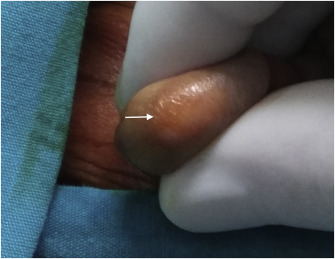
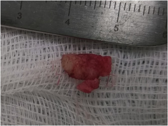
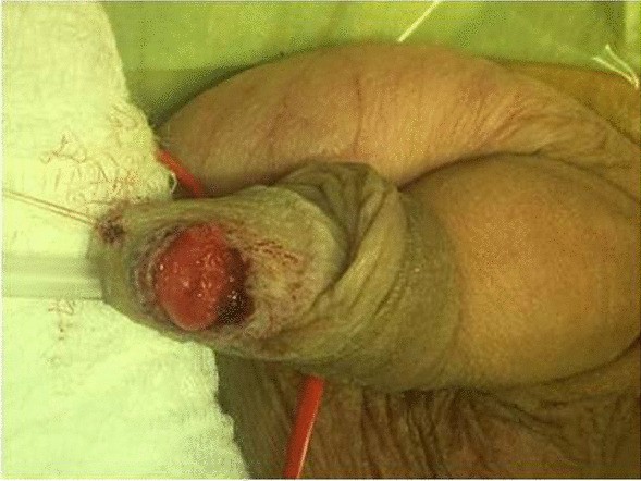
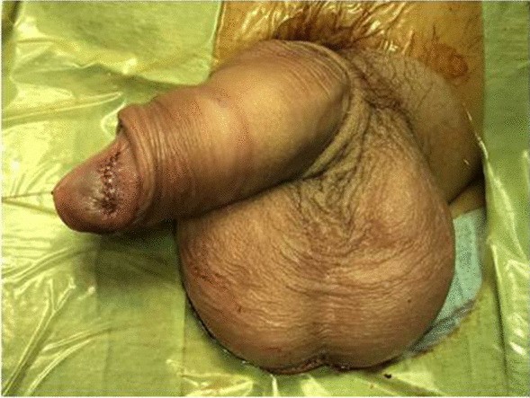
Tanriverdi et al. [13] reported an 11-year-old boy, who was referred to Manisa Celal Bayar University Hospital with a nodule upon the left side of his glans penis. The nodule was detected two weeks preceding his assessment and the patient had not had any symptom except for the finding of the penile mass. The mass was located within the left side of his glans and it had measured 10 mm in size (see figure 1). The penile mass was also immobile and painless. The patient had been circumcised when he was 6 years-old. He did not have any history of any trauma or infection. The results of his laboratory tests were within their normal limits. He had ultrasound scan of his abdomen, urinary tract as well as scrotum which was also normal. The tumour was completely excised totally and his glans penis was sutured primarily. It had an irregular border (see figure 2).
Fig. 1. The nodule in the left side of glans penis (white arrow). Reproduced from: [13] under the Creative Commons Attribution License.
Fig. 2. The nodule excised totally. Reproduced from: [13] under the Creative Commons Attribution License.
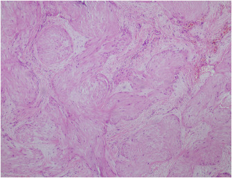
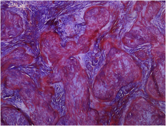
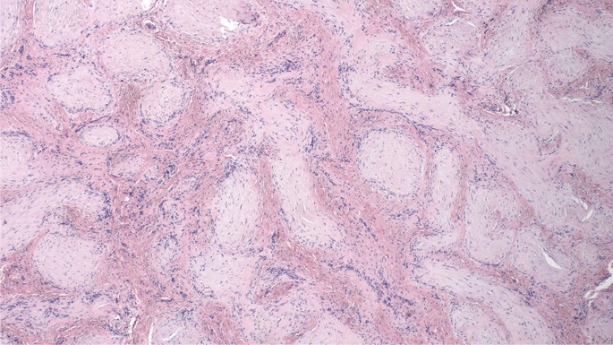
Upon histopathology examination, the mass was found to consist of thick, smooth muscle fibres (see figure 3). There were focal fibrotic/fibromyxoid areas and spindle cell proliferation among them (see figure 4). The neoplastic cells had exhibited strongly positive staining with actin and weakly stained with desmin upon immunohistochemistry staining studies. (see figure 5). All of these aforementioned pathological findings were evaluated as myointimoma. In the first year of follow-up, the patient did not have any problem regarding his penis or other urinary tract organs.
Fig. 3. Thick, smooth muscle bundles (HE, x100). Reproduced from: [13] under the Creative Commons Attribution License.
Fig. 4. Focal fibrotic/fibromyxoid areas among smooth muscle fibers (Masson-trichrome, x100). Reproduced from: [13] under the Creative Commons Attribution License.
Fig. 5. Strongly staining with actin (Actin, x100). Reproduced from: [13] under the Creative Commons Attribution License.
Tanriverdi et al. [13] made the ensuing educative discussion:
Primary benign penile soft tissue tumours are not usual tumours.
It had been iterated that leiomyoma, haemangioma, haemangioendothelioma, myofibroma, neurofibroma, schwannoma may be seen within the penis mostly. [2] [4].
Myointimoma is a rare and recently described benign soft tissue tumour of the penis.
Myointimoma was described by Fetsch and there are a few reported cases of myointimoma. [2] [4] [11-12] [14]
The age distribution of patients afflicted by myointimoma is wide.
Fetsch had presented 10 cases in which the ages of the patients were between 2 years and 61 years. [2]
McKenney had presented 5 cases with patients who were aged between 4 years and 15 years. [4]
The myointimoma tumor usually becomes markedly in a short period of time between 1 month and 2 mounts. [2] [11-12] [14]
Also, the patients do not manifest with any symptoms except for the presence of the penile mass. [4] [11-12] [14]
The lesions were reported to be sized between 0.4 cm and 1.9 cm and they were reported to be located within the glans penis and the corona in different studies. [2] [4] [11-12] [14]
Their patient was 11-years-old. The nodule was detected two weeks earlier and the patient did not manifest with any symptom except for the presence of the recently observed penile mass. The nodule was about 10 mm in size.
Total excision of the myointimoma nodule is enough.
Local recurrence or metastasis had not been reported in the literature. [2] [4] On the other hand, a spontaneous regression of the mass pursuant to biopsy of the mass had been reported. [2]
They had excised the penile mass totally and there was no recurrence within the glans penis for a year.
Typical histopathology examination of myointimoma demonstrates multi-nodular or plexiform intravascular myointimal proliferation of spindle cells within the glans penis. [11] [14]
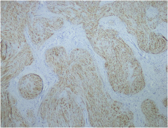
Immunohistochemistry staining studies do demonstrate that myointimal cells exhibit positive immunohistochemistry staining for smooth muscle actin, muscle specific action, calponin and are negative staining for S-100 protein and desmin. [11] [14]
Myointimal proliferation is demonstrated with Verhoeff-van Gieson's stain as elastic fibres encompassing nodules. [11] [14[
In their patient there were thick smooth muscle fibres demonstrated upon pathological examination, which was concordant with myointimoma.
Myointimoma should be differentiated histologically from myofibroma, nodular fasciitis, leiomyoma, plexiform fibrohistiocytic tumour, epithelioid haemangioendothelioma]. [2] [4]
Myoid predominant myofibromas may be nodular, but they do not demonstrate the exclusive intravascular growth like myointimoma. [4]
Also, they do not demonstrate distinctive smooth muscle collarettes.
Fasciitis typically contains intralesional inflammatory cells, mucoid pools and less compact stroma.
Myofibroblastic proliferation in myointimoma tends to be homogeneous and there tends not to be composite lesions with hyalinized and loose stroll patterns of fasciitis. [4]
Leiomyomas do not grow in a barbarizing intravascular pattern. They also have more well-developed fascicular structure and do not typically have a prominent myxoid stroma. [4]
In children, plexiform fibrohistiocytic tumour is important for the differential diagnosis in view of the fact that it might recur and it has a small risk of metastases.
Even though both of the two lesions do have a multi-nodular-plexiform appearance, plexiform fibrohistiocytic tumour is not an intravascular lesion. [4]
Dence myxoid stroma, cytoplasmic eosinophilia and intravascular location of myointimoma may simulate epithelioid haemangioendothelioma.
Immunohistochemistry staining studies could be utilised for the differential diagnosis in cases. [4]
Vascular lesion could be verified with CD31 - CD34 reactivity and absence of staining with smooth muscle actin.
Tanriverdi et al. [13] made the ensuing conclusions:
They had reported a rare benign tumour which is called myointimoma in the glans penis.
Myointimoma must be considered for the differential diagnosis and it is well-known that total excision is sufficient.
Turner et al. [14] reported a case of myointimoma of the penis in a healthy 14-year-old male, who had manifested to the paediatric surgery unit of university of Florida with a firm, non-mobile 1 cm nodule upon the right side of his glans penis, which had been evident over about the preceding one month. His clinical examination was noted to be otherwise normal. An excisional biopsy of the penile lesion was undertaken. Histopathology examination of specimens of the excised penile lesion demonstrated an occlusive myointimal proliferation with complex multi-nodular / plexiform architecture which had involved the vasculature of his glans penis, and which had extended into the tissue margins. The nodular proliferations had comprised of medium to large spindle, and / or stellate cells that had long tapered cytoplasmic process, fine nuclear chromatin, inconspicuous nucleoli, and based upon additional pathology examination features of the lesion, a diagnosis of myointimoma of the penis was made.
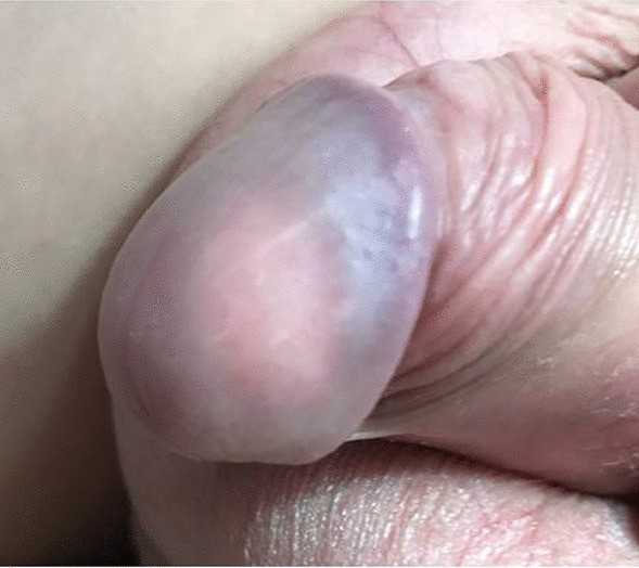
Drlík et al. [1] reported a 15-year-old Caucasian boy, who had manifested with a 6-months history of a slowly growing, palpable firm nodule within his glans penis. Clinically he was completely asymptomatic and had voided freely. He did not report any history of trauma, systemic connective tissue diseases or other autoimmune disorders. During his clinical examination, there was evidence of a palpable, well circumscribed, firm, whitish painless mass, about 1 cm in diameter within his glans penis (see figure 6). His overlying skin was of a normal structure without signs of inflammation. No palpable inguinal lymphadenopathy was identified. The stage of puberty was Tanner III.
Figure 6: Whitish nodule visible under normal overlying skin. Reproduced from: [1] under the Creative Commons Attribution License.
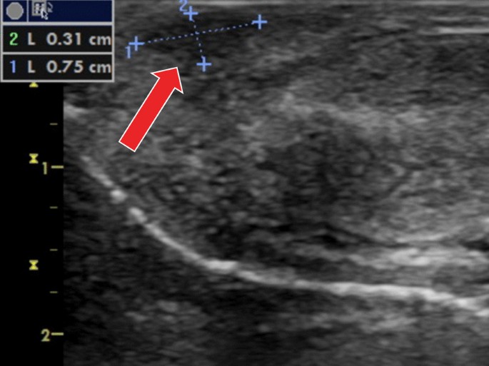
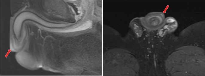
In view of the fact that there were no guidelines concerning penile tumours in his age, the authors [1] adhered to the EAU guidelines for penile cancer in adults and they undertook a penile Doppler Ultrasound scan and MRI (Magnetic Resonance Imaging). The ultrasound scan demonstrated hypoechogenic, hypo-perfused poorly defined area within his glans penis. (see figure 7). The MRI scan did not confirm any other pathology mass inside his glans penis and corpora cavernosa (see figure 8). An excisional biopsy under general anaesthesia with intra-operative pathological evaluation was decided upon. The formation was noted not to have been clearly demarcated from the encompassing glans penis tissues and had reached close to the urethra, without interfering with its wall. The procedure was undertaken at optical magnification, utilising magnifying glasses with particular attention to prevent the injury of the neighbouring urethra (see figure 9). In view of the fact that the intra-operative pathology evaluation had demonstrated a benign nature of the tumour, Drlik et al. [1] simply closed the wound and did not proceed with any more extensive surgery (see figure 10).
Figure 7. Ultrasound finding—a hypoechogenic, hypo-perfused non-well-defined area inside the glans (arrow). Reproduced from: [1] under the Creative Commons Attribution License.
Figure 8.
MRI finding—a single hyperintense mass inside glans (arrow), corpora cavernosa, are normal, sagittal (A) and coronal (B) cut. Reproduced from: [1] under the Creative Commons Attribution License.
Figure 9. Careful excisional biopsy with special attention to protection of the urethra. Reproduced from: [1] under the Creative Commons Attribution License.
Figure 10. Simple wound closure. Reproduced from: [1] under the Creative Commons Attribution License.
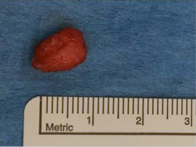
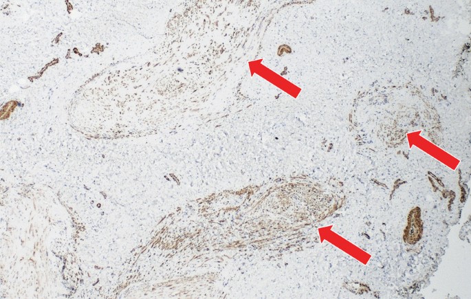
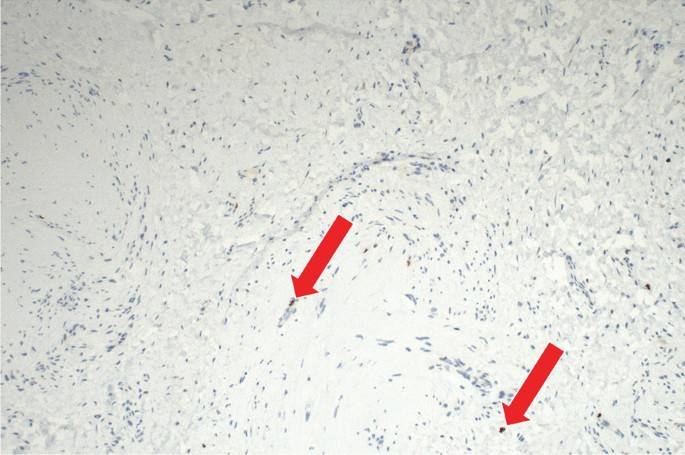
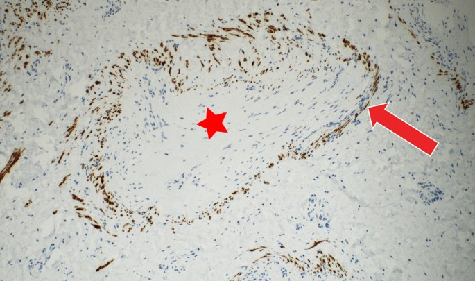
Drlik et al. [1] found the lesion to be a macroscopically pale tissue sample which had measured 10 mm × 8 mm × 5 mm (see figure 11). On the cut surface, the lesion was noted to be light red in colour and had solid consistency. Subsequent detailed histopathology examination of specimens of the lesion demonstrated changes that were diagnostic for myointimoma - nodular intravascular myofibroblastic proliferation involving multiple cavernous spaces (see figure 12). At low power magnification, a complex multinodular architecture was visualised. At higher magnification the myofibroblasts were observed to be uniform, elongated spindle shaped cells with no significant hyperchromasia or pleomorphism, nor any mitotic figures or necrosis. Immunohistochemistry staining studies staining for alpha-smooth muscle actin (αSMA) was noted to be positive within the lesion (see figure 12), the proliferative activity (Ki-67) was low (beneath 1%) (see figure 13). Immunohistochemistry staining for desmin was negative within myofibroblasts, while positive in the pre-existing vessel wall only (see figure 15). No reactivity was visualised for other performed immunohistochemistry staining markers (S100 protein, CD34 and ERG). Microphotographs were taken with Olympus BX41 microscope and processed by QuickPHOTO Software.
Figure 11: Macroscopic appearance of myointimoma. Reproduced from: [1] under the Creative Commons Attribution License.
Figure 12: Nodular intravascular proliferation of spindle myofibroblastic cells presenting typical morphology for myointimoma (hematoxylin-eosin, 100x). Reproduced from: [1] under the Creative Commons Attribution License.
Figure 13: Immunohistochemical expression of alpha-smooth muscle actin (SMA) shows diffuse positivity in intravascular myofibroblastic population (arrow) (200x). Reproduced from: [1] under the Creative Commons Attribution License.
Figure 14: Immunohistochemical expression of Ki-67 (proliferative antigen MIB-1), labelling cells beyond G0 phase of the mitotic cycle, shows expression of sporadic cells (beneath 1%, arrow) (400x). Reproduced from: [1] under the Creative Commons Attribution License.
Figure 15: Immunohistochemical expression of desmin showing positivity in the smooth muscle cells of the pre-existing vessel walls (arrow), tumorous myofibroblastic cells are negative (star) (400x). Reproduced from: [1] under the Creative Commons Attribution License.
In view of the nature of the benign nature of the lesion, Drlik et al. [1] did not undertake staging for distant metastases and simply undertook an outpatient follow-up. Three years pursuant to the excision of the lesion, there was no evidence of local recurrence, no urethral stricture and a cosmetic appearance is good (see figure 16).
Figure 16: Favourable cosmetic outcome 3 years later. Reproduced from: [1] under the Creative Commons Attribution License.
Drlik et al. [1] made the ensuing educative discussion and conclusion:
Their reported case report had presented an additional case to the 10 cases which had previously reported cases of myointimoma in children and adolescents. [4] [13] [14]
In our case, like in all previously reported cases in adolescents and adults [2] [10] [11] [12] [15] [16], the myointimoma affects uniquely the glans penis. Likewise, no reported case was associated with pain, dysuria or signs of lower urinary tract obstruction. In our case, the patient reported a relatively fast-growing mass. The history of initial rapid growth is common in the literature; later however, the formation may remain stable over time. Monsalves [10] described a case of myointimoma that remained unchanged 10 months after an incomplete excision.
Fetsch [2] described the same experience with a 6-month stable residual mass in a patient after an incisional biopsy. In one case, complete regression of myointimoma at 10-years follow-up was described [2]. Local aggressive growth or distant metastases were never reported.
At the time of publication of their article and treatment of their patient, there were no guidelines describing the extent of imaging in adolescents with penile tumours.
The existing literature had not delt with the scope of radiology imaging; both existing series [2] [4] of cases were based upon a retrospective re-evaluation of stored hematoxylin and eosin-stained slides of penile tumours over the preceding decades only. Therefore, they had adhered to EAU guidelines for penile cancer in the adults and undertook penile Doppler Ultrasonography and MRI to exclude corporal invasion. The examinations had confirmed the solid nature of the tumour, and had excluded cystic lesion and multiple involvement of cavernosal tissue.
In a case of penile tumour in adolescents, their main concern was to exclude clinically aggressive conditions, hence, an excisional biopsy was decided upon.
Since the boy was confirmed to have benign findings upon histopathology examination and had clinically normal findings on the inguinal nodes, they did not undertake radiology staging (abdominal, pelvic and thoracic CT).
The diagnosis of myointimoma and its differential diagnosis based upon morphology only might be confusing.
There are many types of mesenchymal tumours that are associated with plexiform or nodular structure.
Immunohistochemistry staining study is a key to the establishment of an exact diagnosis.
Desmin may be absent or exhibit only focal reactivity.
There is no reactivity for S-100 protein, CD31, CD34, ERG, epithelial membrane antigen (EMA) or neuron specific enolase (NSE).
It has been iterated that the plexiform growth pattern could be visualised in plexiform histiocytic tumour (PFHT). [17]
Unlike myointimoma, it contains a mixture of two components: a differentiated spindle fibroblastic/myofibroblastic cells and a round histiocytic cell component containing multinucleated giant cells (osteoclast-like giant cells). Immunohistochemically, the histiocytes and multinucleated giant cell does express CD68, whereas the spindle cells express αSMA.
PFHT might recur and has a low risk of metastases (lymph node, lung). A plexiform or nodular growth pattern could be visualised in some nerve sheet tumours such as plexiform schwannoma [18] or neurofibroma. Immunohistochemical expression for S-100 protein is then helpful in differential diagnosis.
The myointimoma structure might simulate myofibroma, which is a more commonly encountered neoplasm in children. In contrast, it does not exhibit the exclusive intravascular growth; the growth is rather concentric around the small vessels.
The tumour is stated to be composed of oval or spindle myoid cells [19].
Myopericytomas are typified by a distinctive biphasic growth pattern, with central hypercellular zone composed of spindle tumour cells, hyalinization and myoid cell nodules visible towards the periphery of the neoplasm. In contrast to myofibroma, intravascular growth is more common in myopericytoma, but it does not indicate a malignant neoplastic process [20].
Epithelioid haemangioma and haemangioendothelioma could be differentiated from myointimoma by immunohistochemistry staining studies in view of the fact that the endothelial nature of the lesional cells could be confirmed by CD31, CD34 and ERG positivity.
Another structurally similar pathology is a late phase of intravascular fasciitis (intravascular nodular fasciitis). Histologically, intralesional inflammatory cells between spindle myofibroblast cells, mucoid pools, a less compact stroma with more eosinophilic hyalinization, and obvious mitotic figures were visualised [2].
Intravascular spindle cells lesion such as intravascular leiomyoma or leiomyomatosis could be easily differentiated by immunohistochemistry staining studies, with αSMA, desmin and h-caldesmon antibodies, which are typically strongly positive.
Last but not least, the possibility of sarcoma with angio-invasive spread should be excluded during the histopathology examination of the specimen.
Both the clinician and the pathologist should be aware of this rare benign entity.
The key to a correct diagnosis is a careful histopathology examination of the specimen, including immunohistochemistry.
Local excision of myointimoma of the penis is a safe and effective treatment modality.
[B] MISCELLANEOUS NARRATIONS AND DISCUSSIONS FROM SOME CASE REPORTS, CASE SERIES, AND STUDIES RELATE
Myointimoma is a rare, benign soft-tissue tumour which is derived from the intimal cells of blood vessels.
Since little is known about this uncommon tumour entity, they had reported an additional case.
Cito et al. [15] reported a 49-year-old Caucasian man who had presented with a 12-month history of a palpable, firm, solitary lesion involving his glans penis. During his clinical examination, there was a 1 cm palpable, endophytic well-circumscribed nodule, that was located to the left side of his glans penis, close to his coronal sulcus, with disease-free external urethral orifice. The patient underwent complete excisional biopsy. A skin rhombus that measured 1.1 cm × 0.8 cm × 0.3 cm was removed and the biopsy sample, was fixed in 10% formaldehyde, sent to the pathology department. During his 18-month follow-up visit, he was clinically disease free. Histopathology examination of his excised penile lesion demonstrated a multi-nodular intra-vascular proliferation of the corpus spongiosum. The myointimal proliferation had comprised of bland predominantly spindle cells in an abundant fibromyxoid stroma. Immunohistochemistry staining for smooth muscle actin (1A4), cytokeratins (AE1/AE3, CAM5.2), and CD34 were undertaken utilising the avidin-biotin complex (ABC) immunoperoxidase method. The lesional cells had exhibited positive staining for smooth muscle actin and negative staining for cytokeratins and CD34. Cito et al. [15] made the ensuing concluding iterations:
Myointimoma has been confirmed to be a penile benign lesion that may be adequately treated with excisional biopsy.
Even pursuant to incomplete or marginal removal, the penile lesion had been shown to remain stable overtime or regress.
Differential diagnosis is essential to exclude similar histological entities which could be more aggressive or have possible systemic implications.
Casa et al. [6] made the ensuing iterations:
Penile myointimoma is an uncommon, benign tumour which afflicts the corpus spongiosum vasculature of the glans penis.
Up to the time of publication of their article in 2023, there had been twenty-three reported myointimoma tumours in the literature.
Casa et al. [6] reported four additional tumours of this unique myointimal proliferation. Casa et al. [6] summated the results as follows:
The ages of the patients had ranged from 20 years to 68 years and the patients had manifested with a firm mass upon the glans penis.
All four tumours had displayed distinctive morphological features which had consisted of a myointimal proliferation with plexiform architecture of bland myofibroblastic cells within a myxoid background within the corpus spongiosum vasculature.
Typifying cytoplasmic immunoreactivity of the lesional cells with smooth muscle actin in addition to a desmin positive staining had collated with those of native vessel smooth muscle was visualised within all four tumours.
No disease was reported in any of the patients during their last clinical follow-up assessment, which had ranged between 9 years and 15 years, pursuant to their biopsy or excision.
Casa et al. [6] made the ensuing concluding iterations:
Myointimoma is stated to be part of a rare group of mesenchymal tumours which had been recently classified by its distinctive location, morphology, and immunohistochemical reactivity.
For any nodular, spindle cell lesion of the corpus spongiosum, myointimoma should be included in the differential diagnosis given its unique characteristics and favorable clinical outcome.Vadar et al. [12] stated that Myointimoma was a recently described benign tumour, which is regarded to be a rare type of mesenchymal tumour of the penis. Vadar et al. [12] reported a patient, who was a 50-year-old man and who had manifested with a nodule that was located within his glans penis. He had a 2-month history of a mass. An excisional biopsy of the mass was undertaken. The histopathology examination findings revealed a multinodular tumour which was typified by spindle-shaped cells that were located within his intravascular area. Vadar et al. [12] made the ensuing additional iteration:
This case, in addition to 11 cases which had been reported in the literature, had demonstrated that the myointimoma is frequently misdiagnosed upon clinical and pathological grounds in view of its rarity. [14]
Tanriverdi et al. [13] reported an 11-year-old boy, who was referred to Manisa Celal Bayar University Hospital with a nodule upon the left side of his glans penis. The nodule was detected two weeks preceding his assessment and the patient had not had any symptom except for the finding of the penile mass. The mass was located within the left side of his glans and it had measured 10 mm in size (see figure 1). The penile mass was also immobile and painless. The patient had been circumcised when he was 6 years-old. He did not have any history of any trauma or infection. The results of his laboratory tests were within their normal limits. He had ultrasound scan of his abdomen, urinary tract as well as scrotum which was also normal. The tumour was completely excised totally and his glans penis was sutured primarily. It had an irregular border (see figure 2).
Fig. 1. The nodule in the left side of glans penis (white arrow). Reproduced from: [13] under the Creative Commons Attribution License.
Fig. 2. The nodule excised totally. Reproduced from: [13] under the Creative Commons Attribution License.
Upon histopathology examination, the mass was found to consist of thick, smooth muscle fibres (see figure 3). There were focal fibrotic/fibromyxoid areas and spindle cell proliferation among them (see figure 4). The neoplastic cells had exhibited strongly positive staining with actin and weakly stained with desmin upon immunohistochemistry staining studies. (see figure 5). All of these aforementioned pathological findings were evaluated as myointimoma. In the first year of follow-up, the patient did not have any problem regarding his penis or other urinary tract organs.
Fig. 3. Thick, smooth muscle bundles (HE, x100). Reproduced from: [13] under the Creative Commons Attribution License.
Fig. 4. Focal fibrotic/fibromyxoid areas among smooth muscle fibers (Masson-trichrome, x100). Reproduced from: [13] under the Creative Commons Attribution License.
Fig. 5. Strongly staining with actin (Actin, x100). Reproduced from: [13] under the Creative Commons Attribution License.
Tanriverdi et al. [13] made the ensuing educative discussion:
Primary benign penile soft tissue tumours are not usual tumours.
It had been iterated that leiomyoma, haemangioma, haemangioendothelioma, myofibroma, neurofibroma, schwannoma may be seen within the penis mostly. [2] [4].
Myointimoma is a rare and recently described benign soft tissue tumour of the penis.
Myointimoma was described by Fetsch and there are a few reported cases of myointimoma. [2] [4] [11-12] [14]
The age distribution of patients afflicted by myointimoma is wide.
Fetsch had presented 10 cases in which the ages of the patients were between 2 years and 61 years. [2]
McKenney had presented 5 cases with patients who were aged between 4 years and 15 years. [4]
The myointimoma tumor usually becomes markedly in a short period of time between 1 month and 2 mounts. [2] [11-12] [14]
Also, the patients do not manifest with any symptoms except for the presence of the penile mass. [4] [11-12] [14]
The lesions were reported to be sized between 0.4 cm and 1.9 cm and they were reported to be located within the glans penis and the corona in different studies. [2] [4] [11-12] [14]
Their patient was 11-years-old. The nodule was detected two weeks earlier and the patient did not manifest with any symptom except for the presence of the recently observed penile mass. The nodule was about 10 mm in size.
Total excision of the myointimoma nodule is enough.
Local recurrence or metastasis had not been reported in the literature. [2] [4] On the other hand, a spontaneous regression of the mass pursuant to biopsy of the mass had been reported. [2]
They had excised the penile mass totally and there was no recurrence within the glans penis for a year.
Typical histopathology examination of myointimoma demonstrates multi-nodular or plexiform intravascular myointimal proliferation of spindle cells within the glans penis. [11] [14]
Immunohistochemistry staining studies do demonstrate that myointimal cells exhibit positive immunohistochemistry staining for smooth muscle actin, muscle specific action, calponin and are negative staining for S-100 protein and desmin. [11] [14]
Myointimal proliferation is demonstrated with Verhoeff-van Gieson's stain as elastic fibres encompassing nodules. [11] [14[
In their patient there were thick smooth muscle fibres demonstrated upon pathological examination, which was concordant with myointimoma.
Myointimoma should be differentiated histologically from myofibroma, nodular fasciitis, leiomyoma, plexiform fibrohistiocytic tumour, epithelioid haemangioendothelioma]. [2] [4]
Myoid predominant myofibromas may be nodular, but they do not demonstrate the exclusive intravascular growth like myointimoma. [4]
Also, they do not demonstrate distinctive smooth muscle collarettes.
Fasciitis typically contains intralesional inflammatory cells, mucoid pools and less compact stroma.
Myofibroblastic proliferation in myointimoma tends to be homogeneous and there tends not to be composite lesions with hyalinized and loose stroll patterns of fasciitis. [4]
Leiomyomas do not grow in a barbarizing intravascular pattern. They also have more well-developed fascicular structure and do not typically have a prominent myxoid stroma. [4]
In children, plexiform fibrohistiocytic tumour is important for the differential diagnosis in view of the fact that it might recur and it has a small risk of metastases.
Even though both of the two lesions do have a multi-nodular-plexiform appearance, plexiform fibrohistiocytic tumour is not an intravascular lesion. [4]
Dence myxoid stroma, cytoplasmic eosinophilia and intravascular location of myointimoma may simulate epithelioid haemangioendothelioma.
Immunohistochemistry staining studies could be utilised for the differential diagnosis in cases. [4]
Vascular lesion could be verified with CD31 - CD34 reactivity and absence of staining with smooth muscle actin.
Tanriverdi et al. [13] made the ensuing conclusions:
They had reported a rare benign tumour which is called myointimoma in the glans penis.
Myointimoma must be considered for the differential diagnosis and it is well-known that total excision is sufficient.
Turner et al. [14] reported a case of myointimoma of the penis in a healthy 14-year-old male, who had manifested to the paediatric surgery unit of university of Florida with a firm, non-mobile 1 cm nodule upon the right side of his glans penis, which had been evident over about the preceding one month. His clinical examination was noted to be otherwise normal. An excisional biopsy of the penile lesion was undertaken. Histopathology examination of specimens of the excised penile lesion demonstrated an occlusive myointimal proliferation with complex multi-nodular / plexiform architecture which had involved the vasculature of his glans penis, and which had extended into the tissue margins. The nodular proliferations had comprised of medium to large spindle, and / or stellate cells that had long tapered cytoplasmic process, fine nuclear chromatin, inconspicuous nucleoli, and based upon additional pathology examination features of the lesion, a diagnosis of myointimoma of the penis was made.
Drlík et al. [1] reported a 15-year-old Caucasian boy, who had manifested with a 6-months history of a slowly growing, palpable firm nodule within his glans penis. Clinically he was completely asymptomatic and had voided freely. He did not report any history of trauma, systemic connective tissue diseases or other autoimmune disorders. During his clinical examination, there was evidence of a palpable, well circumscribed, firm, whitish painless mass, about 1 cm in diameter within his glans penis (see figure 6). His overlying skin was of a normal structure without signs of inflammation. No palpable inguinal lymphadenopathy was identified. The stage of puberty was Tanner III.
Figure 6: Whitish nodule visible under normal overlying skin. Reproduced from: [1] under the Creative Commons Attribution License.
In view of the fact that there were no guidelines concerning penile tumours in his age, the authors [1] adhered to the EAU guidelines for penile cancer in adults and they undertook a penile Doppler Ultrasound scan and MRI (Magnetic Resonance Imaging). The ultrasound scan demonstrated hypoechogenic, hypo-perfused poorly defined area within his glans penis. (see figure 7). The MRI scan did not confirm any other pathology mass inside his glans penis and corpora cavernosa (see figure 8). An excisional biopsy under general anaesthesia with intra-operative pathological evaluation was decided upon. The formation was noted not to have been clearly demarcated from the encompassing glans penis tissues and had reached close to the urethra, without interfering with its wall. The procedure was undertaken at optical magnification, utilising magnifying glasses with particular attention to prevent the injury of the neighbouring urethra (see figure 9). In view of the fact that the intra-operative pathology evaluation had demonstrated a benign nature of the tumour, Drlik et al. [1] simply closed the wound and did not proceed with any more extensive surgery (see figure 10).
Figure 7.
Figure 7. Ultrasound finding—a hypoechogenic, hypo-perfused non-well-defined area inside the glans (arrow). Reproduced from: [1] under the Creative Commons Attribution License.
Figure 8.
Figure 8.
MRI finding—a single hyperintense mass inside glans (arrow), corpora cavernosa, are normal, sagittal (A) and coronal (B) cut. Reproduced from: [1] under the Creative Commons Attribution License.
Figure 9.
Figure 9. Careful excisional biopsy with special attention to protection of the urethra. Reproduced from: [1] under the Creative Commons Attribution License.
Figure 10.
Figure 10. Simple wound closure. Reproduced from: [1] under the Creative Commons Attribution License.
Drlik et al. [1] found the lesion to be a macroscopically pale tissue sample which had measured 10 mm × 8 mm × 5 mm (see figure 11). On the cut surface, the lesion was noted to be light red in colour and had solid consistency. Subsequent detailed histopathology examination of specimens of the lesion demonstrated changes that were diagnostic for myointimoma - nodular intravascular myofibroblastic proliferation involving multiple cavernous spaces (see figure 12). At low power magnification, a complex multinodular architecture was visualised. At higher magnification the myofibroblasts were observed to be uniform, elongated spindle shaped cells with no significant hyperchromasia or pleomorphism, nor any mitotic figures or necrosis. Immunohistochemistry staining studies staining for alpha-smooth muscle actin (αSMA) was noted to be positive within the lesion (see figure 12), the proliferative activity (Ki-67) was low (beneath 1%) (see figure 13). Immunohistochemistry staining for desmin was negative within myofibroblasts, while positive in the pre-existing vessel wall only (see figure 15). No reactivity was visualised for other performed immunohistochemistry staining markers (S100 protein, CD34 and ERG). Microphotographs were taken with Olympus BX41 microscope and processed by QuickPHOTO Software.
Figure 11.
Figure 11: Macroscopic appearance of myointimoma. Reproduced from: [1] under the Creative Commons Attribution License.
Figure 12: Nodular intravascular proliferation of spindle myofibroblastic cells presenting typical morphology for myointimoma (hematoxylin-eosin, 100x). Reproduced from: [1] under the Creative Commons Attribution License.
Figure 13: Immunohistochemical expression of alpha-smooth muscle actin (SMA) shows diffuse positivity in intravascular myofibroblastic population (arrow) (200x). Reproduced from: [1] under the Creative Commons Attribution License.
Figure 14: Immunohistochemical expression of Ki-67 (proliferative antigen MIB-1), labelling cells beyond G0 phase of the mitotic cycle, shows expression of sporadic cells (beneath 1%, arrow) (400x). Reproduced from: [1] under the Creative Commons Attribution License.
Figure 15: Immunohistochemical expression of desmin showing positivity in the smooth muscle cells of the pre-existing vessel walls (arrow), tumorous myofibroblastic cells are negative (star) (400x). Reproduced from: [1] under the Creative Commons Attribution License.
In view of the nature of the benign nature of the lesion, Drlik et al. [1] did not undertake staging for distant metastases and simply undertook an outpatient follow-up. Three years pursuant to the excision of the lesion, there was no evidence of local recurrence, no urethral stricture and a cosmetic appearance is good (see figure 16).
Figure 16: Favourable cosmetic outcome 3 years later. Reproduced from: [1] under the Creative Commons Attribution License.
Drlik et al. [1] made the ensuing educative discussion and conclusion:
Their reported case report had presented an additional case to the 10 cases which had previously reported cases of myointimoma in children and adolescents. [4] [13] [14]
In our case, like in all previously reported cases in adolescents and adults [2] [10] [11] [12] [15] [16], the myointimoma affects uniquely the glans penis. Likewise, no reported case was associated with pain, dysuria or signs of lower urinary tract obstruction. In our case, the patient reported a relatively fast-growing mass. The history of initial rapid growth is common in the literature; later however, the formation may remain stable over time. Monsalves [10] described a case of myointimoma that remained unchanged 10 months after an incomplete excision.
Fetsch [2] described the same experience with a 6-month stable residual mass in a patient after an incisional biopsy. In one case, complete regression of myointimoma at 10-years follow-up was described [2]. Local aggressive growth or distant metastases were never reported.
At the time of publication of their article and treatment of their patient, there were no guidelines describing the extent of imaging in adolescents with penile tumours.
The existing literature had not delt with the scope of radiology imaging; both existing series [2] [4] of cases were based upon a retrospective re-evaluation of stored hematoxylin and eosin-stained slides of penile tumours over the preceding decades only. Therefore, they had adhered to EAU guidelines for penile cancer in the adults and undertook penile Doppler Ultrasonography and MRI to exclude corporal invasion. The examinations had confirmed the solid nature of the tumour, and had excluded cystic lesion and multiple involvement of cavernosal tissue.
In a case of penile tumour in adolescents, their main concern was to exclude clinically aggressive conditions, hence, an excisional biopsy was decided upon.
Since the boy was confirmed to have benign findings upon histopathology examination and had clinically normal findings on the inguinal nodes, they did not undertake radiology staging (abdominal, pelvic and thoracic CT).
The diagnosis of myointimoma and its differential diagnosis based upon morphology only might be confusing.
There are many types of mesenchymal tumours that are associated with plexiform or nodular structure.
Immunohistochemistry staining study is a key to the establishment of an exact diagnosis.
Desmin may be absent or exhibit only focal reactivity.
There is no reactivity for S-100 protein, CD31, CD34, ERG, epithelial membrane antigen (EMA) or neuron specific enolase (NSE).
It has been iterated that the plexiform growth pattern could be visualised in plexiform histiocytic tumour (PFHT). [17]
Unlike myointimoma, it contains a mixture of two components: a differentiated spindle fibroblastic/myofibroblastic cells and a round histiocytic cell component containing multinucleated giant cells (osteoclast-like giant cells). Immunohistochemically, the histiocytes and multinucleated giant cell does express CD68, whereas the spindle cells express αSMA.
PFHT might recur and has a low risk of metastases (lymph node, lung). A plexiform or nodular growth pattern could be visualised in some nerve sheet tumours such as plexiform schwannoma [18] or neurofibroma. Immunohistochemical expression for S-100 protein is then helpful in differential diagnosis.
The myointimoma structure might simulate myofibroma, which is a more commonly encountered neoplasm in children. In contrast, it does not exhibit the exclusive intravascular growth; the growth is rather concentric around the small vessels.
The tumour is stated to be composed of oval or spindle myoid cells [19].
Myopericytomas are typified by a distinctive biphasic growth pattern, with central hypercellular zone composed of spindle tumour cells, hyalinization and myoid cell nodules visible towards the periphery of the neoplasm. In contrast to myofibroma, intravascular growth is more common in myopericytoma, but it does not indicate a malignant neoplastic process [20].
Epithelioid haemangioma and haemangioendothelioma could be differentiated from myointimoma by immunohistochemistry staining studies in view of the fact that the endothelial nature of the lesional cells could be confirmed by CD31, CD34 and ERG positivity.
Another structurally similar pathology is a late phase of intravascular fasciitis (intravascular nodular fasciitis). Histologically, intralesional inflammatory cells between spindle myofibroblast cells, mucoid pools, a less compact stroma with more eosinophilic hyalinization, and obvious mitotic figures were visualised [2].
Intravascular spindle cells lesion such as intravascular leiomyoma or leiomyomatosis could be easily differentiated by immunohistochemistry staining studies, with αSMA, desmin and h-caldesmon antibodies, which are typically strongly positive.
Last but not least, the possibility of sarcoma with angio-invasive spread should be excluded during the histopathology examination of the specimen.
Both the clinician and the pathologist should be aware of this rare benign entity.
The key to a correct diagnosis is a careful histopathology examination of the specimen, including immunohistochemistry.
Local excision of myointimoma of the penis is a safe and effective treatment modality.
Conclusion
Myofintimoma of the penis is an uncommon benign neoplasm the penis.
Less than 30 cases of myointimoma of the penis has so far been reported in the world literature.
It is important to differentiate myointimoma from more clinically aggressive tumours of the penis.
The key to a correct diagnosis of myointimoma of the penis requires the undertaking of a careful histopathology examination, including immunohistochemistry staining studies of biopsy or excised specimen of the tumour.
Local excision of the myointimoma tumour is a safe and effective treatment option which has been documented to produce good outcome with no subsequent recurrence even in scenarios where there is tumour at the surgical resection margin.
Spontaneous regression of myointimoma of the penis has also been reported in a case of myointimoma of the penis which was diagnosed based upon pathology examination of a biopsy specimen of the tumour and in which the tumour was left alone without excision
All clinicians including urologists, General practitioners, pathologists, and oncologists need to have a high index of suspicion for myointimoma of the penis in order to establish early and correct diagnosis of the neoplasm.
CONFLICT OF INTEREST – Nil
ACKNOWLEDGEMENTS
Acknowledgement to:
Journal of Pediatric Surgery Case Reports and Elsevier under a Creative Commons Attribution License CC BY-NC-ND 4.0 Attribution-Non-Commercial-No-Derivatives 4.0 International. CC BY-NC-ND 4.0
Drlík, M., Gregová, M., Sedláček, J. et al (2022). Myointimoma (angiocentric myofibroblastic tumor) of the glans penis in an adolescent: a case report and review of the literature. BMC Urol. 2022, 22, 186. View
at Publisher |
View
at Google Scholar
Fetsch JF, Brinsko RW, Davis CJ Jr, Mostofi FK, Sesterhenn IA (2000). A distinctive myointimal proliferation ('myointimoma') involving the corpus spongiosum of the glans penis: a clinicopathologic and immunohistochemical analysis of 10 cases. Am J Surg Pathol. 2000 Nov;24(11):1524-30. In 2000 and it was recognized as a distinctive histological entity in the World Health Organization Classification of the Tumours of the Urinary System and Male Genital Organs in 2016 View
at Publisher |
View
at Google Scholar
WHO Classification of tumors of the urinary system. and male genital organs. Moch H, Humphrey PA, Ulbright TM, Reuter V, editors. Lyon: IARC; 2016. View
at Publisher |
View
at Google Scholar
McKenney JK, Collins MH, Carretero AP, Boyd TK, Redman JF, Parham DM (2007). Penile myointimoma in children and adolescents: a clinicopathologic study of 5 cases supporting a distinct entity. Am J Surg Pathol. 2007 Oct;31(10):1622-6. View
at Publisher |
View
at Google Scholar
Mohamed K S, Al-Quran S Z, Zynger D L (Editor), Tretiakova M (Editor) (2023). Myointimoma. Penis & Scrotum Other Tumours Myointimoma PathologyOutlines.com website. Last author update: 16 May 2023 Last staff update: 12 August 2024. Accessed August 31st, 2024. View
at Publisher |
View
at Google Scholar
Casa D, Wang L, Tretiakova M, Cibull T, Pease G (2023). Penile Myointimoma: A Clinicopathologic Study of 4 Tumors. Int J Surg Pathol. 2023 Aug;31(5):675-679. View
at Publisher |
View
at Google Scholar
Dehner LP, Smith BH (1970). Soft tissue tumors of the penis. A clinicopathologic study of 46 cases. Cancer. 1970 Jun;25(6):1431-47. View
at Publisher |
View
at Google Scholar
Redman J F, Liang X, Ferguson A M, Savell V H (2000). Leiomyoma of the glans penis in a child. J Urol. 2000 Sep 1; 164 (3 Part 1): 791. View
at Publisher |
View
at Google Scholar
Val-Bernal JF, Garijo MF (1996). Solitary cutaneous myofibroma of the glans penis. Am J Dermatopathol. 1996 Jun;18(3):317-21. View
at Publisher |
View
at Google Scholar
Monsálvez V, Rodríguez-Peralto JL, Fuertes L, Garrido C, López-Gómez S (2009). Miointimoma: un raro tumor de pene [Myointimoma: a rare tumor of the penis]. Actas Dermosifiliogr. 2009 Jul-Aug;100(6):511-2. Spanish. View
at Publisher |
View
at Google Scholar
Robbins JB, Kohler S (2005). Penile nodule in a 54-year-old man: a case of a myointimoma. J Am Acad Dermatol. 2005 Dec;53(6):1084-6. View
at Publisher |
View
at Google Scholar
Vardar E, Gunlusoy B, Arslan M, Kececi S (2007). Myointimoma of the glans penis. Pathol Int. 2007 Mar;57(3):158-61. View
at Publisher |
View
at Google Scholar
Halil Ibrahim Tanriverdi, Omer Yilmaz, Nalan Nese, Can Taneli, Abdulkadir Genc (2019). Myointimoma of the glans penis, Journal of Pediatric Surgery Case Reports. 2019; 44: 101189, ISSN 2213-5766. View
at Publisher |
View
at Google Scholar
Cito G, Santi R, Gemma L, Galli IC, Cocci A, Carini M, Minervini A, Nesi G. Myointimoma of the penis. Int J Impot Res. 2021 Sep;33(6):583-586. View
at Publisher |
View
at Google Scholar
Cordeiro EZ, Zequi SD, Pinto CA, Santos GC, Lopes A (2007). A Rare Case of Insidious Myointimoma-Case Report. Appl. cancer res. 2007:30-2. View
at Publisher |
View
at Google Scholar
Enzinger FM, Zhang RY (1988). Plexiform fibrohistiocytic tumor presenting in children and young adults. An analysis of 65 cases. Am J Surg Pathol. 1988 Nov;12(11):818-26. View
at Publisher |
View
at Google Scholar
Dear Editorial Team,
Clinical Medical Reviews and Reports.
My experience with the journal was highly positive. The peer-review process was rigorous, constructive, and completed in a timely manner. The reviewers provided valuable comments that helped improve the quality and clarity of our manuscript. The editorial office was professional, responsive, and supportive throughout all stages of the publication process. Communication was clear and efficient, and any questions were addressed promptly. Overall, I found the journal to maintain high scientific standards and an excellent publication workflow. I would be pleased to consider submitting future work to this journal.
Best wishes from,
Elena Popa.
Dr Elena Popa
It was my pleasure to submit my testimonial concerning the Reviewer Board of our Scientific Journal “Brain and Neurological Disorders”. The Reviewers focused on some modifications and their contribution was helpful. The ladies of our Editorial Office were also supported my efforts. It was my honor to have such a co-operation and I am looking forward for more collaboration.
Dr Nikolaos Andreas Chrysanthakopoulos
Dear Grace Pierce,
Editorial Coordinator of Journal of Clinical Research and Reports,
Thank you for the speedy and efficient peer review process. I appreciate the fact that your peer reviewers do not take months to respond like with some other journals. I would also like to thank the editorial office for responding quickly to my questions. It is an excellent journal. I plan to submit more manuscripts in the future.
Best wishes from,
Robert W. McGee
Robert W McGee
Dear Grace Pierce,
Editorial Coordinator of Journal of Clinical Research and Reports,
Working with you and your team on our recent publication in JCRR has been a truly wonderful and enjoyable experience. The responses were prompt, and the reviewers were patient, constructive, and highly professional. One reviewer in particular gave me the feeling that a professor was carefully reading and commenting on my coursework, which was deeply touching. The entire process was straightforward and hassle‑free, with no tedious online forms to complete. I highly recommend this journal.
Best wishes from,
DR Aibing Rao, Head of R&D
Aibing Rao
I Appreciate the Opportunity to Share my Experience with the Journal of Clinical Research and Reports. The peer review process was timely and constructive, and the feedback provided helped improve the quality of our manuscript. The editorial office was professional, responsive, and supportive throughout the process, ensuring smooth communication and efficient handling of the submission. Overall, it was a positive experience collaborating with your team.
Kashani Mehdi
Dear Mercy Grace,
Editorial Coordinator of Obstetrics Gynecology and Reproductive Sciences,
We would like to express our gratitude for your help at all stages of publishing and editing the article. The editors of the magazine answer all the necessary questions and help at every stage.
We will definitely continue to cooperate and publish other works in the Obstetrics Gynecology and Reproductive Sciences!
Best wishes from,
Alla Konstantinovna Politova,