
Case Report | DOI: https://doi.org/10.31579/2641-5194/039
Department of Radiology, Usmanu Danfodiyo University, Sokoto.
*Corresponding Author: Sule MB, Department of Radiology, Usmanu Danfodiyo University, Sokoto.
Citation: Sule MB, Gele IH, Shirama YB, Ribah MM, Aliyu AZ, Abacha M (2021) Multiple Colonic Radiopaque Foreign Bodies in a 7-Year-Old Child: The Plain Radiographic Features and A Case Report. J. Gastroenterology Pancreatology and Hepatobilary Disorders. 5(4) DOI: 10.31579/2641-5194/039
Copyright: © 2021, Sule MB, This is an open access article distributed under the Creative Commons Attribution License, which permits unrestricted use, distribution, and reproduction in any medium, provided the original work is properly cited.
Received: 10 June 2021 | Accepted: 12 June 2021 | Published: 24 July 2021
Keywords: foreign; multiple; bodies; radiopaque
Foreign bodies are uncommon and may be ingested, inserted into a body cavity or deposited in the body by traumatic or iatrogenic injury.
Foreign body ingestion is more common in children with equal incidence in males and females, and has a peak incidence in the ages between six months to three years.
This is a case of a seven-year-old male child with behavioral abnormality and long history of ingestion of foreign bodies who presented with abdominal pain and discomfort with passage of hard solid stone like particles in feaces.
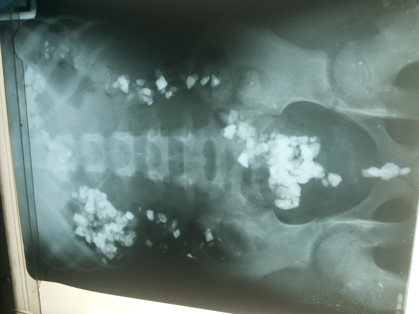
The patient had a conventional abdominal radiograph that showed multiple radiopaque structures of varying sizes, some of which are clump-like in the peripheral abdomen; the large colon and region of the rectum.
Foreign body ingestion is a potentially serious problem that peaks in children aged between six months to three years, has a morbidity of less than one percent and causes about 1500 deaths annually in the united states [1-3].
Most foreign bodies (FB) in the gastrointestinal tract pass spontaneously without complications, some may require either endoscopic or surgical removal in a few children [3, 4].
Children and mentally ill patients commonly swallow foreign bodies, and coins are the most common swallowed foreign body seeking medical attention in the USA [5-7].
The most commonly ingested FBs from the most to least common are coins, magnets, batteries, small toys, jewelry, buttons and bones [5, 8].
Because most FBs pass spontaneously, surgical removal is considered only if no radiographic progression is evident by 3 days following ingestion or if the patient becomes symptomatic[9,10].
Imaging plays an important role in the diagnosis of ingested or aspirated foreign bodies in children and can be crucial in the clinical management of these group of patients [11].
The passage rate of ingested radiopaque FBs is 64.4%, small FBs that have passed the duodenal curve should be managed conservatively via clinical observation and radiographic surveillance [12].
This is a 7-year-old male child with abnormal behavior that was referred from a peripheral health care centre for abdominal radiograph on account of recurrent abdominal pain and discomfort, passage of hard altered colored substance from the anus and history of ingestion of different substances for a long period of time.
The patient subsequently had an abdominal radiograph that showed normal bowel gas pattern, no feature of intestinal obstruction and perforation. The radiograph showed multiple radiopaque materials of varying sizes in the region of the large bowel more in the region of the ascending colon and rectum. These radiopaque materials are either solitary or clump-like, most of which have a diameter of less than 3cm.
We did a complementary abdominal ultrasound that showed normal abdominal organs but marked acoustic shadowing in the region of the large bowel was demonstrated.
We decided to report the radiographic findings of this case due to its peculiar and rare nature in our daily practice.
Foreign bodies in the gastrointestinal tract are often encountered in children and mentally unstable patients; the index case is a child of seven years and happens to be mentally unstable.
No sex predilection is documented in most literatures, some literatures however believe that foreign body ingestion has a female preponderance; the index case is a male child contrary to the literatures with female preponderance.
The commonest presenting symptoms are abdominal pain and discomfort, abdominal distension, vomiting, and passage of hard materials from the anus. Most of these symptoms were found in the index case, however no feature of intestinal obstruction or perforation was documented in this case.
Imaging has played a role in the diagnosis of foreign bodies in the gastrointestinal tract as documented in the literature, this case is not an exception as the diagnosis was made from demonstration of radiopaque materials in the large bowel following abdominal radiography.
No radiopacities were demonstrated centrally or in the region of the small bowel and stomach most likely that they have passed successfully in to the large intestine; these might be because of the shape and size of the foreign bodies and the long history/period of ingestion by the patient. These reasons were those provided and documented in most literatures and might be responsible for not developing any form of intestinal obstruction by the patient.
Basic radiographic imaging like abdominal radiograph should be done for children with complaint of recurrent abdominal pain especially between the ages of six months to three years to rule out the possibility of foreign body in the gastrointestinal tract and to prevent complications that may be life threatening in these group of patients.
Dear Editorial Team, Clinical Medical Reviews and Reports. My experience with the journal was highly positive. The peer-review process was rigorous, constructive, and completed in a timely manner. The reviewers provided valuable comments that helped improve the quality and clarity of our manuscript. The editorial office was professional, responsive, and supportive throughout all stages of the publication process. Communication was clear and efficient, and any questions were addressed promptly. Overall, I found the journal to maintain high scientific standards and an excellent publication workflow. I would be pleased to consider submitting future work to this journal. Best wishes from, Elena Popa.

It was my pleasure to submit my testimonial concerning the Reviewer Board of our Scientific Journal “Brain and Neurological Disorders”. The Reviewers focused on some modifications and their contribution was helpful. The ladies of our Editorial Office were also supported my efforts. It was my honor to have such a co-operation and I am looking forward for more collaboration.

Dear Grace Pierce, Editorial Coordinator of Journal of Clinical Research and Reports, Thank you for the speedy and efficient peer review process. I appreciate the fact that your peer reviewers do not take months to respond like with some other journals. I would also like to thank the editorial office for responding quickly to my questions. It is an excellent journal. I plan to submit more manuscripts in the future. Best wishes from, Robert W. McGee

Dear Grace Pierce, Editorial Coordinator of Journal of Clinical Research and Reports, Working with you and your team on our recent publication in JCRR has been a truly wonderful and enjoyable experience. The responses were prompt, and the reviewers were patient, constructive, and highly professional. One reviewer in particular gave me the feeling that a professor was carefully reading and commenting on my coursework, which was deeply touching. The entire process was straightforward and hassle‑free, with no tedious online forms to complete. I highly recommend this journal. Best wishes from, DR Aibing Rao, Head of R&D

I Appreciate the Opportunity to Share my Experience with the Journal of Clinical Research and Reports. The peer review process was timely and constructive, and the feedback provided helped improve the quality of our manuscript. The editorial office was professional, responsive, and supportive throughout the process, ensuring smooth communication and efficient handling of the submission. Overall, it was a positive experience collaborating with your team.

Dear Mercy Grace, Editorial Coordinator of Obstetrics Gynecology and Reproductive Sciences, We would like to express our gratitude for your help at all stages of publishing and editing the article. The editors of the magazine answer all the necessary questions and help at every stage. We will definitely continue to cooperate and publish other works in the Obstetrics Gynecology and Reproductive Sciences! Best wishes from, Alla Konstantinovna Politova,
