
Case Report | DOI: https://doi.org/10.31579/2690-4861/144
*Corresponding Author: Chithra Ram, Neuroradiologist, Assistant Professor, Department of Radiology, University of Louisville, Kentucky, USA.
Citation: C Ram, H Moss, M Terry, M Kannan, R Sherry. (2021) Multi-Modality Imaging of Craniofacial Fibrous Dysplasia with Orbital Complications and Histopathology Correlation-A case report and short review of literature. International Journal of Clinical Case Reports and Reviews. 7(3); DOI:10.31579/2690-4861/144
Copyright: © 2021 Chithra Ram, This is an open-access article distributed under the terms of the Creative Commons Attribution License, which permits unrestricted use, distribution, and reproduction in any medium, provided the original author and source are credited.
Received: 08 June 2021 | Accepted: 22 June 2021 | Published: 25 June 2021
Keywords: craniofacial fibrous dysplasia; ct of craniofacial fibrous dysplasia; mri of craniofacial fibrous dysplasia; mri perfusion of fibrous dysplasia; fibrous dysplasia imaging; histopathology of fibrous dysplasia
Craniofacial fibrous dysplasia [CF-FD] with orbital complications is a known but rare entity. This is a multi-modality imaging Case Report of extensive CF-FD in a 31-year-old male with right eye pain, swelling, and redness, along with histopathology correlation. In this patient, the CT scan demonstrates the classic ground glass bony appearance in great detail and helps with the diagnosis of FD, while excluding other bony pathology. The patient’s corroborative MRI brain with and without contrast and MRI brain perfusion images are presented to further characterize this pathology along with its orbital and ocular complications. Given the significant mass-effect on the ocular structures, the patient underwent orbital surgery with removal of as much of the lesion as possible. On macroscopic pathology evaluation, the affected bone was rubbery and gritty when sectioned. Microscopically, remnant fragments of woven bone of various size and shapes were seen with lack of an osteoblast rim. The bony fragments had a characteristic curvilinear, trabecular, and/or branching pattern. Post-surgical imaging demonstrated improvement in the mass-effect on orbital structures and proptosis, along with residual bony lesion.
Fibrous dysplasia [FD] forms 7% of benign bone tumors. FD usually presents within the first two decades of life. However, our patient was over 30 years of age. It has no gender predilection. Approximately 85% of cases of fibrous dysplasia are associated with activating missense mutations in GNAS gene with stimulation of adenylyl cyclase and over-expression of cAMP. FD can be monostotic or polyostotic. Monostotic lesions are most common. A very small percent of these lesions can be large enough to cause complications. E.g. involvement of a large portion of the orbital walls with mechanical compression on the orbital structures resulting in related signs and symptoms, etc.Craniofacial involvement may occur both as monostotic or polyostotic fibrous dysplasia. Occasionally it is seen in McCune-Albright syndrome.
A 31-year-old male presented to the emergency department with right eye pain, swelling, and redness that began the prior morning. There was no history of trauma and he denied any visual disturbances. Prior medical history was significant for hypertension. Vital signs were notable for a blood pressure of 179/92. Physical exam showed a large solid lesion in the right fronto-orbital region and proptosis. Neurologic examination showed decreased sensation to light touch on the right side of the face. The rest of the body including the extremities were unremarkable. CT and MRI were obtained to evaluate the right fronto-orbital lesion.
CT head without contrast was performed in a Somatom Definition Edge Siemens scanner [Siemens Medical Systems, USA]. Standard 1mm axial CT soft tissue and bone detail algorithm images were obtained followed by reconstructed images in coronal and sagittal planes.
MRI of the brain was performed on a 3T Siemens Spectra scanner [Siemens Medical Systems, Malvern, Pennsylvania, USA] using a standard head coil. Standard 4 mm thick axial T1, axial T2, fat suppressed axial T2 FLAIR, axial DWI and ADC brain pulse sequences were acquired without contrast. After intravenous administration of 20 ml of Multihance, perfusion imaging was performed along with axial and coronal post contrast T1W sequences.
IMAGING FINDINGS
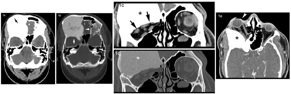
CT examination of the brain showed a large, expansile, sclerotic lesion in the right orbital roof and sphenoid bone with infiltration of the ethmoid sinuses characterized by benign appearing margins and a uniform classic “ground-glass” appearing matrix (Fig.1a,b). The bony contour of the right supraorbital region was smoothly expanded. The expansile right orbital bony lesion compromised the total volume of the right orbit and caused severe proptosis (Fig. 1c, d, e), mass-effect on the extra-ocular muscles particularly lateral rectus and probable optic nerve compression due to the narrowed optic canal.
IMAGING FIGURES: Craniofacial Fibrous dysplasia
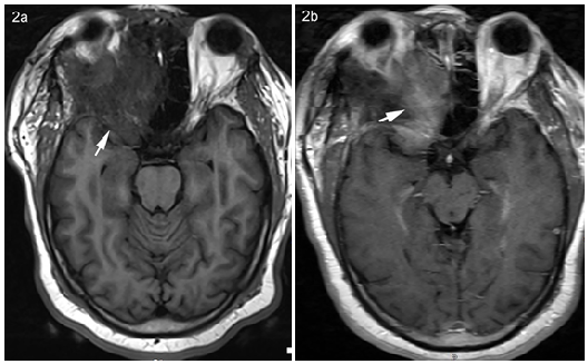
The MRI appearance was in keeping with CT diagnosis of FD. It showed heterogeneous T1 hypointensity and mild diffuse enhancement after contrast administration (Fig. 2). Additional MRI perfusion imaging (Fig. 3a) confirmed that this was not a vascular lesion and helped exclude vascular lesions such as metastasis or meningioma. DWI sequences revealed no restricted diffusion (Fig. 3b, c). Based on the imaging findings and clinical presentation the lesion was felt to be consistent with CF-FD. The patient underwent subtotal resection of the large skull lesion with decompression of the orbit, optic nerve, and superior orbital fissure using microsurgical technique and intraoperative neuronavigation. Thin layers of the lesion were unable to be resected due to their close proximity to the frontal and ethmoid sinuses. Subsequent cranioplasty with titanium mesh was used to create the orbital rim (Fig. 4). Post-operatively he developed moderate vision loss in the right eye. Follow-up postoperative imaging showed no expansion of the fibro-osseous lesion. His most recent 7 years post-op imaging shows stable and unchanged remnants of the fibrous dysplasia and proptosis. No other intervention since initial craniotomy has been necessary.


HISTOPATHOLOGY FINDINGS
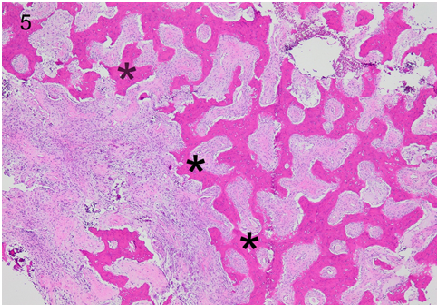
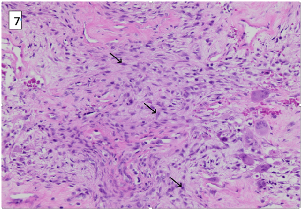
In the 4X low power hematoxylin and eosin (H&E) stained slide of the specimen [Fig.5], a combination of fibrous appearing tissue and scattered bony trabeculae were well seen. The 10X mid power H&E stained slide [Fig.6] demonstrated areas of hemorrhage associated with multinucleated osteoclast-type giant cells. Matrix resembling cementum was found in this craniofacial lesion. 20X High power H&E stained slide [Fig.7] demonstrated moderately cellular stroma with storiform [whirling] and spindle shaped cells. Fibrous dysplasia was diagnosed based on the moderately cellular spindle cells arranged in storiform (cartwheel or whirling) pattern with low mitotic count amidst the woven, discontinuous bone trabeculae.

INTRODUCTION
Fibrous dysplasia [FD] is a well-known benign bony entity. It is a developmental disorder that results from a sporadic mutation that replaces bone with fibrous tissue, resulting in structural instability [1]. FD can be monostotic [involvement of one site] or polyostotic [involvement of 2 or more sites], with monostotic lesions being most common [2,3]. Craniofacial involvement in FD can be monostotic in 10-25% of monostotic cases or polyostotic in up to 50% of polyostotic cases. FD can also exist in conjunction with endocrinopathies (e.g. McCune-Albright Syndrome) [4]. Polyostotic FD is more commonly associated with symptomatic disease and deformity [5]. It's a non-hereditary disorder with expansile lesion(s) caused by a defect in osteoblastic differentiation and maturation. It contains a mixture of fibrous tissue and woven bone, giving rise to the characteristic ground glass appearance. Sarcomatous dedifferentiation can be rarely seen in polyostotic FD with prior radiation therapy.
McCune-Albright Syndrome has a clinical triad of polyostotic FD, hyperfunctioning endocrinopathies and café au lait spots. It has a female preponderance [7]. Mazabraud Syndrome is polyostotic FD with intramuscular myxoma. It has increased risk of malignant transformation. Cherubism is familial FD involving bilateral jaw [8].
CLINICAL PRESENTATION
FD is often asymptomatic and diagnosed incidentally as a well-circumscribed lesion, especially with monostotic disease. Clinical presentation often depends on location and number of lesions. Ribs, femurs, and craniofacial bones are most commonly affected [3,6]. Craniofacial FD presents with asymmetric facial enlargement due to the affected bone, proptosis, atypical facial pain or numbness, headache and cranial neuropathy (diplopia, hearing loss, blindness) [9]. Progression from monostotic disease to polyostotic has not been documented [1]. Craniofacial bones are most commonly implicated in monostotic FD, while the femur is commonly affected in polyostotic FD. Polyostotic disease occurs in a younger age with limp, leg pain, or pathologic fractures. The lesions are commonly unilateral and are located in the same extremity when the appendicular skeleton is affected [8]. Fractures typically occur at the proximal femur with possible “shepherd’s crook” deformity. If the spine is affected, then scoliosis can occur [3,6]. Patients with café-au-lait spots and precocious puberty with FD should be considered for a diagnosis of McCune–Albright syndrome [10]. Association with aneurysmal bone cysts has also been described [11].
IMAGING FINDINGS
Commonly discovered as an incidental finding, lesions of fibrous dysplasia are characterized into three types [12]. Pagetoid lesions are most common, followed by sclerotic and then cystic lesions [7]. In craniofacial FD, more than 1 bone involvement is often seen. Maxilla, orbit, and frontal bones are commonly involved, while ethmoids and sphenoids are rarely involved. CT is most sensitive and specific. CT and MR help define local extent, while bone scintigraphy scan identifies additional lesions [10].
Radiographs: FD is a smooth, well circumscribed, homogeneous ground glass lesion with well-defined borders. Endosteal scalloping and cortical thinning is noted due to the expansive nature of the lesion. It may be sclerotic or lucent (cystic). Sclerotic lesions are homogenously dense. Cystic lesions can show a “rind” sign, where a thick layer of sclerotic reactive bone surrounds a lucent lesion [3,7,14]. This sign is commonly seen in proximal femur.
CT: Radiolucent ground-glass matrix in an expansile bony lesion involving the medulla, is diagnostic especially in pagetoid lesions. Smooth cortical contour is typically maintained. Endosteal scalloping may also be present [13].
MRI: Decreased T2W signal is noted throughout the lesion if it's pagetoid or solid type and only in rim if it's of the cystic type. Increased pathological activity can result in fewer bony trabeculae and hence result in increased T2W signal [3,7]. Decreased T1W signal is usually seen. Enhancement is variable. It can be peripheral, diffuse or non-enhancing.
Bone scan: It's nonspecific with variable radionuclide uptake in perfusion and delayed phases. It's sensitive but not specific in identifying skeletal lesions in polyostotic FD.
PET: FD shows variable uptake on FDG [Fluoro-deoxy glucose] PET. Correlation with CT helps differentiate it from metastasis.
HISTOPATHOLOGY
The histopathologic features of CF-FD consists primarily of normal bone that is replaced and distorted by bland fibrous tissue [15]. Grossly, the affected bone is rubbery and gritty when sectioned [15]. Microscopically, remnant fragments of woven bone of various size and shapes are seen. These bony fragments characteristically lack an osteoblast rim, and may seamlessly interface with adjacent cancellous bone or cortex [15,16]. As a result, there may be no clear demarcation between lesion and adjacent normal bone on imaging [17]. The bony fragments are curvilinear, trabecular, and/or branching, which gives a characteristic “Chinese letter” or “alphabet soup” appearance [15]. This manifests as diffusely distributed opacities on imaging. The bone fragments are typically trabecular or woven, although lamellar bone is occasionally present if the lesion has been present for many years [15-17]. The fibrous tissue is relatively cellular and contains spindled or stellate fibroblasts with associated collagen. Mitotic figures are scarce or absent [15]. Cases with abundant cartilage, named fibrocartilaginous dysplasia, have been described [15]. Synonyms that are no longer recommended for use include osteitis fibrosa, fibrous osteoma, and unilateral von Recklinghausen disease. Approximately 85% of cases of fibrous dysplasia are associated with activating missense mutations in GNAS gene with stimulation of adenyl cyclase and over-expression of cAMP [15,18].
PROGNOSIS AND TREATMENT
Prognosis is good, many cases spontaneously resolve with aging as the lesions become inactive after puberty, and the condition is considered benign. Asymptomatic cases can be managed with patient education and clinical observation [19]. The maintenance of bone density is important during management, especially for prevention of fractures [1]. Malignant transformation can rarely occur and is more common in polyostotic rather than monostotic FD [7]. No treatment methods are currently available to slow or alter the disease course. Bisphosphonates can control bone pain [19]. Polyostotic patients should be carefully monitored to optimize function and ambulation as they can result in pathological fractures, which may affect mobility. Surgery with grafting is an option to correct deformities or otherwise symptomatic lesions and large monostotic lesions can be resected [3,19]. Aggressive resection is reserved for visual loss and severe deformity of the cranium.
FD is a benign congenital condition that results in replacement of normal bone architecture with fibro-osseous tissue. The condition can be monostotic or polyostotic and can be in association with endocrine abnormalities in the context of McCune-Albright Syndrome and Mazabraud Syndrome. Disruption of bone structure predisposes these patients to deformity and fracture, but the overall prognosis is good. Radiologists should be aware of the potential for some of these lesions to rarely undergo malignant transformation in polyostotic FD cases. The characteristic ground-glass appearance on CT is pathognomonic. In this patient the MRI perfusion images were useful in confirming the avascular nature of the lesion and in helping to exclude potential vascular lesions such as Paget’s disease, metastatic disease and invasive meningioma. Most patients can be managed conservatively, with surgical options reserved for those with extensive deformity, fracture, or malignant transformation. In the patient reported here, surgical intervention was necessitated by the severe mass-effect on the ocular structures from the expansile bony lesion with resultant proptosis, eye pain, redness and potential for optic nerve compression.
ABBREVIATIONS
FD - Fibrous dysplasia, CF-FD – Craniofacial Fibrous dysplasia, CT - Computerized Tomography, MRI - Magnetic Resonance Imaging. PET – Positron Emission Tomography.
CONFLICT OF INTEREST: None.
FUNDING: None.
Dear Editorial Team, Clinical Medical Reviews and Reports. My experience with the journal was highly positive. The peer-review process was rigorous, constructive, and completed in a timely manner. The reviewers provided valuable comments that helped improve the quality and clarity of our manuscript. The editorial office was professional, responsive, and supportive throughout all stages of the publication process. Communication was clear and efficient, and any questions were addressed promptly. Overall, I found the journal to maintain high scientific standards and an excellent publication workflow. I would be pleased to consider submitting future work to this journal. Best wishes from, Elena Popa.

It was my pleasure to submit my testimonial concerning the Reviewer Board of our Scientific Journal “Brain and Neurological Disorders”. The Reviewers focused on some modifications and their contribution was helpful. The ladies of our Editorial Office were also supported my efforts. It was my honor to have such a co-operation and I am looking forward for more collaboration.

Dear Grace Pierce, Editorial Coordinator of Journal of Clinical Research and Reports, Thank you for the speedy and efficient peer review process. I appreciate the fact that your peer reviewers do not take months to respond like with some other journals. I would also like to thank the editorial office for responding quickly to my questions. It is an excellent journal. I plan to submit more manuscripts in the future. Best wishes from, Robert W. McGee

Dear Grace Pierce, Editorial Coordinator of Journal of Clinical Research and Reports, Working with you and your team on our recent publication in JCRR has been a truly wonderful and enjoyable experience. The responses were prompt, and the reviewers were patient, constructive, and highly professional. One reviewer in particular gave me the feeling that a professor was carefully reading and commenting on my coursework, which was deeply touching. The entire process was straightforward and hassle‑free, with no tedious online forms to complete. I highly recommend this journal. Best wishes from, DR Aibing Rao, Head of R&D

I Appreciate the Opportunity to Share my Experience with the Journal of Clinical Research and Reports. The peer review process was timely and constructive, and the feedback provided helped improve the quality of our manuscript. The editorial office was professional, responsive, and supportive throughout the process, ensuring smooth communication and efficient handling of the submission. Overall, it was a positive experience collaborating with your team.

Dear Mercy Grace, Editorial Coordinator of Obstetrics Gynecology and Reproductive Sciences, We would like to express our gratitude for your help at all stages of publishing and editing the article. The editors of the magazine answer all the necessary questions and help at every stage. We will definitely continue to cooperate and publish other works in the Obstetrics Gynecology and Reproductive Sciences! Best wishes from, Alla Konstantinovna Politova,
