
short communication | DOI: https://doi.org/10.31579/2642-973X/059
1 Candidate of biological science, assistant professor of pathophysiology department named D.A. Maslakov, Grodno State Medical University, Grodno State Medical University, Belarus
2 State Medical University, Grodno, Republic of Belarus
*Corresponding Author: Lizaveta I. Bon, Candidate of biological science, assistant professor of pathophysiology department named D.A. Maslakov, Grodno State Medical University, Grodno State Medical University, Belarus
Citation: Lizaveta I. Bon, N.Ye. Maksimovich, S.M. Zimatkin, O.V. GrasevichGrodno, (2023), Morphological Disorders of Rat Hippocampal Neurons in Mechanical Asphixia, J. Brain and Neurological Disorders, 6(4): DOI:10.31579/2642-973X/059
Copyright: © 2023, Lizaveta I. Bon. This is an open-access article distributed under the terms of The Creative Commons Attribution License, which permits unrestricted use, distribution, and reproduction in any medium, provided the original author and source are credited.
Received: 17 April 2023 | Accepted: 27 April 2023 | Published: 04 May 2023
Keywords: asphyxia; neurons; hippocampus
Brain damage to one degree or another develops in persons who have suffered respiratory arrest. The study of the hippocampus of rats under conditions of mechanical asphyxia revealed the presence of structural changes in both studied periods (after 30 and 60 minutes): a decrease in the area and a change in the shape (loss of sphericity and an increase in elongation) of cells, as well as a change in the degree of chromatophilia, which was manifested by the disappearance of normochromic neurons with simultaneous appearance of hyperchromic and hyperchromic shriveled neurons. To the greatest extent, these changes were expressed during 60 minutes of mechanical asphyxia.
Asphyxia caused by exposure to an external mechanical factor is called mechanical asphyxia. It is characterized by severe disorders of the central nervous system, cardiovascular system and respiratory organs, which occur due to a lack of oxygen in the blood and tissues (hypoxemia and hypoxia) and the accumulation of carbon dioxide (hypercapnia).
Brain damage to one degree or another develops in persons who have experienced respiratory arrest [1-3]. Previous studies have studied changes in neurons in the cortex of the parietal lobe of the rat brain [4,5]. Along with this, such a brain structure as the hippocampus deserves special attention [6]. This is due to its importance in the life of the body. The hippocampus serves as an important memory center, provides orientation in space. In acute oxygen starvation of the hippocampus, Korsakov's syndrome occurs, in which memory for current events is lost, with comparative preservation of traces of long-term memory.
To date, the processes of neuronal damage to the hippocampus due to global anoxia caused by mechanical asphyxia of varying duration remain poorly understood.
Purpose - to study the morphological disorders of neurons in the hippocampus of rats with mechanical asphyxia.
The study was carried out on outbred white rats (18 males, weight 240±20 g), divided into 3 groups (n=6) in compliance with the requirements of the Directive of the European Parliament and Council No. 2010/63/EU of 22.09.2010 on the protection of animals used for scientific purposes.
The control group consisted of sham-operated rats (group 1).
Simulation of mechanical asphyxia was performed by tying the trachea of rats below the cricoid cartilage of the larynx with a ligature for 30 minutes (group 2) and 60 minutes (group 3) [5].
The studies were carried out under conditions of intravenous anesthesia (sodium thiopental, 40 mg/kg). The brain was removed and fixed in Carnoy's fluid, after which serial frontal paraffin sections 7 μm thick were made and stained according to the Nissl method. The location of the field CA1 of the hippocampus was determined using a stereotaxic atlas [7].
Visual and morphometric assessment of neurons was performed using the ImageWarp image analysis program (Bitflow, USA).
In each animal, 30 neurons of the pyramidal layer of the field CA1 of the hippocampus were evaluated with a study of their size and shape [8]. In histological preparations, various types of neurons were determined by the degree of staining of their cytoplasm (chromatophilia).
The obtained quantitative continuous data were processed using the methods of nonparametric statistics, the licensed computer program Statistica 10.0 for Windows (StatSoft, Inc., USA). The data are presented as Me (LQ; UQ), where Me is the median, LQ is the value of the lower quartile; UQ is the value of the upper quartile. Differences between the indicators of the control and experimental groups were considered significant at p less than 0.05 (MannWhitneyU-test) [9].
In the control group, normochromic cells accounted for 100% of the neuron population in the hippocampus. Perikaryons had a rounded shape, distinct even contours of the cellular and nuclear surfaces. The perikaryon area was 76.2 (72.9; 79.5) µm2, form factor 0.9 (0.9; 0.9) units, elongation factor 1.2 (1.1; 1.3) units (Table 1).
Groups | Indicators | ||
area (mcm2) | form factor (units) | elongation factor (units) | |
| control | 76,2 (72,9; 79,5) | 0,9 (0,9; 0,9) | 1,2 (1,1; 1,3) |
| mechanical asphyxia 30 min | 67,1 (62,7; 71,5)* | 0,7 (0,6; 0,7)* | 2,1 (2,0; 2,1)* |
| mechanical asphyxia 60 min | 59,3 (56,9; 61,6)*# | 0,6 (0,6; 0,7)* | 2,2 (2,1; 2,3)* |
Table 1: Indicators of the size and shape of neurons in the hippocampus of the brain of rats with mechanical asphyxia (Me;LQ;UQ)
Note: – * – differences are significant (p less thsn 0.05) compared with the control group;
– # – differences are significant (p less than 0.05) compared with the group "mechanical asphyxia 30min".
In both studied time periods of mechanical asphyxia, morphological changes in hippocampal neurons manifested themselves in changes in the area and shape of neurons, and in the intensity of staining of their cytoplasm (Figure 1, 2, 3, 4).
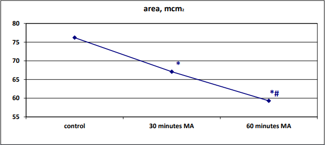
Figure 1: Dynamics of changes in the area of neurons in the pyramidal layer of the field СА1 гиппокампа после 30 минут и 60 минут механической асфиксии (МА)
Note: – * – differences are significant (p less than 0.05) compared with the control group;
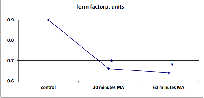
Figure 2: Dynamics of changes in the form factor of neurons in the pyramidal layer of the field CA1 of the hippocampus after 30 minutes and 60 minutes of mechanical asphyxia (MA).
Note: – * – differences are significant (p less than 0.05) compared with the control group.

Figure 3: Dynamics of changes in the elongation factor of neurons in the pyramidal layer of the field CA1 of the hippocampus after 30 minutes and 60 minutes of mechanical asphyxia (MA)
Note: – * – differences are significant (p less than 0.05) compared with the control group.

Figure 4: Graphic display of morphometry parameters of neurons (size and shape) of the pyramidal layer of field CA1 of the hippocampus of rats with anoxia
Note: – * – differences are significant (p less than 0.05) compared with the control group.
– # – differences are significant (p less than 0.05) compared with the group «mechanical asphyxia 30 min»
After 30 minutes of mechanical asphyxia, the area of neurons decreased by 12% (p less than 0.05) compared to the control group, the form factor decreased by 27% (p less than 0.05), and the elongation factor increased by 76% (p less than 0.05), which reflects the loss of sphericity and, at the same time, an increase in their elongation. After 60 minutes of mechanical asphyxia, the neuronal area decreased by 22% (p less than 0.05) compared to the control group, the form factor decreased by 29% (p<0>0.05).
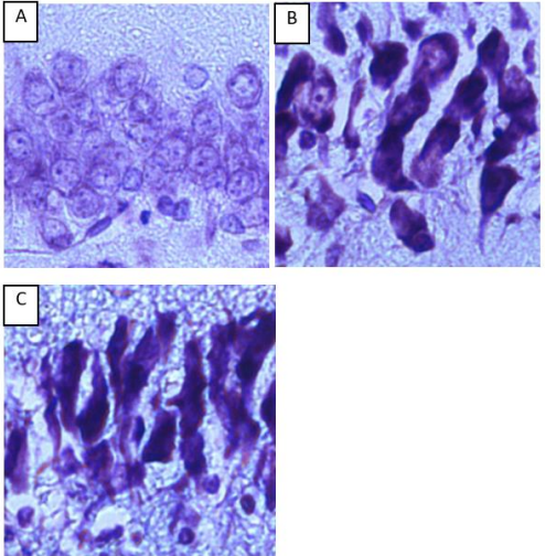
Figure 5: Neurons of the pyramidal layer of the CA1 field of the rat hippocampus. Digital micrograph. Nissl stain.
А – control group (normochromic neurons)
B - after 30 minutes of mechanical asphyxia (hyperchromic and hyperchromic shriveled neurons)
C - after 60 minutes of mechanical asphyxia (hyperchromic and hyperchromic shriveled neurons); magnification x 40.
In both experimental groups of rats with mechanical asphyxia, normochromic neurons completely disappeared (Fig. 5). Most of the cells in the groups of rats with 30 and 60 minutes of mechanical asphyxia were hyperchromic and hyperchromic shriveled neurons.
Thus, mechanical asphyxia in dynamics led to anoxic damage to the neurons of the pyramidal layer of the field CA1 of the hippocampus of rats, manifested in the form of a decrease in the area and deformation of the perikaryons, an increase in the degree of chromatophilia of the neuronal cytoplasm. After 60 minutes of mechanical asphyxia, there was an aggravation of the decrease in the size of hippocampal neurons, without changing their shape and intensity of staining of the cytoplasm compared with a 30-minute period of mechanical asphyxia.
Structural disorders of neurons in the hippocampus of the brain of rats with mechanical asphyxia were unidirectional in nature with the previously obtained results on changes in the parietal cortex. In contrast to the parietal cortex, morphological changes were noted earlier and were more pronounced, namely, a decrease in the area and a change in the shape of neurons in combination with an increase in the degree of chromatophilia [4,5].
Dear Editorial Team, Clinical Medical Reviews and Reports. My experience with the journal was highly positive. The peer-review process was rigorous, constructive, and completed in a timely manner. The reviewers provided valuable comments that helped improve the quality and clarity of our manuscript. The editorial office was professional, responsive, and supportive throughout all stages of the publication process. Communication was clear and efficient, and any questions were addressed promptly. Overall, I found the journal to maintain high scientific standards and an excellent publication workflow. I would be pleased to consider submitting future work to this journal. Best wishes from, Elena Popa.

It was my pleasure to submit my testimonial concerning the Reviewer Board of our Scientific Journal “Brain and Neurological Disorders”. The Reviewers focused on some modifications and their contribution was helpful. The ladies of our Editorial Office were also supported my efforts. It was my honor to have such a co-operation and I am looking forward for more collaboration.

Dear Grace Pierce, Editorial Coordinator of Journal of Clinical Research and Reports, Thank you for the speedy and efficient peer review process. I appreciate the fact that your peer reviewers do not take months to respond like with some other journals. I would also like to thank the editorial office for responding quickly to my questions. It is an excellent journal. I plan to submit more manuscripts in the future. Best wishes from, Robert W. McGee

Dear Grace Pierce, Editorial Coordinator of Journal of Clinical Research and Reports, Working with you and your team on our recent publication in JCRR has been a truly wonderful and enjoyable experience. The responses were prompt, and the reviewers were patient, constructive, and highly professional. One reviewer in particular gave me the feeling that a professor was carefully reading and commenting on my coursework, which was deeply touching. The entire process was straightforward and hassle‑free, with no tedious online forms to complete. I highly recommend this journal. Best wishes from, DR Aibing Rao, Head of R&D

I Appreciate the Opportunity to Share my Experience with the Journal of Clinical Research and Reports. The peer review process was timely and constructive, and the feedback provided helped improve the quality of our manuscript. The editorial office was professional, responsive, and supportive throughout the process, ensuring smooth communication and efficient handling of the submission. Overall, it was a positive experience collaborating with your team.

Dear Mercy Grace, Editorial Coordinator of Obstetrics Gynecology and Reproductive Sciences, We would like to express our gratitude for your help at all stages of publishing and editing the article. The editors of the magazine answer all the necessary questions and help at every stage. We will definitely continue to cooperate and publish other works in the Obstetrics Gynecology and Reproductive Sciences! Best wishes from, Alla Konstantinovna Politova,
