
Research Article | DOI: https://doi.org/10.31579/2690-1897/175
Grodno State Medical University, Republic of Belarus, 230009, Grodno, Gorky St., 80.
*Corresponding Author: Bon E.I. Grodno State Medical University, Republic of Belarus, 230009, Grodno, Gorky St., 80.
Citation: Maksimovich N.E, Bon E.I, Vishnevskaya E.I, Bon E.I. (2024), Morphological Changes in rats' Cerebral Cortex Neurons during Strangulation Asphyxia, J, Surgical Case Reports and Images 7(1); DOI:10.31579/2690-1897/175
Copyright: © 2024, Bon E.I. This is an open access article distributed under the Creative Commons Attribution License, which permits unrestricted use, distribution, and reproduction in any medium, provided the original work is properly cited.
Received: 07 February 2024 | Accepted: 15 February 2024 | Published: 20 February 2024
Keywords: asphyxia; neurons; occipital cortex; brain; rats
Objective: To study the peculiarities of morphological changes of neurons of the occipital lobe of rats with strangulation asphyxiation.
Methods: The study was carried out on the native white rats (24 males, weighing 240+20 g), divided into a control group and three experimental groups with a strangulation asphyxia of 6 individuals in each group.
Results: Morphological changes in the occipital cortex neurons were observed in experimental animal groups during the study periods, in the form of changes in the area, shape of the neurons and intensity of cytoplasm staining.
Conclusion: It was concluded that the strangulation asphyxia caused anoxic damage to the neurons of the occipital lobe of rats, manifested in the form of changes in the size, shape of neurons, intensity of coloration of their cytoplasm.
Acute oxygen deficiency can cause severe and often irreversible changes in brain tissue and even lead to its death [3]. Brain asphyxia occurs in many diseases, pathological conditions, and due to environmental factors. Specifically, oxygen deficit can result from impaired air passage in the respiratory tract due to external mechanical factors (mechanical asphyxia) [4]. The occipital lobe cortex, responsible for visual information perception and processing, and orientation in new environments, deserves special attention. Hypoxia in this brain region can lead to loss of visual function - cortical blindness.
Previous studies have examined morphological changes in the brain cortex under total ischemia caused by decapitation [1, 2].
To date, the specific damage to neurons in the occipital lobe cortex due to respiratory-genesis anoxia caused by external mechanical factors has not been studied.
The aim is to study the morphological changes in neurons of the occipital lobe cortex in rats with strangulation asphyxia.
The study was conducted on non-breed white male rats (24 individuals, weight 240±20 g), divided into a control and three experimental groups with strangulation asphyxia, six animals in each group.
The control group consisted of sham-operated rats without strangulation asphyxia (group 1, n=6).
Strangulation asphyxia was modeled under thiopental anesthesia (intravenous, 50 mg/kg) by applying a ligature to the trachea 1.0 cm below the cricoid cartilage for 30 minutes (group 2), 60 minutes (group 3), and 24 hours (group 4) [7].
The experiment was conducted in compliance with the requirements of the European Parliament and Council Directive No. 2010/63/EU of 22.09.2010 on the protection of animals used for scientific purposes.
The brain was extracted in the cold and fixed in Carnoy's solution. Serial frontal paraffin sections 7 µm thick were prepared and stained with thionin using the Nissl method. The location of the occipital lobe cortex was established using a stereotaxic atlas [6].
In each animal, 30 neurons of the fifth layer of the occipital lobe cortex were studied, determining their area, shape, and chromophilic degree. Changes in the area and shape (form factor, elongation factor) of neurons were assessed using the ImageWarp image analysis software (Bitflow, USA) [5].
Obtained quantitative continuous data were processed using non-parametric statistical methods and the licensed computer program Statistica 10.0 for Windows (StatSoft, Inc., USA). Data are presented as Me(LQ;UQ), where Me – median, LQ – lower quartile value; UQ – upper quartile value. Differences between the control and experimental groups were considered significant at p<0>
Research results: In the experimental groups, morphological changes in the neurons of the occipital lobe cortex were observed during the studied periods in terms of changes in area (table 1, fig.2), neuron shape (table 1, fig.1,3), and the intensity of their cytoplasm staining (figure. 4).
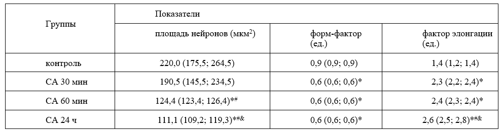
Table 1 - Parameters of size (area) and shape (form factor and elongation factor) of neurons in the occipital lobe cortex of rats with strangulation asphyxia (SA), Me(LQ;UQ)
Note: - * - differences are significant (p<0>
- # - differences are significant (p<0>
- & - differences are significant (p<0>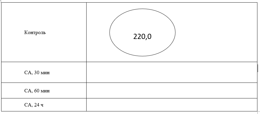
Figure 1. Graphical representation of morphometric parameters of neurons (size, µm², and shape) in the occipital lobe cortex of rats with strangulation asphyxia (SA)
After 30 minutes of asphyxia, there were no changes in the area of neurons compared to the control group, but a decrease in the form factor by 29% (p<0>
After 60 minutes of asphyxia, the area of neurons decreased by 40% compared to the control group (p<0>
Additionally, in rats with 60 minutes of asphyxia, there was a 35
Thus, strangulation asphyxia led to anoxic damage to neurons in the occipital lobe cortex of rats, manifested in changes in the size, shape of neurons, intensity of staining of their cytoplasm. With 30 minutes of asphyxia, neurons acquired an elongated shape, losing roundness, and their sizes did not change. At 60 minutes of asphyxia, there was a significant decrease in neuron area, which worsened by 24 hours of asphyxia, while no change in neuron shape was observed. During all studied periods of asphyxia, there was an increase in the number of hyperchromic shrunken neurons with a decrease in the number of normochromic neurons and their complete disappearance by 24 hours of asphyxia.
Dear Editorial Team, Clinical Medical Reviews and Reports. My experience with the journal was highly positive. The peer-review process was rigorous, constructive, and completed in a timely manner. The reviewers provided valuable comments that helped improve the quality and clarity of our manuscript. The editorial office was professional, responsive, and supportive throughout all stages of the publication process. Communication was clear and efficient, and any questions were addressed promptly. Overall, I found the journal to maintain high scientific standards and an excellent publication workflow. I would be pleased to consider submitting future work to this journal. Best wishes from, Elena Popa.

It was my pleasure to submit my testimonial concerning the Reviewer Board of our Scientific Journal “Brain and Neurological Disorders”. The Reviewers focused on some modifications and their contribution was helpful. The ladies of our Editorial Office were also supported my efforts. It was my honor to have such a co-operation and I am looking forward for more collaboration.

Dear Grace Pierce, Editorial Coordinator of Journal of Clinical Research and Reports, Thank you for the speedy and efficient peer review process. I appreciate the fact that your peer reviewers do not take months to respond like with some other journals. I would also like to thank the editorial office for responding quickly to my questions. It is an excellent journal. I plan to submit more manuscripts in the future. Best wishes from, Robert W. McGee

Dear Grace Pierce, Editorial Coordinator of Journal of Clinical Research and Reports, Working with you and your team on our recent publication in JCRR has been a truly wonderful and enjoyable experience. The responses were prompt, and the reviewers were patient, constructive, and highly professional. One reviewer in particular gave me the feeling that a professor was carefully reading and commenting on my coursework, which was deeply touching. The entire process was straightforward and hassle‑free, with no tedious online forms to complete. I highly recommend this journal. Best wishes from, DR Aibing Rao, Head of R&D

I Appreciate the Opportunity to Share my Experience with the Journal of Clinical Research and Reports. The peer review process was timely and constructive, and the feedback provided helped improve the quality of our manuscript. The editorial office was professional, responsive, and supportive throughout the process, ensuring smooth communication and efficient handling of the submission. Overall, it was a positive experience collaborating with your team.

Dear Mercy Grace, Editorial Coordinator of Obstetrics Gynecology and Reproductive Sciences, We would like to express our gratitude for your help at all stages of publishing and editing the article. The editors of the magazine answer all the necessary questions and help at every stage. We will definitely continue to cooperate and publish other works in the Obstetrics Gynecology and Reproductive Sciences! Best wishes from, Alla Konstantinovna Politova,
