
Case Report | DOI: https://doi.org/10.31579/2642-9756/140
1 Emeritus Professor, Obstetrics Gynaecology, Ex. Dean,Mahatma Gandhi Institute of Medical Sciences, Sevagram.Chief Executive Officer, Akanksha ShishuKalyan Kendra, Sevagram.Officer On Special Duty, Dr. SushilaNayar Hospital, Amravati, Kasturba Health Society, Sewagram,Wardha, Maharashtra, India.
2 Senior Resident Obstetrics Gynaecology, Mahatma Gandhi Institute of Medical Sciences, Sevagram.
3Assistant Professor Obstetrics Gynaecology, Mahatma Gandhi Institute of Medical Sciences, Sevagram.
*Corresponding Author: Chhabra S, Emeritus Professor, Obstetrics Gynaecology, Ex. Dean, Mahatma Gandhi Institute of Medical Sciences, Sevagram. Chief Executive Officer, Akanksha Shishu Kalyan Kendra, Sevagram. Officer On Special Duty, Dr. Sushila Nayar Hospital, Amravati, Kastu
Citation: Chhabra S, Mishra U, Thombre., (2023), Mixed Malignant Mullerian Tumor of Uterine Corpus In a Treated Case of Advanced Cervical Cancer. J. Women Health Care and Issues. 6(2); DOI:10.31579/2642-9756/140
Copyright: © 2023, Chhabra S. This is an open access article distributed under the Creative Commons Attribution License, which permits unrestricted use, distribution, and reproduction in any medium, provided the original work is properly cited.
Received: 28 February 2023 | Accepted: 09 March 2023 | Published: 30 March 2023
Keywords: malignant mixed mullerian tumor (mmmt); carcinosarcoma;metaplastic carcinoma
An unusual case of advanced mixed malignant mullerian tumor in a case, treated for cervical cancer (stage IIB) one year back is presented Patient had debulking surgery and postoperative chemotherapy. She did well for a year after completion of therapy but succumbed to her illness after 1 year without any workup.
Mixed malignant mullerian tumor (MMMT): carcinoma metaplastically changed into sarcoma [1], an uncommon malignancy, comprise only 1–2% of uterine neoplasms . Beside uterus, it has been identified, in decreasing order of frequency in vagina [2] cervix [3], ovary [4], and most rarely the fallopian tube [5]. According to be current knowledge, MMMT is a biphasic female genital tract tumour made up of both epithelial and mesenchymal components [6]. Present case is of MMMT uterus.
Case Summary:
A 64 years old woman with 3 full term normal births, postmenopausal for 15 years, presented with pain and lump in abdomen with difficulty in micturition since 1 month, 1 year after completion of radiochemotherapy for cervical cancer stage IIB . She had a mass arising from pelvis of approximately 26 weeks of pregnancy with variegated consistency going above umbilicus with restricted mobility. Vaginal examination was not possible, due to cicatrisation of vagina post chemoradiation. However rectal examination was suggestive of mostly cystic mass (in retropubic region) of 14 weeks size of uterus with fluid inside, suggestive of pyometra with over all mass of around 26 weeks size of pregnancy with no demarcation of margins.
Magnetic resonance imaging (MRI) revealed a well defined large solid cystic mass of around 13 x 12 x 11 cm (CC x TR x AP) in pelvis extending upto lower abdomen involving whole of the cervix, uterus and vagina. The lesion was involving perineal fibro-fatty tissues. Superiorly there was a focal breach in the serosal lining and loss of fat planes with the bowel loops superiorly, urinary bladder anteriorly and rectum posteriorly. Uterus and bilateral ovaries were seen partly separately from the mass. Loculated collection in adnexa with mild ascites in abdomen and pelvis.
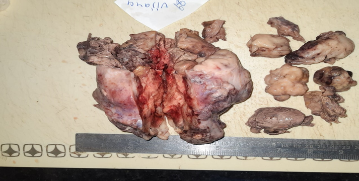
After necessary work up exploratory laparotomy was performed. Intraoperative there was 28 weeks size mass overall, 400ml encysted tumor fluid with Uterus having multiple cauliflower like growth of variegated consistency and multiple solid and cystic areas present (Figure1), Tumor was removed as much as possible. Patient did well postoperative.
Histopathological findings were suggestive of Mixed malignant mullerian tumor of uterine corpus infiltrating into cervix. Tumor was also infiltrating into deep myometrium and replacing endometrium. Both ovaries and tubes were unremarkable uninvolved. Both parametrium were involved. Section from omentum showed inflammation but negative for tumor infiltration. She received 6 cycles of chemotherapy (Paclitaxal and Carboplatin). Patient had follow up, but died one year of completion of therapy without any further investigations .
MMMT represent less than 5% of all uterine tumors [1], but are believed to be responsible for 16.4% of all deaths caused by a uterine malignancy (7).Most common age of presentation is sixth and seventh decade [8], as was in the present case. Risk factors include nulliparity, advanced age, obesity, exposure to exogenous estrogens and long-term use of tamoxifen and are comparable to those for endometrial carcinoma [9]. Exposure to the human papilloma virus, is also reported ,as a aetiological variable linked to the development of MMMT [10]. Present case had advanced cervical cancer and had received chemo-radiotherapy also. It is reported that 5% to 30% of patients with carcinosarcoma have history of irradiation for pelvic pathology. But usually present after long interval interval [11]. Present case had cervical cancer and received radiotherapy too but had short interval. A typical presentation of carcinosarcoma includes pyometra with vaginal bleeding, bloody or watery discharge, abdominal pain, or as a polypoid mass in an older, postmenopausal woman [12] In the present case complaints were lower abdomen pain with lump and difficulty in micturition. Fluid in uterus gave feel of pyometra but on opening polypoidal growth was seen with fluid under and around uterus. Upto 53% of carcinosarcoma patients present with advanced-stage disease [13] as was present case.The macroscopic histological appearance of uterine carcinosarcoma typically resembles a single polypoid mass with areas of haemorrhage and necrosis protruding into the uterine cavity [14]. In the present case there were multiple polypoidal masses with haemorrhage and necrosis.
Inspite of most common presentation as advanced disease the role of vigorous cytoreductive surgery and the best chemotherapy regimen in stage IV disease is uncertain. Similarly it is unknown how multimodal therapy can be used in such cases. Present case is unusual as was advanced mixed malignant mullerian tumor of uterine corpus after a short interval of treatment of advanced cervical cancer II B.
A advance case of mixed malignant mullerian tumor of uterine corpus after having treatment of advance cervical cancer, presented with a big lump, did well almost 1.5 year after treatment but succumbed.
Dear Editorial Team, Clinical Medical Reviews and Reports. My experience with the journal was highly positive. The peer-review process was rigorous, constructive, and completed in a timely manner. The reviewers provided valuable comments that helped improve the quality and clarity of our manuscript. The editorial office was professional, responsive, and supportive throughout all stages of the publication process. Communication was clear and efficient, and any questions were addressed promptly. Overall, I found the journal to maintain high scientific standards and an excellent publication workflow. I would be pleased to consider submitting future work to this journal. Best wishes from, Elena Popa.

It was my pleasure to submit my testimonial concerning the Reviewer Board of our Scientific Journal “Brain and Neurological Disorders”. The Reviewers focused on some modifications and their contribution was helpful. The ladies of our Editorial Office were also supported my efforts. It was my honor to have such a co-operation and I am looking forward for more collaboration.

Dear Grace Pierce, Editorial Coordinator of Journal of Clinical Research and Reports, Thank you for the speedy and efficient peer review process. I appreciate the fact that your peer reviewers do not take months to respond like with some other journals. I would also like to thank the editorial office for responding quickly to my questions. It is an excellent journal. I plan to submit more manuscripts in the future. Best wishes from, Robert W. McGee

Dear Grace Pierce, Editorial Coordinator of Journal of Clinical Research and Reports, Working with you and your team on our recent publication in JCRR has been a truly wonderful and enjoyable experience. The responses were prompt, and the reviewers were patient, constructive, and highly professional. One reviewer in particular gave me the feeling that a professor was carefully reading and commenting on my coursework, which was deeply touching. The entire process was straightforward and hassle‑free, with no tedious online forms to complete. I highly recommend this journal. Best wishes from, DR Aibing Rao, Head of R&D

I Appreciate the Opportunity to Share my Experience with the Journal of Clinical Research and Reports. The peer review process was timely and constructive, and the feedback provided helped improve the quality of our manuscript. The editorial office was professional, responsive, and supportive throughout the process, ensuring smooth communication and efficient handling of the submission. Overall, it was a positive experience collaborating with your team.

Dear Mercy Grace, Editorial Coordinator of Obstetrics Gynecology and Reproductive Sciences, We would like to express our gratitude for your help at all stages of publishing and editing the article. The editors of the magazine answer all the necessary questions and help at every stage. We will definitely continue to cooperate and publish other works in the Obstetrics Gynecology and Reproductive Sciences! Best wishes from, Alla Konstantinovna Politova,
