
Review ariticle | DOI: https://doi.org/10.31579/2690-4861/669
Department of Obstetrics and Gynecology, University Clinics of Schleswig Holstein (UKSH), Arnold Heller Strasse 3, Haus C, 24105 Kiel, Germany.
*Corresponding Author: Liselotte Mettler, Department of Obstetrics and Gynecology, University Clinics of Schleswig Holstein (UKSH), Arnold Heller Strasse 3, Haus C, 24105 Kiel, Germany.
Citation: Liselotte Mettler, Ibrahim Alkatout and Andreas Schmutzler, (2025), Minimal Invasive Surgery (MIS) and Artificial Reproductive Technologies (ART) over 60 years, International Journal of Clinical Case Reports and Reviews, 23(4); DOI:10.31579/2690-4861/669
Copyright: © 2025, Liselotte Mettler. This is an open-access article distributed under the terms of the Creative Commons Attribution License, which permits unrestricted use, distribution, and reproduction in any medium, provided the original author and source are credited.
Received: 27 December 2024 | Accepted: 08 January 2025 | Published: 19 February 2025
Keywords: artificial reproductive technologies (ART); current realities in mis and art and future aspects on molecular basic structures in mis and art; minimal invasive surgery (MIS); molecular genetic basis (MGB)
The focus of this review is based on molecular targets for MIS and ART. Molecular imaging technologies are increasingly used to diagnose, monitor, and guide treatment in various gynecological performances for the treatment of infertility, cancer and many others. In this review, the history, current status and future prospects of the use of molecular technologies as instrument to help realize precision treatment with Minimal Invasive Surgery - MIS - and Artificial Reproductive chnologies -ART - are summarized.
With 60 years of development, both of these technologies are well established and ready to be improved by applying molecular imaging and molecular genetic technologies. The surgery is addressed with focus on the main components that form the conceptual basis of intraoperative molecular imaging. Paramount for successful interventions is the relevance and accessibility of surgical targets. Preimplantation genetic testing is commonly performed in conjunction with IVF. It refers to various genetic assays performed on embryos before transfer to identify possible genetic disorders. For example, preimplantation genetic testing for aneuploidy (PGT-A) screens for whole chromosome abnormalities, whereas preimplantation genetic testing for monogenic disorders (PGT-M) screens for single-gene disorders in high-risk patients. In addition, selection of the correct combination of imaging agents and modalities is critical to visualize both microscopic and bulk disease sites with high affinity and specificity. In this context developments within engineering/imaging physics continue to drive the improvement of Image-Guided Surgery and Artificial Reproductive Technologies
The development of Minimal Invasive Surgery (MIS) started multidisciplinary in medicine over 200 years ago. However, the real ground stone was set by Georg Kelling in 1903 at the Natural Scientists meeting in Hamburg, Germany, when he performed the world’s first endoscopic exploration looking into the stomach of a dog, using air insufflation and Nitze’s cystoscope [1,2]. Later in the 20th century, French and German gynecologists, Raoul Palmer, Hans Frangenheim, Kurt Semm, Hans Lindemann and Jacques Hamou, set milestones for laparopscopy and hysteroscopy [3]. As a contemporary witness, it is my pleasure to share with you some features of endoscopic surgery, what later became known as Minimal Invasive Surgery (MIS). This surgical approach soon spread from gynecology to all medical disciplines, particularly into general surgery in the late 1980s. As gynecologists, we performed the first laparoscopic appendectomy in Kiel in 1981 [4,5]. Later, general surgeons, urologists and all surgical medical specialties widened the field in a glorious way. The technical development in instrumentation, apparatuses, optics, lenses and lens systems, energy sources, suturing, minimalization, digitalization and artificial intelligence led to today’s open field of endoscopic surgery in procedures of benign cases as well as in malignancies [6] Kurt SEMM went from diagnostic to operative gyne-endoscopic surgery and described hysterectomies in the same time frame as Harry Reich in the USA. Camran Nezhat was the first to publish on video laparoscopy [7].
Today we have long time entered into adding molecular imaging technologies increasingly into the field to diagnose, monitor, and guide treatment specifically in cancer cases. In this direction, the current status and future prospects of the use of molecular imaging as an instrument to help realize precision surgery is addressed with focus on the main components that form the conceptual basis of intraoperative molecular imaging. Very important for successful interventions is the relevance and accessibility of surgical targets. In addition, selection of the correct combination of imaging agents and modalities is critical to visualize both microscopic and bulk disease sites with high affinity and specificity. In this context developments within engineering/imaging physics continue to drive the growth of image-guided surgery. Particularly important herein is enhancement of sensitivity through improved contrast and spatial resolution, features that are critical if sites of cancer involvement are not to be overlooked during surgery. By facilitating the connection between surgical planning and surgical execution, digital surgery technologies such as computer-aided visualization successfully complement these technologies. Diagnostic and operative hysteroscopy started with Hans Lindemann, Kurt Semm and Jacques Hamou [8-10], also in the 1980s.
Endoscopic surgery started with a bent position of the surgeon, looking directly through a laparoscope, to video laparoscopy in a comfortable standing position and more recently to robotic assisted laparoscopic surgery in a sitting position with more to come like automatic surgery with high precision and artificial intelligence support, always following the rules of our Greek doctor and philosopher Hippokrates [11] who said, be courageous in surgery but do not hurt.
Artificial Reproductive Technologies (ART)
This fascinating field started with multinational publications in the 1960s, mainly from our British colleagues, Robert Edwards and Patrick Steptoe. The following citations are mentioned in this special issue directly in the text, as they express best the long preparation towards increasing the understanding for this technology (1965-1978):
After the British successes in the years 1980-1981, IVF pregnancies were described around the world (England, Australia, USA and India). In September 1981, Patrick Steptoe and Robert Edwards invited their friends, clinicians and researchers from around the world to the “First Bourne Hall Meeting” with 25 participants from around the world
Five concepts were developed that guide IVF protocols and procedures until today:
However, ART has revolutionized the field of reproductive medicine by offering new possibilities for individuals and couples to conceive and have children of their own. For example, ART can help individuals overcome issues such as blocked fallopian tubes, male factor infertility (including low sperm count or motility), and age-related fertility decline. By using ART, individuals and couples can increase their chances of achieving a successful pregnancy and experiencing the joys of parenthood.
Facts and developments of Endoscopic Surgery / Minimal Invasive Surgery in Gynecology and Artificial Reproductive Technologies (ART)
Minimal Invasive Surgery (MIS)
Based on the milestones in the development of endoscopic surgery, today’s modern surgical achievements can be well understood and are being continuously further developed. All the following colleagues have already passed away, but their achievements still guide us today:
The last 60 years - a rocky road, what do I know about it and who are the present and future giants in our field? [6].
There arises the question what gives life and a certain professional field a meaning? It is always only the connection to human beings in our field, colleagues, co-workers and technical advisers that make our work meaningful. Just as life is full of adventures, sadness and joy, also the field of endoscopic surgery experienced its ups and downs over 60 years before finally finding the wide acceptance of today. It was a rocky road, and many surgeons had to accept blasphemy, professional inconveniences and mistrust but of course also a lot of understanding, joy, success and acceptance from patients, colleagues, hospitals, governments and families. Unlike in any other field the international family of colleagues and friends in this field has grown rapidly and it is interesting how everyone has given new contributions into this evolution.
Still active in our profession today are : Harry Reich, Jacques Hamou, Jacques Donnez, Denis Querleu, Mark Possover, Arnaud Wattiez, Christopher Sutton, Ray Garry, Ellis Downes, Frank Loffer, Tom Lyons, Camran Nezhat, Farr Nezhat, Ceana Nezhat, Robert Zuravin, Linda Bradley, Karl-Werner Schweppe, Diethelm Wallwiener, Hans Tinneberg, Ivo Meinhold-Heerlein, Ibrahim Alkatout, Günter Noe, Goentje Peters, Johannes Ackermann, Bernd Holthaus, Thilo Wedel, Bruno van Herendael, Prashant Mangeshikar, Prakash Trivedi, Bhaskar Goolab, Artin Ternamian, Chi Long Lee, Hirohiko Yamada, Robert O’Shea, Adel Shervin, Lotte Clevin, Linda Michels, Maurice Abrao, Michelle Canis, Ted Anderson and many others. Kurt Semm dedicated his life to the development of endoscopic surgery. Today endoscopic surgeons form a big medical technical family with increasing success in the treatment of patients.
In the 21st century according to the rapid development in the past century 80% of gynecological surgical procedures are performed by Minimal Invasive Surgery
MIS, which is still often referred to wrongly as keyhole surgery, allows u to perform procedures with smaller incisions but better vision. It results in less pain, faster recovery times and lower complication rates for patients. We have already made significant advances in gynecological practice, particularly in the treatment of conditions such as, endometriosis, fibroids, ovarian and adnexal tumors and in the performance of hysterectomies in laparoscopy and in the treatment of polyps, fibroids, adhesions and congenital uterine anomalies (CUA) in hysteroscopy. The ability to operate with just a few small incisions has not only improved the patient’s positive experience but also increased the efficiency of these procedures. Several areas in gynecological malignancies, such as radical hysterectomies and lymphadenectomies, have been performed for 20 years by MIS but are still under evaluation. MIS is already part of the medical student’s curriculum with surgical procedures in several countries globally. Laparoscopy training on the pelvi-trainer was established as an integral part of the medical curriculum in many Medical Schools. The ongoing positive evaluations of students also support the maintenance of these courses in the future. It was possible to show that it is possible to implement a concept of this nature sustainably and permanently. The multilevel training concept could be easily implemented. Modern media permit exchange and comparisons between students and teachers. The integration of MIS training programs in medical curricula will expand and intensify the scope of medical education as well as improve the skills of future surgeons [6].
Many national and global societies have developed to guide training performances, such as the European Society of Gynecological Endoscopy (ESGE), the American Society of MIS and Robotic Surgery (AAGL), the Asian Pacific Association of Gynecological Endoscopy (APAGE) and the International Society of Gynecological Endoscopy (ISGE).
Artificial Reproductive Technologies
Controlled ovarian hyperstimulation has been addressed by many authors [12-30]. What has happened in the last 4-5 decades (1980-2025) in ART?
1980 - 1981: The first IVF pregnancies were described around the world and the beginning of international experience exchange - ESHRE, IVF Annual meetings, ASRM, etc. Between 1982- 1988 transvaginal ultrasound guided follicular puncture was developed. Initial trials described transvesical follicular aspiration, but quickly the transvaginal way was found by Wikland, M. et al.: Collection of human oocytes by the use of sonography. Fertil Steril 1983,39,603-608. There were also many other publications, e.g. on Gamete Intra Fallopian Tube Transfer and Zygote Intra Fallopian Tube Transfer (GIFT, ZIFT). However, the breakthrough came with Sub Zonal Sperm Injection and Intra Zytoplasmatic Sperm Injection (SUSI/ICSI), developed in 1986 (Palermo).
1990 - 2000: Pharmaceutical advancement, better fertilization techniques, ultrasound guided embryo transfer, vitrification, trophectoderm biopsy for genetic screening with PCR, sex determination of the fetus. Handyside, A et al. Pregnancies from human preimplantation embryos sexed by Y specific DNA amplifications Nature 1990, 344,768-770. MESA, microsurgical epididymal sperm aspiration, TESE (Testicular Sperm Extraction) and micro TESE, in vitro maturation (IVM) was developed, GnRH antagonists, rec. FSH and LH.
2000 - 2010: Technological improvements and international knowledge transfer, protocol improvements, donor oocytes and donor embryos, surrogacy, cryoconservation of ovarian and testicular tissue.
2010 - 2025: Here we talk about the freezing of embryos for transfer in a consecutive cycle (freeze all concept). Pregnancy rates are better as we transfer without hyperstimulation.
Next Generation Sequencing (NGS) and other technologies continue. We learn more about inheritance of disease by ART and the medical tourism and third-party involvement is growing.
We nearly overcame COVID 19, AIDS and HIV but are still confronting cases. There is a lot of talk about artificial gametes and artificial placenta. Stem cell concepts are continuously discussed and developed. Finally, let us be aware of what we have learned for good practice in ART. It is important to know the ovarian reserve, understand controlled ovarian hyperstimulation and perform a safe transvaginal follicular puncture of oocytes. The embryo transfer is performed under ultrasound control with a filled bladder into the uterine cavity at about 1cm distance to the uterine fundus. We understand successful embryology, preimplantation genetics and screening (PGTA and PGTS) and preimplantation genetic testing is almost routinely performed in modern IVF centers.
Current Situation of MIS and ART
State of MIS in 2024
Good surgery. has goals that are identical whether laparotomy, laparoscopy or endoscopic procedures are performed; regardless of the angle, location or means of access. They are identical for all fields of endoscopic surgery reaching today into robotic single and multiple port surgery [6]
Robotic tools, three-dimensional vision and force feedback are essential features in surgery. They make surgery more precise and easier. The surgical goals remain as:
While moving towards image guided augmented reality surgery with increasing molecular recognition of tissue surgeons continue along the path of minimal trauma, maximum vision and good tactile feeling to come to ensure optimal surgical success until the time of better image-guided surgery, the application of “magic bullets” to destroy cancer without side effects becomes reality and better molecular-genetic recognition of disease makes extensive surgeries unnecessary in the future. Tissue extraction in contained bags or as homogenized tissue, to be genetically and by proteomics examined after its extraction, will hopefully finally be easily possible. Surgery with minimal bleedings, with articulated and robotic instruments with multiple degrees of liberty and precision coagulation is necessary. Computer-assisted instruments tips will allow the surgeon to position the angles to the desired tissue planes and give tactile feedback.
Only with good knowledge of “clinical anatomy” endoscopic surgical procedures (Figure 1) can be understood. The training of laparoscopic surgery on body donors definitely enriches all learning efforts (Figures 2 and 3). When teaching on body donors there is no time pressure, which - of course - at life surgical demonstrations has to be discussed. Figure 4 shows the final surgical situation after SLH (Subtotal Laparoscopic Hysterectomy) performed in a body donor compared to a patient.

Figure 1: Clinical anatomy of the minor pelvis.
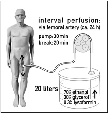

Figure 2: 2a: Interval perfusion mechanism 2b: Interval perfusion mechanism in body donors via femoral artery. on body donors with ethanol glycerol.

Figure 3: Ibrahim Alkatout and Johannes Ackermann operating in the Kiel Anatomy Department on a body donor by laparoscopy.


Figure 4: Cervical stump after a body donor Laparoscopic Subtotal Hysterectomy (LSH) (4a) in comparison to a patient LSH (4b)
From a chemical perspective, most of the efforts towards designing disease specific imaging agents find their origins in nuclear medicine and its subdiscipline of radiochemistry. Radiology guided surgery applications for sentinel nodes (radiocolloids, somatostatin receptor overexpressing lesions (peptides, and prostate specific membrane antigen (PSMA) expressing lesions (small molecules, have established. This is primarily driven by the common availability of tracer generators in clinics world-wide. This further focused tracer design, with recent examples of widely implemented agents is currently investigated in detail [31]. Besides the application-specific design of radiotracers there are various attempts to use off-the-shelve PET tracers for image guidance by exploring gamma rays [32,33], and beta particles Advantages of the use of radiotracers are that they can be applied under a micro-dosing regime, are compatible with quantitative biodistribution studies and support non-invasive pre-operative imaging approaches such as scintigraphy, single photon emission computed tomography (SPECT) and PET.
State of ART in 2024
Ten million babies and more have now been born around the world with the help of ART, worldwide 1-5 % of babies are born after an ART treatment. At ESHRE 2024 in Amsterdam, the European Society of Human Reproduction and Embryology held its 40th anniversary annual meeting from July 7 to 10, 2024. With more than 1120 posters and oral presentations, ESHRE 2024 celebrated successes from across the reproduction and embryology field.
At the first ESHRE meeting in Bonn, Germany in 1984 about 40 delegates from around the world gathered and had big dreams. Bob Edwards stated, ESHRE will be great one day" and here we are. At many press conferences in Germany and worldwide ART was established and defended (Figures 5 and 6).

Figure 5: Patrick Steptoe and Lilo Mettler in an interview at the first ESHRE meeting in Bonn in 1984.

Figure 6: Bob Edwards and Lilo Mettler in 1982 after the announcement of the first IVF baby born in Kiel.
Some of the highlights from the 2024 ESHRE conference were:
Specifically, the ongoing pregnancy rate at ten weeks post-embryo transfer was 46.3% for OXO-001 users, compared to 35.7% for those on the placebo. The live birth rate was also higher for OXO-001 at 42.6%, compared to 35.7% for the placebo. Although these results were not statistically powered to confirm significance, they provide good reason to continue to a Phase 3 trial.
Future visions and logical features come with technological development, Artificial Intelligence (AI) and ethical acceptance
MIS in the next 25 years (2025-2050)
The future is based on the history of laparoscopy which is a unique mixture of various trends in different fields, spurred by the activities of established societies as well as opportunities of their publication and influenced by the world’s progress, recession, war, piece and the love of the individuals for life. The influence of industry, which has kept pace and actively supported this development for years, is the driving force besides the heroes of doctors and engineers that introduce new ideas. Without suitable technology, this dissemination would not have been possible. Endoscopic development and its future do depend on new inventions, on the audacity of leading heroes, their input into this field but also on their management of life to continue to survive and in a healthy and successful cooperation with the medical technical industry and the governments of our countries which grant us the peace and freedom for new achievements.
The integration of robotics into surgery has further advanced this development. Robot-assisted systems provide precise control and allow us to perform complex procedures with a level of accuracy that is often unattainable with traditional methods. The 3D visualization and improved mobility of the instruments allow us to get into hard-to-reach areas of the body while sparing the surrounding tissue. This is particularly important in gynecology, where the anatomy is often complex and variable. Another advantage of robotic surgery is the ability to improve ergonomics during the procedure. A surgeon can work in a more comfortable sitting position which reduces fatigue and maintains concentration during longer operations. This not only leads to better results for the patients, but also to greater satisfaction for the surgeons.
The future of minimally invasive and robotic surgery will also be shaped by technological innovation, and artificial intelligence (AI). AI will play an increasingly important role by helping us plan procedures and analyze real-time data during surgery. AI-supported systems can recognize patterns in patient data that help us to create individual treatment plans based on the specific needs of each patient. This personalized approach could significantly improve outcomes and further reduce complication rates. In addition, AI will be able to perform precise analysis during surgery by evaluating image data in real time. This could help us to identify potential problems early and make immediate adjustments. The combination of robotic surgery and AI will take the precision and safety of our procedures to a new level. Another exciting aspect is the possibility of remote surgery. With the advancement of robotics and communication infrastructure, surgeons already are able to perform procedures remotely, which could be of great benefit to domestic or underserved areas in particular. This would greatly improve access to specialized surgical procedures and could help to make healthcare more equitable around the world.
In summary, the future of minimally invasive and robotic surgery is bright, and the combination of innovative technologies, AI and robotic systems will not only revolutionize the way we operate, but also significantly improve patient experience and outcomes. All surgeons are looking forward to being part of this exciting development and taking full advantage of the opportunities that the future offers. Continuous training and adaptation to new technologies will be crucial to ensure that we can provide the best possible care to our patients.
ART New achievements and challenges 2025-2050
It is absolutely not the intention of this paper to go into a discussion on the future of fertility with ideas of revolutionizing human reproduction as stated by Emily Witt in 2023, and many others where she leads a discussion on researchers that are attempting to produce oocytes without human ovaries. This discussion on sperm cells is already around a bit longer. Technically in oocyte retrieval a more or less automated and very exact follicular puncture, can be foreseen, automated laboratory processment of gametes and embryos and an automated embryo transfer, based on previous imaging measurements.
Like the first successful IVF outcome with the delivery of Louise Brown in 1978, the Japanese experiment of reproductive biologists, Katsuhiko Hayashi and Mitinori Saitou in 2016 may challenge the science of human reproduction. They published in Nature that they produced oocytes from skin of the tip of a mouse tail, reprogrammed them into stem cells and then turned those stem cells into egg cells (oocytes). The eggs, once fertilized, were transferred to the uteruses of female mice, which gave birth to ten pups; some of the pups went on to have babies of their own. Hayashi and Saitou provided the first proof of what’s known as in-vitro gametogenesis (IVG), the production of gametes outside the body. The mice that descended from the lab-made egg cells were described as “grossly normal” [34].
The introduction of AI in ART procedures will revolutionize reproductive techniques, but it will definitely need a cautious and thoughtful approach, particularly when drafting legislative and regulatory frameworks solidly grounded in ethics precepts and core values, prioritizing human dignity and upholding fundamental rights to privacy, data protection, and equality. Continuous publications detail latest findings and discuss possible dangers [35-39]. Ultimately, such goals can only be achieved by preserving human control in order to make AI meet our needs, while at the same time operating transparently and achieving equitable outcomes.
Only a few factors are essential to a successful aspiration of healthy Metaphase-2 oocytes (egg retrieval). Priority has the controlled ovarian hyperstimulation as detailed in a 2024 publication on the subject [39].
Secondly, technical details have to be observed like:
With exact imaging, AI and robotic control of the movements, automated egg retrieval will come soon.
Molecular basis for MIS and ART. Where have we come from and where are we going?
Multi-omic experimental approaches are revolutionizing the field of biological and medical sciences, opening new possibilities for understanding complex diseases and improving health care. These technologies analyze multiple types of biological information, such as genomics, transcriptomics, proteomics, metabolomics, etc, to gain a comprehensive understanding of biological systems. In MIS with all the robotic assistance advances and in ART with all the achieved success rates new approaches are expected, which ensure automated, programmed MIS, follicular punctures, embryo transfers as well as genetically based ovarian hyperstimulation. An advanced three-dimensional machine vision system is essential in making intelligent surgical robots smarter and safer. As the medical field moves towards more laparoscopic approaches for surgeries, it will be important to have an automated robotic system designed to assist such procedures.
Dear Editorial Team, Clinical Medical Reviews and Reports. My experience with the journal was highly positive. The peer-review process was rigorous, constructive, and completed in a timely manner. The reviewers provided valuable comments that helped improve the quality and clarity of our manuscript. The editorial office was professional, responsive, and supportive throughout all stages of the publication process. Communication was clear and efficient, and any questions were addressed promptly. Overall, I found the journal to maintain high scientific standards and an excellent publication workflow. I would be pleased to consider submitting future work to this journal. Best wishes from, Elena Popa.

It was my pleasure to submit my testimonial concerning the Reviewer Board of our Scientific Journal “Brain and Neurological Disorders”. The Reviewers focused on some modifications and their contribution was helpful. The ladies of our Editorial Office were also supported my efforts. It was my honor to have such a co-operation and I am looking forward for more collaboration.

Dear Grace Pierce, Editorial Coordinator of Journal of Clinical Research and Reports, Thank you for the speedy and efficient peer review process. I appreciate the fact that your peer reviewers do not take months to respond like with some other journals. I would also like to thank the editorial office for responding quickly to my questions. It is an excellent journal. I plan to submit more manuscripts in the future. Best wishes from, Robert W. McGee

Dear Grace Pierce, Editorial Coordinator of Journal of Clinical Research and Reports, Working with you and your team on our recent publication in JCRR has been a truly wonderful and enjoyable experience. The responses were prompt, and the reviewers were patient, constructive, and highly professional. One reviewer in particular gave me the feeling that a professor was carefully reading and commenting on my coursework, which was deeply touching. The entire process was straightforward and hassle‑free, with no tedious online forms to complete. I highly recommend this journal. Best wishes from, DR Aibing Rao, Head of R&D

I Appreciate the Opportunity to Share my Experience with the Journal of Clinical Research and Reports. The peer review process was timely and constructive, and the feedback provided helped improve the quality of our manuscript. The editorial office was professional, responsive, and supportive throughout the process, ensuring smooth communication and efficient handling of the submission. Overall, it was a positive experience collaborating with your team.

Dear Mercy Grace, Editorial Coordinator of Obstetrics Gynecology and Reproductive Sciences, We would like to express our gratitude for your help at all stages of publishing and editing the article. The editors of the magazine answer all the necessary questions and help at every stage. We will definitely continue to cooperate and publish other works in the Obstetrics Gynecology and Reproductive Sciences! Best wishes from, Alla Konstantinovna Politova,
