
Research article | DOI: https://doi.org/10.31579/2642-973X/082
1 Neurosurgery Service of the Maciel Hospital, Montevideo, Uruguay.
2 Department of Anatomy, Faculty of Medicine, University of the Republic, Montevideo, Uruguay.
*Corresponding Author: Alejandra JAUME, Neurosurgery Service of the Maciel Hospital, Montevideo, Uruguay.
Citation: Alejandra Jaume, Alejandro RUSSO, (2023), Methodology and Anatomical Study of Microsurgical Tracts White Substance in Human Brain. J. Brain and Neurological Disorders. 6(6): DOI:10.31579/2642-973X/082
Copyright: © 2023, Alejandra Jaume. This is an open-access article distributed under the terms of The Creative Commons Attribution License, which permits unrestricted use, distribution, and reproduction in any medium, provided the original author and source are credited.
Received: 18 September 2023 | Accepted: 27 September 2023 | Published: 04 October 2023
Keywords: white sustancia tractos; Klingler´s technology; tractografía; blanca sustancia distribución, tractography of white matter distribution
Introduction: The studio of white sustancia fibers has been reported since 1930 with the object of a correct neurosurgical technique. In 1934 Klingler´s developed an excellent technique for the tract studio. The misma is based on the freezing of the cerebral tejido, allowing a greater difference between white and gray substance. This work proposes a practical training system, the steps must be taken to obtain a correct and ordered anatomical understanding of the white sustancia tracts.
Materials and Methods: 4 hemispheres obtained from adult corpses are used in a formal solution, and without macroscopic neurologic pathology. The mismos are displayed using Klingler's technology, using the Olympus 10x optical zoom microscope for magnification.
Results: in each hemisphere, we designate the tracts, of systematic manner, comenzando por la cara lateral: fasciculo longitudinal superior, capsula extrema, fasciculo fronto-occipital, fasciculo uncinado, capsula externa, comisura blanca anterior, capsula interna, y corona radiada. It is said at the level of the internal body: fascia of the cíngulo, cuerpo calloso with forces major menor y tapetum, fornix, y pedúnculo talamico (anterior, superior, posterior and inferior). Finally, the basal cara level is determined by the lower longitudinal fascia.
Discussion: Mediate the results obtained, we propose to carry out an orderly and systematic working protocol, for the methodological analysis of the white sustancia fibers. The discussion is continued with an updated bibliography on the topic.
Conclusions: the good knowledge of the morphological anatomy, topography, and, the distribution of the fibers of association, and, projection in each of the hemispheres; It is fundamental for a good interpretation of imagenological studies and a correct neurosurgical approach.
From 1934, with the important contribution of Klingler´s, that uncovering a freezing technique for the study of white sustancia, began to study with greater depth the provision of different tracts of white sustancia, allowing a great neuroanatomical advance, y su correcta neurosurgical application [4].
White fibers can be systematized for your studio in large groups: association fibers, and projection fibers. The fibers of association, its aquellas that asocian structures teleencefálicas, y, can be divided into: 1) fibers of association intrahemisfericas, that its fibers of communication between a mismo hemisferio: y 2) fibers of association interhemisfericas or comisurales, the fibers of communication between sectors homotopics between ambos hemisferios. The projection fibers are defined as fibers that communicate with other encéfalo sectors (diencéfalo, mesencéfalo, romboencéfalo or spinal sector). [6- 20].
This system of white sustancia tractors can only be carried out at the same level as the telephone, so that, in this order, only it can be carried out at this level. This work proposes a practical training system, the steps must be taken to obtain a correct and ordered anatomical understanding of the white sustancia tracts.
Se utilizaron 4 hemispheres obtainedidos de cadáveres adultos formalados sin pathología neurológica macroscópica. Initially it will freeze in 10% formaldehyde for 4 weeks, then remove the arachnoid capa, and freeze at 16 degrees of temperature for one week. Once the process has been completed, after removing the frizer, within 24 hours, it will begin to be recognized by Klingler's technology, using different size blades (4 and 2mm), and the Olympus optical zoom microscope 10x for magnification.
The dissection is carried out in the very cerebral surfaces, starting with the lateral surface, then the medial surface, and finally the basal surface.
At the level of the hemispherical lateral cara, initially it is necessary to recognize surcos, y, giros; to start removing the corteza, from the superior temporal surco, in the form of C hasta the inferior frontal surco. Read the cortex from the lower temporal surface to the upper frontal surface, and then successively remove the cortex from the lateral surface to observe the coral arrangement of the white fibers.
Once you have completed the initial phase, follow the orderly description of the fascicles that are located on the lateral side, from the surface to the depth: a) upper longitudinal fascia, which is the superficial mass, to remove the fibers in U cortas , b) extrema capsula, the one that was observed to be disecado the insular corteza (Figura 1), c) uncinado fasciculo, fronto-occipital fasciculo, and externa capsula, los cuales se encuentran al mismo vel, una vez removedado la capsula extrema y the claustro, (Figura 2), d) comes to white anterior, is topografía in a sector more deep, to disecar the fasciculo uncinado and fronto-occipital (Figura 3), ye) internal capsule, and corona radiada, that its fibers From topography deep to level of the lateral surface, observe and remove the putamen (Figure. 4).

Figure 1: Lateral view of the hemisphere is observed: the extrem capsule is observed (CE), one is exposed to the insular cortex, with its limits: surco insular superior (SIS), surco insular anterior (SIA), and surco insular posterior (SIP)

Figure 2: Vista lateral hemisférica izquierda: it is observed capsule externa (CEx), fasciculo uncinado (FU), y fascículo occipital front (FFO), as described in the text.

Figure 3: Left hemispheric lateral view: seen anterior white commissure (CBA), and putamen (Pu).

Figure 4: Left lateral view: capsule is observed internal (CI), corona radiata (CR), globe pale (GP), and CBA.
It is shown at the level of the internal surface, from the surface area to the depth of the following tracts: a) fascia of the cíngulo (Figura 5), b) cuerpo calloso con sus four sectors en su vista medial: rostrum (Ros), rodilla ( Rod), cuerpo (CueC), y esplenio (ECC); as if the level anterior and posterior of the major force (FM) and menor (Fm) (Figura 6), c) fornix in addition to four sectors: fimbria (FF), crura (CrF), core (CuF), and column (CoF ), where it ends at the level of the breast bone (CM), where it originates from the mámilo-tálamico fascia (FMT) which is directed to the anterior sector of the tálamo, and then to the cíngulo, constituyendo el circuito de Papéz (Figuras 7 y 8 ), d) where the calloso a nivel del tapetum (TCC) (Figura 9), ye) pedúnculo tálamico, una vez withdrawado el caudado, con sus sectors: anterior (PTA), superior (PTS),

Figure 5: Left hemispheric medial view: seen cingulate fasciculus (CF) as described in the text.

Figure 6: Left hemispheric medial view: seen corpus callosum: with Ros, Rod, CueC, ECC, Fm,and FM.
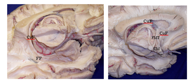
Figure 7 and 8: Left hemispheric inferior and medial view: FF is observed, CrF, CuF, CoF, CM, and FMT, mentioned in the text.
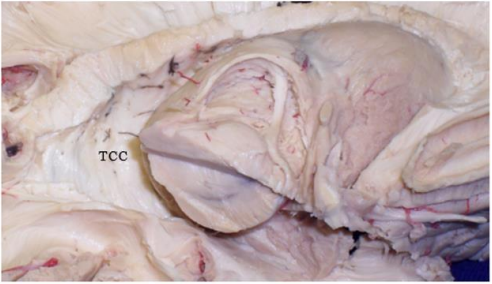
Figure 9: Vista medial hemisférica izquierda: se observa TCC.

Figure 10: Vista medial hemisférica izquierda: se observaPTA; PTS; PTP; y PTI. Finally, at the level of the basal surface, the inferior longitudinal fasciculus (ILF) was dissected, removing the cortex of the fusiform gyrus (Figure 11).

Figure 11: Top view of hemispheric basal fase left: FLI is observed.
As previously mentioned, at the telephone level there are large types of fibers: association fibers (intra and interhemisfericas), and, projection fibers. The intrahemispheric association fibers can be divided into groups: a) U-shaped fibers, each one in a circle with the other center of the lobe; constituting the extrema capsule, a set of short U-shaped fibers, that an island enters if, and the island with the fronto-parieto-temporal operculums, y, b) long U-shaped fibers, the ones that have a lobe with the other, We can distinguish 5 major tracts: upper longitudinal fasciculus, lower fasciculus, fronto-occipital fasciculus, cíngulo fasciculo, and lower longitudinal fasciculus. The interhemisferic or comisural association fibers, one in one hemisphere with the other, can be observed as follows: a) white anterior area, including the upper part of the lower part of the eye, one of the anterior sectors of the temporal lobes; b). yc) hippocampal composition, topografiado at the level of the crura del fórnix, uniendo ambos lóbulos límbicos.
The projection fibers connect the telescope to other encipheral sectors, and they can be connected in: a) corona radiada, defined as the projection fibers located topographically at the beginning of the putamen nucleus, and b) internal capsule, that its fibers are topographically medial y subyacente al putamen (1, 2, 5, 6, 7, 9, 13, 19, 29).
To log an ordered and systematic study, it is necessary to begin the dissection in each hemisphere, taking into account the very surface areas of the brain (6, 7, 20). The object is to log the visualization of the layout, from the surface to the depth, and the three-dimensional understanding, of each white sustancia capa, in each sector of the encephalon. Therefore, it must be started by dissecting the lateral side, then the medial side, and finally the basal side (5, 6, 7, 8, 9, 20).
In the lateral face, remove the cortex, and first observe its association fibers in U cortas. Aligning the U-shaped fibers, the upper longitudinal fasciculus is topographical, as is the fronto-parieto-occipito-temporal lobes. There is a form of C, where the Fisura Silviana, y, can be distinguished between three sectors: a) vertical sector, one of the parietal lobe with the temporal, b) horizontal sector, one of the frontal lobe with the parietal, y, c) fibers that rodean the island, connecting the posterior temporal region with the prefrontal area, it fascinates the arch.
At the level of the island, once the cortex withdraws, fibers are individualized into U cortas that constitute the extreme capsule, which is topographical between the island, and the claustro. The claustro is a gray nucleus, divided into two sectors: caudal claustro (fina lamina of gray sustancia between the extrema capsula and the externa capsula); and ventral claustro (group of gray substance fragmented by the uncined and fronto-occipital fasciculus) (1).
When disecar the claustro, we observed a group of fibers of white sustancia: the external capsule, which was topografía between the claustro and the putamen, así like the uncinado fasciculo and the fronto-occipital fasciculo, in the ventral sector. The external capsule connects the fronto-mesial region with the temporo-mesial region.The uncined fasciculus, one of the temporal lobes with the orbito-frontal region, is part of the ventral portion of the external capsule, which is difficult to individualize each fascicle, as well as the fronto-occipital fasciculus (7).
When it comes to the uncined and fronto-occipital fasciculus, we observe the anterior white comisura, which plays an important interhemispheric role, between ambos temporal lóbules (visual, auditory, olfactory, and gustatory). It has a transverse direction, united with the anterior sector of the temporal lobes. On its path cursa by the anterior face of the pale globe, there is an impression called the Gratiolet channel (5). When it comes white, during its course, it divides the gray nuclei into the basal region. The innominated substance, is topografía delante y debajo de la comisura blanca anterior, y, encima de la sustancia perforada anterior, de nonde se situúan las nucleos basales de Meynert (main input colinergico del córtex). Medially, the innominated substance continues with the septal region, the latter, represented in the cortical surface, septal nuclei. This region is made up of the paraterminal gyro, the posterior paraolfatorio, the paraolfatorio giro, and the anterior paraolfatorio. In the septal region, there are mainly three types of fibers: prefornicial fibers, medial olfactory estría, and amigadlo-septal fibers. At the septal level, the acumbens nucleus is also topographical (intermediate nucleus between the limbic system and the extrapiramidal system). The next nucleus is also located on the ventral side of the neck, which corresponds to the anterior sector of the caudado and put amen, located by the release of the anterior white core (7). medial olfactory estría, y amigadlo-septales fibers. At the septal level, the acumbens nucleus is also topographical (intermediate nucleus between the limbic system and the extrapiramidal system). The next nucleus is also located on the ventral side of the neck, which corresponds to the anterior sector of the caudado and put amen, located by the release of the anterior white core (7). medial olfactory estría, y amigadlo-septales fibers. At septal level, the acumbens nucleus is also topographical (intermediate nucleus between the limbic system and the extrapiramidal system). The next nucleus is also located on the ventral side of the neck, which corresponds to the anterior sector of the caudado and put amen, located by the release of the anterior white core (7).
How to map the map, it's easy to understand, it's easy to understand, it has the difference in the pale globe. This is important, because the external spinal cord, which separates the putamen from the pale globe, is difficult to identify during the dissection. Once you have removed the putamen, you will be able to identify the projection fibers, which are deeper than the hemispheric lateral sector. The radiated corona begins at the cortical level and is directed vertically up to the superior border of the putamen, where it continues with the number of internal capsules. The internal capsule can be divided into two sectors: a) lateral sector, where the cortico-mesencephallic, cortico-romboencephalic, and cortico-spinal fibers are topographical; y, b) medial sector, where the topografían of the cortico-dience fibers, llamados also pedúnculos talamicos. The lateral sector of the internal capsula is where it is visualized from the lateral hemispheric cara, and, the medial sector, is observed from the internal hemispheric cara. For the capsule studio, in antero-posterior direction, the misma can be divided into 5 sectors: anterior arm, rodilla, posterior arm, sublenticular sector, and retrolenticular sector. The anterior arm is topografía between the head of the caudado nut, and the putamen. At the level of the anterior arm pasan: in the lateral sector, fronto-ponto-cerebral fibers, and in the medial sector, the anterior tálamico pedúnculo (frontal cortex and cíngulo). The rod of the internal capsule, is identified in the vertex that exists between the putamen, head of caudado, and tálamo, at the level of the foramen of Monro. In this sector pasan: laterally, cortico-nuclear fibers, and, medially, the anterior portion of the upper peduncle. The rear arm is located between the putty and the foot. Now: in the lateral sector, fronto-ponto-cerebral fibers, cortico-spinal fibers, cortico-reticular fibers, y, cortico-rubricas fibers; y, in the medial sector, the upper peduncle (premotor, motor, and sensitive cortex). The sublenticular sector, is topografía by the bottom of the putamen, and has the level found: in the lateral sector, temporo-ponto-cerebellar fibers, and, in the medial sector, inferior peduncle thalamus (temporal cortex, and auditory radiation). The retrolenticular sector, located behind the putamen, is where it is located: in the lateral sector, parieto-ponto-cerebelosa fibers, occipito-fronto-cerebelosa fibers; y, in the medial sector, the posterior tálamico pedúnculo (parietal cortex, occipital, and temporal, including optical, and auditory radiation). Finally, we should mention that in the lateral aspect of the hemisphere, constituting the lateral aspect of the temporal bone and the ventricular atrium, it is topografía the sagittal strato. This layer, it is formed by a set of intermixed fibers during its tray, which cannot be individualized each bundle separately. This tract is composed by: Meyer Loop fibers of optical radiances, uncined fasciculus, occipito-frontal fasciculus, pedunculo talamico inferior y posterior, comisura blanca anterior, capsula extrema, fibras temporo-pontinas, cortico-tectales, cortico-tegmentales, y fibras occipito-pontinas (2 , 6, 7, 9, 10, 13, 14, 19, 20).
Sistematizando la cara lateral, we can decide that from the surface to the depth, in contramos: fibers of association intrahemisfericas cortas en U (capsula extrema); long U-shaped intrahemispheric association fibers (superior longitudinal fasciculus, uncined fasciculus, and fronto-occipital fasciculus); fibers of association between hemispheres (comisura blanca anterior); y projection fibers (corona radiada, y, lateral sector of the internal capsule).
On the medial side, first remove the corteza del giro del cíngulo, expose the fasciculo of the cíngulo, and cual connecta lóbulo límbico. When I say this fascinator, I observe the calloso's head. Rodeando la cara superior del cuerpo calloso, se topografía la estría longitudinal lateral (ELL) y medial (ELM), así como una capa de sustancia gris, llamada indicium grisium (IG), que ma parte de la formation mación hipocampal, la cual, en su descenso en el desarrollo embriológico, deja dicha capa (6, 7, 20).
The callous head connects the homotopic shape of the frontal, parietal, occipital and posterior temporal lobes. You can expose the callous head, in all of its sectors: head, head, head and back, it comes from the description of the device. The fórnix can be considered, as interhemisferic association fibers, because at the level of the crura of the fórnix it is topografía the hippocampal composition, and, also can be classified as projection fibers, there are tele-fálica connections. In four sectors we distinguish: a) fimbria, is a tract of white sustancia, formed by the combination of the white sustancia that rode to the hipocampo, called alveus, constitutes the main eferencia of the hipocampo. The fimbria extends from the coroidal fissure up to the point where it separates from the toothed girdle, at the level of the joy of the heart calloso; b) crura del fórnix, es la continuación de la fimbria a nivel del esplenio calloso, y terminata en el point, en que se acuentra con el contralateral fórnix; c) Cuerpo, es el sector que comienza donde ambos fórnix se unen, y terminar a nivel del foramen de Monro, del se vulven a separar; yd) columna, es el sector final, que comienza en el foramen de Monro, y terminan, en su major parte, en los breasts mamilares, teniendo also precomisural fibers que terminan en la region septal. From the breast bones, the mámilo-thalamico fascicle originated, which ended in the anterior tálamico nuclei. From the floor, it goes to the cíngulo, constituting the circuit of Papez, previously mentioned, which has a fundamental role in memory (7, 20). is the continuation of the fimbria at the level of the callous splendor, ending at the point, where it joins with the contralateral horn; c) Cuerpo, es el sector que comienza donde ambos fórnix se unen, y terminar a nivel del foramen de Monro, del se vulven a separar; yd) columna, es el sector final, que comienza en el foramen de Monro, y terminan, en su major parte, en los breasts mamilares, teniendo also precomisural fibers que terminan en la region septal. From the breast bones, the mámilo-thalamico fascicle originated, which ended in the anterior tálamico nuclei. From the floor, it goes to the cíngulo, constituting the circuit of Papez, previously mentioned, which has a fundamental role in memory (7, 20). is the continuation of the fimbria at the level of the callous splendor, ending at the point, where it joins with the contralateral horn; c) Cuerpo, es el sector que comienza donde ambos fórnix se unen, y terminar a nivel del foramen de Monro, del se vulven a separar; yd) columna, es el sector final, que comienza en el foramen de Monro, y terminan, en su major parte, en los breasts mamilares, teniendo also precomisural fibers que terminan en la region septal. From the breast bones, the mámilo-thalamico fascicle originated, which ended in the anterior tálamico nuclei. From the floor, it goes to the cíngulo, constituyendo el circuito de Papez, previously mentioned, which has a fundamental role in memory (7, 20). in which it is connected with the contralateral horn; c) Cuerpo, es el sector que comienza donde ambos fórnix se unen, y terminar a nivel del foramen de Monro, del se vulven a separar; yd) columna, es el sector final, que comienza en el foramen de Monro, y terminan, en su major parte, en los breasts mamilares, teniendo also precomisural fibers que terminan en la region septal. From the breast bones, the mámilo-thalamico fascicle originated, which ended in the anterior tálamico nuclei. From the floor, it goes to the cíngulo, constituting the circuit of Papez, previously mentioned, which has a fundamental role in memory (7, 20). in which it is connected with the contralateral horn; c) Cuerpo, es el sector que comienza donde ambos fórnix se unen, y terminar a nivel del foramen de Monro, del se vulven a separar; yd) columna, es el sector final, que comienza en el foramen de Monro, y terminan, en su major parte, en los breasts mamilares, teniendo also precomisural fibers que terminan en la region septal. From the breast bones, the mámilo-thalamico fascicle originated, which ended in the anterior tálamico nuclei. From the floor, it goes to the cíngulo, constituting the circuit of Papez, previously mentioned, which has a fundamental role in memory (7, 20). que comienza en el foramen de Monro, y terminan, en su major parte, en los breasts mamilares, teniendo also precomisural fibers que terminan en la region septal. From the breast bones, the mámilo-thalamico fascicle originated, which ended in the anterior tálamico nuclei. From the floor, it goes to the cíngulo, constituting the circuit of Papez, previously mentioned, which has a fundamental role in memory (7, 20). que comienza en el foramen de Monro, y terminan, en su major parte, en los breasts mamilares, teniendo also precomisural fibers que terminan en la region septal. From the breast bones, the mámilo-thalamico fasciculus originated, which ended in the anterior tálamicos nucleos. From the floor, it goes to the cíngulo, constituting the circuit of Papez, previously mentioned, which has a fundamental role in memory (7, 20).
At the level of the lateral wall of the ventricular atrium, to remove the ependicular, we observe the tapetum, which constitutes the sector of the callous heart, which forms the lateral wall of the atrio, and the temporal region. To finalize the dissection of the medial hemispheric cara, you must disecar the caudado nucleus, to be able to observe the medial sector of the internal capsule, or pedúnculos talamicos (6,7, 20). So, in the internal hemisphere shape, from the surface to the depth, observe: intrahemisferic association fibers short in U, intrahemisferic association fibers long in U (fascículo del cíngulo), interhemisférica association fibers (cuerpo calloso, y fórnix); y, projection fibers (medial sector of the internal capsule or pedunculos talamicos).
In the basal or lower hemisphere shape, according to the corteza del giro fusiforme, the lower longitudinal fascia is topographical. This is a long intrahemispheric association fascicle, which connects the temporal lobe with the occipital. Sistematizando, we can conclude, that in the three hemispheric surfaces, it is comienza the disección of the surface to the depth, observing successively, fibers of association intrahemisféricas, fibers of association interhemisféricos, and fibers of projection.
The good knowledge of the morphological anatomy, topografia, and; the distribution of the association fibers, and, projection, in each of the hemispheres; It is fundamental for a good interpretation of imagenological studies, and, a correct approach to neurology.
Dear Editorial Team, Clinical Medical Reviews and Reports. My experience with the journal was highly positive. The peer-review process was rigorous, constructive, and completed in a timely manner. The reviewers provided valuable comments that helped improve the quality and clarity of our manuscript. The editorial office was professional, responsive, and supportive throughout all stages of the publication process. Communication was clear and efficient, and any questions were addressed promptly. Overall, I found the journal to maintain high scientific standards and an excellent publication workflow. I would be pleased to consider submitting future work to this journal. Best wishes from, Elena Popa.

It was my pleasure to submit my testimonial concerning the Reviewer Board of our Scientific Journal “Brain and Neurological Disorders”. The Reviewers focused on some modifications and their contribution was helpful. The ladies of our Editorial Office were also supported my efforts. It was my honor to have such a co-operation and I am looking forward for more collaboration.

Dear Grace Pierce, Editorial Coordinator of Journal of Clinical Research and Reports, Thank you for the speedy and efficient peer review process. I appreciate the fact that your peer reviewers do not take months to respond like with some other journals. I would also like to thank the editorial office for responding quickly to my questions. It is an excellent journal. I plan to submit more manuscripts in the future. Best wishes from, Robert W. McGee

Dear Grace Pierce, Editorial Coordinator of Journal of Clinical Research and Reports, Working with you and your team on our recent publication in JCRR has been a truly wonderful and enjoyable experience. The responses were prompt, and the reviewers were patient, constructive, and highly professional. One reviewer in particular gave me the feeling that a professor was carefully reading and commenting on my coursework, which was deeply touching. The entire process was straightforward and hassle‑free, with no tedious online forms to complete. I highly recommend this journal. Best wishes from, DR Aibing Rao, Head of R&D

I Appreciate the Opportunity to Share my Experience with the Journal of Clinical Research and Reports. The peer review process was timely and constructive, and the feedback provided helped improve the quality of our manuscript. The editorial office was professional, responsive, and supportive throughout the process, ensuring smooth communication and efficient handling of the submission. Overall, it was a positive experience collaborating with your team.

Dear Mercy Grace, Editorial Coordinator of Obstetrics Gynecology and Reproductive Sciences, We would like to express our gratitude for your help at all stages of publishing and editing the article. The editors of the magazine answer all the necessary questions and help at every stage. We will definitely continue to cooperate and publish other works in the Obstetrics Gynecology and Reproductive Sciences! Best wishes from, Alla Konstantinovna Politova,
