
Research Article | DOI: https://doi.org/10.31579/2641-0419/214
*Corresponding Author: : Cobo Daniel L, Av. Brigadeiro Faria Lima, 5416, Vila São Pedro, CEP: 15.090-000, São José do Rio Preto, SP, Brasil.
Citation: Cobo Daniel L., Sciarra Adília M.P., Batigália Fernando., Monteiro Vinicius H.F., Neves Ricardo A., De Paula, Jaynne L.. (2021) Mapping and Epidemiological analysis of types of Congenital Malformations in a Teaching Hospital. J. Clinical Cardiology and Cardiovascular Interventions, 4(15); Doi:10.31579/2641-0419/214
Copyright: © 2021 Cobo Daniel L, This is an open-access article distributed under the terms of the Creative Commons Attribution License, which permits unrestricted use, distribution, and reproduction in any medium, provided the original author and source are credited.
Received: 16 August 2021 | Accepted: 13 September 2021 | Published: 17 September 2021
Keywords: congenital malformations; anatomical variations and congenital abnormalities
Introduction: The World Health Organization (WHO) defines as congenital malformation, all anomalies triggered by the alteration of normal development resulting in a deficient formation from the initial stage of the fetus still in intrauterine, whose origin occurs before birth, having causes genetic, environmental or unknown, whether structural or functional, these disorders can be seen in prenatal care, childbirth, or even manifested in childhood.
Background: To map and analyze epidemiological data on congenital malformations in children at a Teaching Hospital in the interior of the State of São Paulo.
Materials and Methods: Data were collected from children with congenital malformations between the years 2010 and 2017, searched through the International Disease Code (ICD 10) with codes from Q00 to Q89. During the analysis of the medical records, 30,309 male and female children with congenital malformations were identified, aged between 1 day and 12 years.
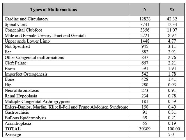
Results: 30,309 male and female children with congenital malformations were identified (16,956 male or 54.75% and 13,713 female or 45.25%) aged between 1 day and 12 years (19,587 from 0 to 4 years or 64.63 %; 5,780 from 5 to 8 years or 19.07% and 4,942 from 9 to 12 years or 16.30%). The main types of congenital malformations were found: cardiac and circulatory or 42.32%; spinal cord or 12.34%; congenital clubfoot or 11.07%; male and female genitals and urinary tracts or 8.97% which add up to 74.70%.
Conclusion: Congenital malformations are rare anomalies and this study concluded that they are mostly associated with male gender, early age and with a predominance of cardiac and circulatory alterations.
The World Health Organization (WHO) defines as congenital malformation, all anomalies triggered by the alteration of normal development resulting in a deficient formation from the initial stage of the fetus still in intrauterine, whose origin occurs before birth, having genetic causes, environmental or unknown, whether structural or functional, these disorders can be seen in prenatal care, childbirth, or even manifested in childhood. We have as congenital malformations those of the nervous system, musculoskeletal, digestive, chromosomal or genetic abnormalities, cleft lip and palate, circulatory, respiratory, urinary and genitourinary, hemangiomas and lymphangiomas. [1]
Congenital malformations can be classified as major, which would be severe anatomical, aesthetic and functional changes that can lead to death, or minor, which are mild mutations. About 5% of live births (LB) have some developmental anomaly, determined, in whole or in part, by genetic factors, and may last into adulthood, difficultly influencing the lives of individuals, families, health systems and society, requiring surgical procedures of an aesthetic or functional nature. [2,3]
Congenital anomalies accounted for 510,400 deaths worldwide in 2010. In Brazil, in 2008, they corresponded to approximately 19% of mortality in children under one year of age, being responsible for the second leading cause of death in this age group. In the city of São Paulo from 2007 to 2011, the prevalence of congenital anomalies was 1.2% of live births. In South America, Gil et al. assessed the prevalence rate of births with congenital anomalies, and found that indicators of low socioeconomic conditions, such as low maternal education, age, infectious diseases and use of medications during pregnancy were detected as risk factors within these regions. Among the cleft lip anomalies with or without cleft palate, the ventricular septal defect presented a significantly higher risk in the lowest socioeconomic level. [5,6,7,8]
In many cases, congenital malformations are not fully attributed to a specific factor, but we can mention some factors that may be linked and justify certain malformations such as genetic factors through genes inherited from the sex chromosome or normal XY chromosome, triggering some type of morphological change , parental homogeneous blood type, environmental factors, socio-demographic factors (attributed by the possible lack of access to healthy foods), low health acuity such as prenatal care and limitations to information, infectious factors, low maternal weight and prematurity. The cardiovascular system is the most affected by congenital malformations, associated or not with other malformations. Congenital heart diseases account for 40% of all congenital defects, consisting of a structural change in the heart affecting the chambers, septa and/or large intrathoracic vessels, with important functional repercussions, requiring palliative or corrective surgical procedures. [9,10]
The study aims to map and analyze epidemiological data on congenital malformations in children at a Teaching Hospital in the interior of the State of São Paulo.
Data collection was performed randomly in the computerized system regarding the care of children with congenital malformations at an outpatient or hospital level between 2010 and 2017, searched through the International Disease Code (ICD 10) with codes from Q00 to Q89 referring to the types of congenital malformations. During the analysis of the medical records, 30,309 male and female children with congenital malformations attended to in consultation or hospitalization, aged between 1 day and 12 years, were identified. All medical records of children who underwent evaluations, consultations, admissions and procedures at the Children and Maternity Hospital of São José do Rio Preto were included in the study, and the records that were repeated during the analysis and research were excluded.
Descriptive analyzes of each single variable were obtained using the Excel software and tool (version 2016).11 The variables considered in the study are categorical and include: types of malformations, age, sex. In the databases, the following descriptors were used: congenital malformations, anatomical variations and congenital anomalies, which were addressed together in the research. From the analysis of the references, only publications relevant to the present study were selected and, after consulting the databases, the articles were cataloged and analyzed, taking into account the data collected at the Children's and Maternity Hospital.
A total of 30,309 male and female children with Congenital Malformations attended to consultations or hospitalizations were identified (16,956 male or 54.75% and 13,713 female or 45.25%) aged between 1 day and 12 years (19,587 from 0 to 4 years or 64.63%; 5,780 from 5 to 8 years or 19.07% and 4,942 from 9 to 12 years or 16.30%). The main types of congenital malformations were found: cardiac and circulatory or 42.32%; spinal cord or 12.34%; congenital clubfoot or 11.07%; male and female genitals and urinary tracts or 8.97% which add up to 74.70% and the other congenital malformations 25.30%.


Congenital malformations are rare anomalies, mostly associated with male gender, early age and with a predominance of cardiac and circulatory alterations.
Mendes et al, described that a study identified 1.2% of live births in the city of São Paulo with congenital malformations, with males having the highest incidence (50.9%) Thus, Catarino et al, identified 1,086,139 live births with congenital malformations; among these, 345 (4.3%) had congenital heart disease, representing a frequency of 3.18/10 thousand live births, 42.3% were girls, 56.4% boys and 1.3% had an unknown record regarding to sex. The most common malformations were: 3.2% unspecified polydactyly, 2.9% Down syndrome, 2.8% interatrial communication, 2.7% supernumerary finger(s) and 2.4% non-malformation specified of the heart. [5,12]
Cosme et al, reported 14,657 congenital anomalies of 819,018 live births, the most frequent malformations of the osteoarticular system, mainly polydactyly and foot deformities, followed by malformations of the cardiovascular system and of the head and neck, are associated with greater findings in osteoarticular malformations for easier identification at the time of birth. However, Hurtado et al in their study in Risaralda Colombia, heart disease ranked first, followed by lip and palate, abdominal wall defects, skeletal dysplasia, hydrocephalus, polydactyl syndrome and Down syndrome. [7,13]
Murray et al report that oral clefts are craniofacial anomalies that require rehabilitation ranging from surgical intervention to nutritional, dental, speech therapy, medical and psychological guidance. Its occurrence is approximately 1 in 700 newborns worldwide, and it may vary according to geographic area and socioeconomic status. Most cleft lip and palate are the result of multiple factors, genetic and non-genetic, each causing a minor developmental disturbance.[14]
Congenital brain malformations affect about 1.94%. According to Alberto et al, the brain is part of the central nervous system, contained in the skull cavity, and encompassing the brain, cerebellum, pons and medulla. Due to the complexity of its embryological development, its abnormal development in humans is not uncommon. Malformations of the nervous system, the driving force and coordinator of all vital manifestations, namely, the intellectual, the sensitive and the vegetative, were countless diseases. [15]
Horovitz et al, describe osteogenesis imperfecta (OI) as a rare disease, which affects 1.8% of cases, is characterized by bone fragility, recurrent fractures with secondary deformities, early deafness, bluish sclera and dentinogenesis imperfecta. Rodovalho et al, report that bone fragility and deformities, associated with recurrent fractures and short stature are characteristics of OI. These outcomes are attributed to mutations in genes responsible for the synthesis of type 1 collagen, being the central point of its pathophysiological mechanism. [16,17]
Picado-Vaquero et al report that hip instabilities have a prevalence of 1% to 1.5% in newborns, with females having the highest incidence of 13:1000 live births. These congenital hip changes encompass a wide spectrum of manifestations, such as acetabular dysplasia, hip subluxation and dislocation. According to Babock et al, clinical examination and imaging resources, such as radiography or ultrasound, contribute to the differential diagnosis of Developmental Hip Dysplasia (DHD). The Ortolani and Barlow tests, as well as the analysis of the size of the segments of the lower limbs, skinfold heights and family history, in addition to the type of delivery, are part of the clinical examination, guided by the pediatric orthopedist. [18,19]
Neurofibromatosis affects 0.91% of cases, it is an autosomal dominant disease. Lucchese et al report that it is a multisystemic affection with the possibility of ophthalmological, musculoskeletal, cardiovascular, endocrine, central and peripheral nervous system involvement. According to Friedman et al, neurofibromatosis is inherited from one of the parents in about 50% of cases. [20,21]
Maranhão et al state that congenital anomalies of the upper urinary tract, including milder forms, are not rare. In 0.98% newborns, there is some abnormality of the kidneys and ureters, with abnormalities in the shape and position of the kidneys being the most common. Congenital anomalies of the upper urinary tract imply morphofunctional changes with a variable clinical spectrum, from asymptomatic manifestations to renal failure and incompatibility with life. [22]
Congenital multiple arthrogryposis, 0.59% are part of the cases. Saccani et al, report that it was first described by Otto, in 1841 and believed that the pathological process was due to a congenital myodystrophy, however Stern, in 1923, called it "congenital multiple arthrogryposis". It is a rare syndrome that constitutes a heterogeneous group of congenital malformations of unknown etiology, multifactorial, characterized mainly by severe joint contractures and may be part of a complex of multisystemic congenital anomalies, such as musculoskeletal, genitourinary, cardiovascular, gastrointestinal, otorhinolaryngological and ophthalmological have been associated with congenital multiple arthrogryposis. [23]
Ehlers-Danlos Syndrome, Marfan, Klipell-Feil and Abdomen in Prune account for 0.49% of the cases. Espósito et al refer to Ehlers-Danlos as a term used for a group of relatively rare connective tissue pathologies. It is based on hereditary alterations in genes that affect the synthesis of different forms of collagen, without a predominance of race or gender. Differential diagnosis includes joint hypermobility syndrome, Marfan syndrome, osteogenesis imperfecta, among others. For Araújo et al, Marfan syndrome is also a connective tissue disease, which mainly involves the cardiovascular, musculoskeletal and visual systems. The most serious problems include aortic root dilation and dissection. [24,25]
According to Mizuta et al, the Klippel-Feil syndrome was described by Maurice Klippel and Andre Feil, in 1912. It is a rare congenital disease, belonging to the group of so-called craniocervical joint malformations. [26]
Santos et al, describe that Gastroschisis affects 0.31% and is a malformation characterized by a defect in the closing of the abdominal wall associated with the exteriorization of intra-abdominal structures, mainly the fetal intestine. Martillotti et al report that gastroschisis is observed with a failure of continuity in the abdominal wall, usually to the right of the umbilical cord, through which the intestinal loops are exteriorized in the amniotic cavity, sometimes accompanied by other organs. Due to the absence of a membrane covering the herniated content, it is free and in direct contact with the amniotic fluid. [27,28]
Epidermolysis bullosa affects 0.21% of cases and, according to Ângelo et al, it is a rare hereditary dermatosis, characterized by the development of blisters in the cutaneous-mucosal region of the entire body, in response to minimal trauma, heat or no apparent cause, which may manifest at birth or during the first years of life. According to Gonçalves et al, there are prospects for the development of gene therapies in the future, care should start from birth. Clinical support aims to prevent and treat blisters, infections, retractions and synechiae. In cases of tissue adhesion caused by excessive blisters, such as syndactyly of the hands, feet and esophageal stenosis, surgical corrections are performed. [29,30]
Achondroplasia, the least affected congenital malformation in children aged 0 to 12 years, with 0.19%. Uemura et al report that the disease is known as Parrot's Disease and is the main cause of genetic dwarfism, being a classic dominant disease with low incidence. In addition to short stature, the patient presents as clinical signs: spinal deformities, macrocephaly, hydrocephalus and hands with short and thick fingers in a trident and as oral manifestations: maxillary atresia or hypoplasia, open bite, soft palatal cleft and dental changes in number and shape. [31]
Congenital malformations are rare anomalies and this study concluded that they are mostly associated with male gender, early age and with a predominance of cardiac and circulatory alterations. The mapping of these malformations can contribute to improvements in the specific hospital sector and investment in technology in equipment and professional training in surgical procedures and techniques.
Dear Editorial Team, Clinical Medical Reviews and Reports. My experience with the journal was highly positive. The peer-review process was rigorous, constructive, and completed in a timely manner. The reviewers provided valuable comments that helped improve the quality and clarity of our manuscript. The editorial office was professional, responsive, and supportive throughout all stages of the publication process. Communication was clear and efficient, and any questions were addressed promptly. Overall, I found the journal to maintain high scientific standards and an excellent publication workflow. I would be pleased to consider submitting future work to this journal. Best wishes from, Elena Popa.

It was my pleasure to submit my testimonial concerning the Reviewer Board of our Scientific Journal “Brain and Neurological Disorders”. The Reviewers focused on some modifications and their contribution was helpful. The ladies of our Editorial Office were also supported my efforts. It was my honor to have such a co-operation and I am looking forward for more collaboration.

Dear Grace Pierce, Editorial Coordinator of Journal of Clinical Research and Reports, Thank you for the speedy and efficient peer review process. I appreciate the fact that your peer reviewers do not take months to respond like with some other journals. I would also like to thank the editorial office for responding quickly to my questions. It is an excellent journal. I plan to submit more manuscripts in the future. Best wishes from, Robert W. McGee

Dear Grace Pierce, Editorial Coordinator of Journal of Clinical Research and Reports, Working with you and your team on our recent publication in JCRR has been a truly wonderful and enjoyable experience. The responses were prompt, and the reviewers were patient, constructive, and highly professional. One reviewer in particular gave me the feeling that a professor was carefully reading and commenting on my coursework, which was deeply touching. The entire process was straightforward and hassle‑free, with no tedious online forms to complete. I highly recommend this journal. Best wishes from, DR Aibing Rao, Head of R&D

I Appreciate the Opportunity to Share my Experience with the Journal of Clinical Research and Reports. The peer review process was timely and constructive, and the feedback provided helped improve the quality of our manuscript. The editorial office was professional, responsive, and supportive throughout the process, ensuring smooth communication and efficient handling of the submission. Overall, it was a positive experience collaborating with your team.

Dear Mercy Grace, Editorial Coordinator of Obstetrics Gynecology and Reproductive Sciences, We would like to express our gratitude for your help at all stages of publishing and editing the article. The editors of the magazine answer all the necessary questions and help at every stage. We will definitely continue to cooperate and publish other works in the Obstetrics Gynecology and Reproductive Sciences! Best wishes from, Alla Konstantinovna Politova,
