Malignant Peripheral Nerve Sheath Tumour (MPNST) of the Prostate Gland: A Review and Update

Review Article | DOI: https://doi.org/10.31579/2640-1053/131

Malignant Peripheral Nerve Sheath Tumour (MPNST) of the Prostate Gland: A Review and Update

  • Anthony Kodzo-Grey Venyo *

North Manchester General Hospital, Delaunays Road, Crumpsall, M8 5RB. Manchester. United Kingdom. 

*Corresponding Author: Anthony Kodzo-Grey Venyo, North Manchester General Hospital, Delaunays Road, Crumpsall, M8 5RB. Manchester. United Kingdom

Citation: Anthony Kodzo-Grey Venyo, (2023) Malignant Peripheral Nerve Sheath Tumour (MPNST) of the Prostate Gland: A Review and Update, J. Cancer Research and Cellular Therapeutics, 7(1) DOI: 10.31579/2640-1053/131

Copyright: © 2023 Anthony Kodzo-Grey Venyo, This is an open access article distributed under the Creative Commons Attribution License, which permits unrestricted use, distribution, and reproduction in any medium, provided the original work is properly cited.

Received: 29 November 2022 | Accepted: 26 December 2022 | Published: 13 January 2023

Keywords: malignant peripheral nerve sheath tumour; mpnst; prostate gland; radical prostatectomy; radical radiotherapy; chemotherapy; metastases

Abstract

Even though adenocarcinoma of the prostate gland is the commonest malignant tumour of the prostate gland that is encountered in all countries globally, other rare tumours of the prostate gland do exist and because of their rarity most clinicians would not have ever encountered such rare tumours during their training and their practices. Serum prostate specific antigen tests tend to be undertaken globally in screening for early detection of adenocarcinoma of the prostate gland globally because most adenocarcinomas of the prostate gland tend to be associated with high serum prostate specific antigens in comparison with lower serum PSA levels that tend to be found in majority of individuals of the same age group who do not have adenocarcinoma of the prostate gland. Malignant peripheral nerve sheath tumour (MPNST) of the prostate gland is a rare malignant that tends to be sporadically diagnosed and quite often the tumour to be diagnosed at an advanced stage or locally advanced stage as well as the tumour has tended to portend an aggressive biological behaviour and poor prognosis. MPNST can be diagnosed in some patients who are younger than the ages of individual adenocarcinomas but the tumour can also be found in individuals whose ages are similar to ages of individuals who have adenocarcinoma of the prostate gland. MPNST may be diagnosed incidentally in some individuals who do not have any symptoms, and they can also be diagnosed in individuals who have lower urinary tract symptoms of recent onset that is progressive, and they could also manifest with urinary tract infections, retention of urine, or symptoms related to their metastases or obstruction of the ureter or difficulty in opening their bowels. The serum PSA of patients who have MPNST tends to be low even if digital rectal examination of the patients, have demonstrated features of an abnormal prostate mass that could be firm or nodular or large and in individuals who have radiology image evidence of metastases from MPNST also tend to have low serum PSA levels but the serum PSA level could be raised when there is a contemporaneous synchronous adenocarcinoma associated with the MPNST. Radiology images that have tended to be used in assessing the prostate gland of individuals who have MPNST in various parts of the word including developing countries include: trans-rectal ultrasound scan of the prostate gland, Computed tomography (CT) scan of the prostate, magnetic resonance imaging (MRI) scan of the prostate gland. Diagnosis of MPNST tends to be made sporadically, in association with neurofibromatosis or pursuant to radiotherapy. Diagnosis of MPNST is made based upon the pathology examination features of prostate biopsies. Gross examination of specimens of MPNSTs had tended to demonstrate the ensuing features: Fusiform to globoid, pseudo-encapsulated tumour which often tend to be associated with gross evidence of necrosis. If the MPNST tumour is found to be arising from a nerve, an attached medium or large nerve tends to be evident upon gross examination. The frozen section examination features of MPNST had tended to demonstrate the following features: With regard to high grade tumours, overt features of malignancy tend to be readily found, including nuclear pleomorphism, brisk mitotic activity as well as areas of geographic necrosis; The diagnosis of low-grade MPNST lesions have tended to be difficult in frozen section examinations of the tumours. With regard to NF1, mitotic activity, increased cellularity and nuclear atypia within a neurofibroma do raise concern for the diagnosis of MPNST. Histopathology examination of specimens of MPNST have been summated to include the following: Upon low power microscopy pathology examination of MPNST, marbled appearance tends to be seen due to alternating hypocellular and hypercellular areas with perivascular accentuation tend to be seen; Uniform spindle cells with hyperchromatic, thin, wavy, or focally buckled nuclei tend to be seen; MPNST, could have uniform cellularity with fibrosarcoma-like fascicular growth, which would raise the differential diagnosis of synovial sarcoma; Specimens of MPNST could have foci of myxoid stroma and hyalinization; Epithelioid morphology can be seen upon microscopy examination in cases of MPNST, In MPNST specimens, precursor lesion, such as neurofibroma, could be identifiable; In cases of MPNST, Nuclear palisading tends to be uncommon; In cases of MPSNT, heterologous differentiation could be visualized upon microscopy examination of the specimen which might include chondrosarcomatous, osteosarcomatous and rhabdomyosarcomatous components (malignant triton tumour). With regard to the immunohistochemistry staining features of MPNST, It has been stated that specimens of MPNST tend on immunohistochemistry staining studies to exhibit positive staining for the ensuing tumour markers with the following features; Upon immunohistochemistry staining of MPNST specimens for S100 and SOX10, the specimens usually tend to exhibit patchy or focal and only seen in up to 50% of cases; Immunohistochemistry staining studies of MPNST specimens tend to exhibit positive staining for Desmin, myogenin, as well as MyoD1 within the rhabdomyosarcomatous elements of the MPNST. There is no consensus opinion on the treatment of MPNST, however, it has been stated that with regard to the treatment of MPNST, an aggressive surgical resection which is ensued by radiotherapy to achieve local control is the treatment of choice. It had also been pointed out that treatment options for the management of metastatic MPNST are limited; as well as that conventional chemotherapy had usually been limited to patients who have metastatic disease. Considering the MPNT of the prostate gland is an aggressive tumour and the fact that serum PSA levels tend to be low in such tumours, a high index of suspicion is required to establish early diagnosis of MPNST at a lower stage by undertaking thorough assessment of all patients who manifest with severe lower urinary tract symptoms with benign, firm prostates and low serum PSAs by undertaking radiology image biopsies of the prostates early whilst treating them with medications including tamsulosin to reassure patients they have benign disease or MPNST of the prostate gland or a different type of prostate cancer. A global multi-centre trial of radical prostatectomy with complete excision of the tumour plus a combination of radiotherapy and chemotherapy for localized tumours would be recommended as well as a global multi-centre trial of various combination chemotherapy options would be required for the treatment of metastatic MPNSTs as well as development of new effective chemotherapy medications would be recommended for the treatment of MPNSTs of the prostate gland..

Introduction

Hsieh et al. [1] iterated that prostate cancer is one of the commonest malignancies in males, which is important to identify male patients who have enlarged prostate. Serum prostate-specific antigen (PSA) level is stated to be one of the key examinations to screen the malignancy potential. [1] The majority of prostate cancer is adenocarcinoma, but there are still some uncommon malignancies developed from other origins [2] Hsieh et al. [1] also stated that in view of their non-acinar origin, the serum PSA level is typically not elevated in cases of malignant peripheral nerve sheath tumour (MPNST) of the prostate gland. Based upon this a high index of suspicion is said to be required as well a thorough examination of prostate biopsy specimens is required in order to establish a diagnosis of this rare tumour and because of the rarity of the tumour, it would be envisaged that majority of clinicians would not have encountered a case of the rare tumour before during their training and in their practices and they would also tend not to be familiar with the clinical and diagnostic features as well as the treatment and treatment outcome of the tumour. The ensuing review of the literature on MPNST of the prostate gland has been divided into two parts: (A) Overview and (B) Miscellaneous Narrations and Discussions Related to Some Case Reports, Case Series and Studies Related to MPNSTs of the Prostate Gland.

Aims 

To Review and Update the Literature on MPNST of the prostate gland. 

Methods

Internet data bases were searched including: Google; Google Scholar; PUBMED; and Yahoo. The search words that were used included: Malignant peripheral nerve sheath tumour of the prostate gland and prostatic malignant nerve sheath tumour. Sixty-five (65) references were identified which were used to write the article on MPNST of the prostate gland that has been divided into two parts: (A) Overview and (B) Miscellaneous Narrations and Discussions Related to Some Case Reports, Case Series and Studies Related to MPNSTs of the Prostate Gland.

Results

[A] Overview 

Definition / general [3]

  • MPNST refers to malignant neoplasm which does arise from peripheral nerve [3]
  • It has been iterated that MPNST could arise from a pre-existing nerve sheath tumour in neurofibromatosis type 1 (NF1) or in the setting of prior radiotherapy [3]
  • It has been documented that in the absence of an association with NF1 or radiotherapy, the diagnosis of MPNST tends to be challenging and based upon histology and immunohistochemical staining features that suggest Schwannian differentiation [3] [4] 
  • Variants: variants of MPNST have been classified as follows: [3]
    • Epithelioid MPNST
      • Is stated to be a rare subtype, which usually tends not to be associated with NF1 [5] 
      • It has been stated that rare cases of epithelioid MPNST could arise in epithelioid Schwannoma [3]
      • It has been iterated that epithelioid MPNSTs typically have diffuse and strong immunohistochemistry [3]
      • expression of S100 and SOX10 [5]
      • Distinct molecular features from conventional MPNSTs and harbour SMARCB1 gene inactivation and INI1 loss in up to 40 - 67% of cases [3] [5] [6] [7] 
    • MPNST with heterologous rhabdomyoblastic differentiation (Malignant Triton tumour) [3]
      • It has been iterated that Malignant triton tumour had been reported to be associated with adverse clinical behaviour [8] 

Essential features [3]

Sarcoma with peripheral nerve sheath differentiation with typically aggressive behaviour [3]

Can occur in the following settings: [3]

  • Sporadic (~ 50%) [3]
  • In neurofibromatosis type 1 (40 - 50%) [3]
  • In the setting of prior radiation therapy (10%) [9] 
    • Sporadic occurrence which does occur in approximately 50% of cases of MPNST

Morphology: marbling at low magnification (alternating areas of hypocellularity and hypercellularity) with perivascular accentuation, uniform cytologic features [3]

Heterologous differentiation in 10 - 15% of cases [3]

Rhabdomyoblastic differentiation associated has been stated to be associated with adverse clinical behaviour. [8] 

SOX10 and S100 IHC only seen in 50% of cases [3]

Terminology

It has been iterated that the terminologies that have been utilized for MPNST include the following [3]

  • Malignant peripheral nerve sheath tumour (MPNST)
  • Obsolete: neurofibrosarcoma, malignant schwannoma, neurogenic sarcoma

Epidemiology

The epidemiology of MPNST had been summated as follows: [3]

It has been iterated that MPNST could occur within the ensuing settings: [3]

  • MPNST does tend to occur in neurofibromatosis type 1 in about 40% to 50% of cases
  • MPNST does occur within the setting of previous radiotherapy which does tend to occur in 10% of cases

Plexiform neurofibroma is a common precursor lesion in patients who have NF1 [3]

  • Patients who have NF1 have an 8 % to 13% lifetime risk for the development of MPNST
  • Plexiform neurofibromas tend to be seen with regard to about 50% of patients [10] 
  • 10% to 15% of plexiform neurofibromas do tend to transform to MPNST [11] 

It had been pointed out that other nerve sheath tumours (schwannoma, ganglioneuroma) could rarely give rise to secondary MPNST. [3] [12] [13] [14] [15] 

Sites [3]

  • It has been documented that MPNST could arise within virtually any anatomic location of the human body [3] 
  • It has been iterated that the commonest sites for the development of MPNSTs do include the trunk and extremities, which is followed by head and neck region. [3] [4]
    • Transformation process tends to be ensued or accompanied by progressive genomic changes that involve NF1, CDKN2A / CDKN2B and PRC2 [3] [17]

Pathophysiology [3]

It has been iterated that Germline mutations in NF1 does predispose to the development of peripheral nerve sheath neoplasms in patients who have type 1 neurofibromatosis [3] [16] 

It has been iterated that with regard to the setting of NF1, lesions often tend to arise from plexiform neurofibroma [3] [16] 

  • Radiotherapy does tend to predispose to the development of secondary sarcomas via the process of repeated DNA damage and defective repair [3]

Clinical features [3]

It has been pointed out that there is no sex predilection for the development of MPNST. [3] 

It has been iterated that patients who have NF1 tend to be typically younger in comparison than their sporadic and radiotherapy-associated counterparts [3] [18] 

Diagnosis [3]

With regard to the diagnosis of MPNST, it has been iterated that histopathology Histologic evaluation is necessary but not always specific and requires correlation with clinical and radiologic findings [3]

It has been pointed out that helpful features for the diagnosis of MPNST do include close association with peripheral nerves and a history of NF1 or precursor lesions [3]

Radiology description features [3] 

Association with a large peripheral nerve or neurofibroma on MRI [3]

FDG PET avid; not specific but can help distinguish MPNST from neurofibroma [19] [20]

Prognostic factors [3]

The prognostic features of MPNST had been summated as follows: [3] 

  • MPNST generally tends to portend an aggressive biological behaviour with the development of frequent metastatic disease and local recurrence of the tumour
  • It has been stated that FNCLCC grading of MPNST, is controversial, and that high grade could be associated with aggressive clinical and biological behaviour, [21] [22] 
  • It has been pointed out that MPNST of the trunk has the worse prognosis in comparison with the extremities, [22] [23] 
  • It has also been iterated that NF1 and radiotherapy-associated MPNST do have worse prognosis. [4] [24] 
  • It has additionally been iterated that Rhabdomyoblastic differentiation of MPNST (malignant triton tumour) has tended to be associated with aggressive clinical and biological behaviour [3] [8] 

Assessment and Investigations 

Haematology Blood Tests 

Full blood count and ISR

All individuals who have lower urinary tract symptoms including all individuals who have MPNST of the prostate gland do undergo routine haematology blood tests of full blood count and ISR, and the results could tend to be normal but those who have anaemia would be treated for anaemia and the cause of the anaemia would be thoroughly investigated.

Biochemistry Blood tests 

Serum Prostate Specific Antigen. 

Serum Prostate Specific Antigen (PSA) levels are undertaken in all cases of prostatism due to the fact that majority of prostate cancers are adenocarcinomas which tend to be associated with high PSA levels, and hence serum PSA levels are utilized in the screening for prostate cancer and individuals who have raised serum PSAs do undergo prostate biopsies to ascertain the pathology features of the prostate including confirming that the individual has adenocarcinoma of the prostate gland. 

There are rare cancers of the prostate gland that tend to be associated with low serum PSA levels and hence clinicians need to bear this information in mind in order to investigate patients who have abnormal digital examination findings of the prostate with low PSA, those who have had radiotherapy as well as those individuals who have neurofibromatosis and lower urinary tract symptoms so that they can let such selected patients as well as younger patients who have severe lower urinary tract symptoms undergo prostate biopsy in order to diagnose MPNST at an early stage.

The level of PSA does not predict presence or absence of MPNST of the prostate gland. Nevertheless, an individual who has contemporaneous, synchronous adenocarcinoma of the prostate gland in addition to MPNST could have raised levels of serum PSA. 

All individuals who have lower urinary tract symptoms including all individuals who have MPNST of the prostate gland do undergo routine biochemistry blood tests including CRP, Serum urea and electrolytes, eGFr, liver function tests, bone profile, and blood glucose, and the results could tend to be normal but those who have abnormal results would be investigated, and treated to provide improvement in their biochemistry functions. And those who have obstruction of their ureters would have insertion of nephrostomy or ureteric stents. Those who have urinary retention would be catheterised and some of them could undergo trans-urethral resection of prostate (TURP) to enable spontaneous voiding. .

Microbiology

Urine 

  • Urinalysis, urine microscopy and culture tends to be done for patients who have MPNST of the prostate gland, and with regard to most patients, there would be no evidence of urinary tract infection, but with regard to the few patients who have urinary tract infection, they would be effectively treated based upon the antibiotic sensitivity pattern of the cultured organism. 

Radiology

Chest radiography 

  • Chest radiograph tends to be undertaken in patients who have MPNST, to ascertain if they have any pulmonary infection or metastasis within the lung and has tended to be combined with ultrasound scan of the prostate, renal tract as well as abdomen and pelvis in the initial assessment of patients who have primary MPNST of the prostate for initial staging of the tumour as well as in the follow-up assessment the patients following their treatment but this has been superseded in developed countries by the undertaking of computed tomography (CT) scan of the thorax, abdomen and pelvis and / or magnetic resonance imaging (MRI) scan of thorax, abdomen, and pelvis. 

Ultrasound scan 

Trans-rectal ultrasound scan of prostate

  • Trans-rectal ultrasound scan of prostate and prostate biopsies had been the traditional way of obtaining prostate biopsies and pathology examination of the specimens had demonstrated features of MPNST, and this procedure has continued to be undertaken in developing areas where facilities for computed tomography (CT) scan and magnetic resonance imaging (MRI) scans are not yet available.  

Ultrasound scan of renal tract, abdomen and pelvis

  • Ultrasound scan of renal tract, abdomen and pelvis is a common radiology image procedure which tends to be undertaken in the initial assessment of individuals who have MPNST to ascertain the size of the prostate and imaging features of the prostate as well as presence or absence of lymph node metastasis or metastasis elsewhere in the abdomen and pelvis. 
  • Ultrasound scan of the abdomen and pelvis, and renal tract also tends to be undertaken in the follow-up assessment of individuals who have undergone treatment for MPNST of the prostate gland. 

Computed Tomography Scan 

Computed tomography (CT) scan of renal tract, abdomen and pelvis is a common radiology image procedure which tends to be undertaken in the initial assessment of individuals who have MPNST to ascertain the size of the prostate and imaging features of the prostate as well as presence or absence of lymph node metastasis or metastasis elsewhere in the abdomen and pelvis. 

CT scan of the thorax, abdomen and pelvis, and renal tract also tends to be undertaken in the follow-up assessment of individuals who have undergone treatment for MPNST of the prostate gland in order to ascertain progress of the disease and to establish if there is development or progress of metastasis.

CT-scan guided-biopsies tend to be undertaken in many places in the world but these days this has been superseded by the undertaking of MRI scan of the thorax, abdomen, and pelvis within a number of developed countries that have facilities for the undertaking of MRI scan. 

Magnetic Resonance Imaging (MRI) scan 

Magnetic Resonance Imaging (MRI) scan of renal tract, abdomen and pelvis is a common radiology image procedure which tends to be undertaken in the initial assessment of individuals who have MPNST to ascertain the size of the prostate and imaging features of the prostate as well as presence or absence of lymph node metastasis or metastasis elsewhere in the abdomen and pelvis. 

MRI scan of the thorax, abdomen and pelvis, and renal tract also tends to be undertaken in the follow-up assessment of individuals who have undergone treatment for MPNST of the prostate gland in order to ascertain progress of the disease and to establish if there is development or progress of metastasis.

MRI-scan guided-biopsies tend to be undertaken in some developed areas with facilities for MRI scan

MRI scan of the prostate gland does tend to provide more detailed information about the prostate gland in comparison with ultrasound scan and computed tomography scan of the prostate and in many well established Urology Departments in developed countries MRI scan of the prostate gland is utilized to scrutinize the prostate gland so as to undertake template biopsies of the prostate which does include biopsies being taken from targeted lesions found in areas where prostate cancer does not usually develop. 

Isotope Bone Scan 

Isotope bone scan is a routine radiology imaging procedure that tends to be undertaken globally to ascertain whether there is bone metastasis or not in cases of prostate cancer including MPNST.

Positron Emission Tomography Computed Tomography (PET-CT) Scan 

PET-CT scan is a radiology imaging procedure that is undertaken to ascertain if there is development of recurrence of tumour or metastases and this radiology imaging procedure tends to pick up recurrent and metastatic tumours that are too small to be identified by conventional CT scan of thorax, abdomen, and pelvis (CT-TAP). 

Treatment [3]

The treatment localized and metastatic MPNST have been summated as follows: [3]

  • It has been iterated that with regard to the treatment of MPNST, an aggressive surgical resection which is ensued by radiotherapy to achieve local control is the treatment of choice. [25] [26] 
  • It had been pointed out that treatment options for the management of metastatic MPNST are limited; as well as that conventional chemotherapy had usually been limited to patients who have metastatic disease [27] 
  • It has also been stated that dramatic response to vemurafenib had been reported in a single case harbouring BRAF V600E mutation. [28] 

Macroscopy (Gross) description [3]

It has been iterated that macroscopy examination of specimens of MPNSTs had demonstrated the ensuing features: [3]

Fusiform to globoid, pseudo-encapsulated tumour which often tend to be associated with gross evidence of necrosis If the tumour is found to be arising from a nerve, an attached medium or large nerve tends to be evident upon gross examination

Frozen section description [3]

The frozen section examination features of MPNST had been summarized as follows: [3]]

  • With regard to high grade tumours, overt features of malignancy tend to be readily found, including nuclear pleomorphism, brisk mitotic activity as well as areas of geographic necrosis
  • The diagnosis of low-grade MPNST lesions has tended to be difficult in frozen section examinations of the tumours.
  • With regard to NF1, mitotic activity, increased cellularity and nuclear atypia within a neurofibroma do raise concern for the diagnosis of MPNST

Microscopic (histopathology) description [3]

During low power microscopy pathology examination of MPNST, marbled appearance due to alternating hypocellular and hypercellular areas with perivascular accentuation tend to be seen. [3]

Uniform spindle cells with hyperchromatic, thin, wavy, or focally buckled nuclei [3]

MPNST, could have uniform cellularity with fibrosarcoma-like fascicular growth, which would raise the differential diagnosis of synovial sarcoma [3]

Specimens of MPNST could have foci of myxoid stroma and hyalinization

Epithelioid morphology can be present in cases of MPNST [3]

In MPNST specimens, precursor lesion, such as neurofibroma, might be identifiable [3]

In cases of MPNST, Nuclear palisading tends to be uncommon [3]

In cases of MPSNT, heterologous differentiation could be visualized upon microscopy examination of the specimen which might include chondrosarcomatous, osteosarcomatous and rhabdomyosarcomatous components (malignant triton tumour) [3]

In cases of MPNFT, glandular elements tend to be exceedingly rare upon microscopy examination of the specimens [3] [29] 

It has been iterated that proposed nomenclature for the spectrum of NF1 associated nerve sheath tumours had been summated to include the ensuing: [3] Atypical neuro-fibromatous neoplasm of uncertain biologic potential (ANNUBP): [3] In ANNUBP, at least 2 of the ensuing features tend to be found: cytologic atypia, loss of neurofibroma architecture, hypercellularity, > 1/50 HPF and < 3>

Cytology description [3]

Some of the cytology examination features of specimens of MPNST have been summated as follows: [3]

  • Cytology examination of specimens of MPNST does demonstrate highly cellular smears of uniform spindled cells singly as well as in clusters [3] [30] 
  • It has been pointed out that there tends to be cytomorphologic overlap between MPNST tissues and with other sarcomas which 
  • is significant [3]

Positive immunohistochemistry stains [3]

It has been stated that specimens of MPNST tend on immunohistochemistry staining studies to exhibit positive staining for the ensuing tumour markers with the following features: [3]

  • Upon immunohistochemistry staining of MPNST specimens for S100 and SOX10, they specimens usually tend to exhibit patchy or focal and only seen in up to 50% of cases. [3] [31] 
  • Immunohistochemistry staining studies of MPNST specimens tend to exhibit positive staining for Desmin, myogenin, as well as MyoD1 within the rhabdomyosarcomatous elements of the MPNST. [3]
    • This has been stated to occur within the context of NF1 [3]

Negative immunohistochemistry stains [3]

It has been iterated that immunohistochemistry staining for MPNST specimens does demonstrate negative staining for various tumour markers as follows; [3]

There tends to be loss of nuclear H3K27me3 within high grade sporadic and radiotherapy-associated tumours [3]

The sensitivity tends to be lower in low grade and NF1 associated- MPNST and complete loss has been reported in 88 out of 122 tumours that amounted 72% of the tumours and mosaic / partial loss within 23 out of 122 tumours that amounted to 19% of the tumours, loss of expression in 47 out of 68 tumours that amounted to 69% of the tumours [32] [33] 

Loss of nuclear IN11immunoreactivity had been reported in a large subset of epithelioid MPNST which included 3 out of 6 tumours that amounted to 50% of the tumours, and in 35 out of 52 tumours that amounted to 67% of tumours. [3] [6] [7] 

Molecular / cytogenetics description [3]

The molecular and cytogenetics examination features of MPNST have been summated as follows: [3]

MPNSTs are Cytogenetically complex sarcomas [3]

Inactivating mutations in CDKN2A / CDKN2B and PRC2 had been found in MPNSTs [3] [34] [35] 

Germline NF1 mutations in the setting of NF1 had been documented.

As precursor lesions transform, they have been stated to tend to acquire additional mutations [3] Point mutations in BRAF V600 in a subset of cases of MPNSTs had been documented. [3] [36] [37] [38]

Differential diagnoses [3]

Some of the differential diagnoses of MPNST had been summated to include the following: [3] 

Atypical neurofibroma [3]

  • It has been stated that this can be distinguished from low grade MPNST based upon lower mitotic activity of less than 3 per 10 high power fields, and lack of prominent cytologic atypia, as well as preserved architecture and lack of necrosis [3] [17] 

Monophasic synovial sarcoma: [3]

  • It has been pointed out that this tumour does demonstrate monotonous cytology features
  • This tumour does tend to exhibit patchy expression of keratin or EMA. 
  • This tumour does tend to depict SYT gene rearrangements

Dedifferentiated liposarcoma: [3]

  • This tumour does exhibit MDM2 amplification

Spindle cell melanoma: [3]

  • This tumour does exhibit diffuse expression of S100 and quite often with expression of mature melanocytic markers

Leiomyosarcoma: [3]

  • This tumour on pathology examination tends to be found to contain blunt ended cigar-shaped nuclei with bright pink eosinophilic cytoplasm
  • This tumour does exhibit positive immunohistochemistry staining for SMA, desmin, as well as for h-caldesmon. 

Neurofibroma: [3]

  • This tumour does upon pathology examination demonstrate no mitoses, less cellularity, no nuclear atypia
    • This tumour does exhibit low grade cytologic features

Low-grade fibromyxoid sarcoma.: [3]

  • This tumour does upon immunohistochemistry staining exhibit positive staining for MUC4.
  • FUS-CREB gene rearrangement

Cellular Schwannoma: [3]

  • This tumour upon pathology examination is found to contain foamy macrophages and hyalinized vessel walls
  • This tumour upon immunohistochemistry staining does exhibit intense S100 protein expression

[B]Miscellaneous Narrations and Discussions from some case reports, case series and studies related to MPNST of the Prostate Gland

Hsieh et al. [1] made the ensuing iterations:

  • Carcinoma of the prostate gland is one of the most common malignancies that tend to be encountered in males, which is important in order to identify male patients who have enlarged prostate glands. 
  • Serum prostate-specific antigen (PSA) level is one of the key laboratory tests that tend to be undertaken in the screening of to ascertain the malignancy potential of their prostate glands. 
  • The majority of cases of prostate cancer has been known to be adenocarcinoma, of the prostate gland, nevertheless, it has been pointed out that there are still some uncommon malignancies of the prostate gland which have developed from other origins. [2]
  • In view of the non-acinar origin of MPNST of the prostate gland, the serum PSA level typically tends not to be elevated in cases of pure MPNST. 

Hsieh et al. [1] reported a young man who had presented to their urology outpatient department because he was having lower urinary tract symptoms. The results of his prostate cancer survey demonstrated a markedly enlarged prostate gland that was associated with at very low serum PSA level. Pathology examination of specimens of his prostate biopsy was reported to have demonstrated features of very-rare cancer: malignant peripheral nerve sheath tumour (MPNST) of the prostate the prostate gland. Detailed report of the case was narrated as follows:   

Hsieh et al. [1] reported a 44-year-old man, who did not have any systemic underlying disease who had manifested with painless visible haematuria, that was combined with urine retention some days. He had lower urinary tract symptoms (LUTS) for a number of years that had become worse recently preceding his presentation. During his digital rectal examination, a stony-hard prostate gland with markedly enlarged size was found, but his serum PSA level was found to be within the normal range (total PSA: 0.42 ng/mL). He had trans-rectal ultrasound scan of his prostate (TRUS) which demonstrated multiple nodules within his prostate gland. The largest prostate nodule measured 6.3 cm and the whole prostate gland measured about 300 grams. Architecture derangement of prostate, possible central necrosis with haemorrhage, and enlarged right internal iliac lymphadenopathy were demonstrated upon prostate magnetic resonance imaging (MRI) (see figure 1). No bony metastasis was identified upon isotope bone scan. Trans-rectal ultrasound scan-guided prostate biopsy (TRUS-guided prostate biopsy) was undertaken but massive gross haematuria ensued days pursuant to the procedure. Thus, cystoscopy for checking bleeding was undertaken and the specimen resected from the prostate gland was reported to have demonstrated features of a malignant peripheral nerve sheath tumour (see figure 2). For further staging, the patient underwent chest computed tomography (CT), which showed a solitary 0.9 cm nodule within the right upper lobe of the prostate gland. 

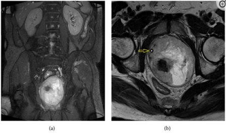
Figure 1: Prostate MRI with coronal (a) and axial (b) views showing marked enlargement of prostate gland with architecture derangement. Urethra with Foley (arrow) was shifted to the right side. 

Figure 2: Microscopic findings of the transurethral resection biopsy specimen. (a) Low-power view shows spindle cells arranged in tightly packed fascicles, with alternating hypo- and hypercellular areas and geographic necrosis (H&E stain, ×40). (b) High-power view shows spindle-shaped tumor cells with hyperchromatic nuclei and frequent mitoses. Some entrapped prostatic glands are also seen (H&E stain, ×400). (c) By immunohistochemistry, the tumor cells demonstrate focal immunopositivity of S-100 protein (Immunoperoxidase, ×200) and (d) no immunoreactivity with CDK4 (Immunoperoxidase, ×200). Reproduced 

Hsieh et al. [1] iterated that taking into consideration the difficulty of radical surgery due to huge size of the prostate gland, they had prescribed neoadjuvant chemotherapy, which had comprised of:5-Fluorouracil + Cisplatin + Ifosfamide for 5 cycles. Hsieh et al. [1] reported that regression of the patient’s previous lung nodule was found on his follow-up chest CT scan. The patient underwent pelvic MRI scan which demonstrated residual tumour lesion within the visible prostate gland, whose size had reduced to 83 grams. Radical cystoprostatectomy was undertaken, and invasion of tumour into the urinary bladder was found during the operation. Bilateral pelvic lymph node dissection was also undertaken and no metastasis was found. Pathology examination of the cystoprostatectomy specimen was reported as consistent with the diagnosis of MPNST. Pursuant to the cystoprostatectomy operation, Hsieh et al. [1] prescribed adjuvant concomitant chemoradiation therapy (Cisplatin + 5-Fluorouracil + Ifosfamide with external beam radiation therapy 6600 cGy, 33 fractions). The patient had a follow-up CT scan of abdomen 3 months pursuant to his operation which showed multiple ill-defined poorly enhanced nodules within his liver and a 3.5 cm gallbladder mass. Target therapy, of Pazopanib 400 mg per day, was given for palliative treatment due to suspicion of progression of his disease. Nevertheless, his clinical condition went downhill and he died three months later.

Hsieh et al. [1] made the ensuing summating discussions: 

  • MPNST is malignant tumour which is derived from peripheral nerves or which does demonstrate peripheral nerve differentiation. 
  • It has been iterated that about half of the patients who have PMNST, tend to be associated with neurofibromatosis I (NF-I) and the others tend to be sporadic or radiotherapy-induced. 
  • Some articles had reported prostate gland involvement in patients who have NF-I, however, majority of the tumours had tended to be benign lesions and they had been documented to originate from other pelvic organs [39] 
  • MPNST which has developed from prostate is iterated to be extremely rare. 
  • In their review, they had noted that Rames and Smith had reported the second case in 1999 [40], and no other case was reported up to the time of the report of their patient.
  • The commonest clinical manifestation is mass effect, which does tend to result from rapidly expanding tumour. 
  • The size of the tumour has tended to be independent of the site of the tumour, nevertheless, the average size of the tumour has tended to be greater than 5 cm.
  • Besides, up to 50% of patients tend to be diagnosed as having metastasis status, in which the most involved organ has tended to be the lung. 
  • With regard to their patient, progressive obstructive LUTS were the only symptom found. Besides, they had noticed a small lung nodule during the time of the initial diagnosis, which was considered suspicious metastasis and which had regressed pursuant to treatment with neoadjuvant chemotherapy. 
  • Liver metastasis was also suspected in their patient. Hence, it is important to survey distant metastasis prior to and pursuant to treatment in view of a high metastasis rate.
  • Akin to other sarcomas, complete surgical resection of the tumour does tend to represent the treatment of choice. 
  • No randomized study had strongly suggested the option of chemotherapy to an MPNST patient. Nevertheless, in one systematic meta-analysis study, adjuvant chemotherapy for localized resectable soft-tissue sarcoma reported marginal survival benefit, [41] which did imply that chemotherapy could have a role in selected MPNST patients.
  • The benefit of utilizing neoadjuvant chemotherapy was also noted in their reported case. Surgical approach became feasible in view of the regressive effect pursuant to neoadjuvant chemotherapy.
  • In recent years preceding the report of their case, many studies had researched in molecular pathway related sarcomas. Many clinical trials related to target therapy to sarcoma and MPNST had been completed or were ongoing, the results of which were exciting. [42]
  • In PALETTE study, utilization of Pazopanib as treatment had demonstrated a 3-month benefit in progression-free survival in patients who had non-adipocytic sarcomas. [43] 
  • They had prescribed Pazopanib to their patient for palliative reasons in view of the progression, and he expired about 2 months after receiving Pazopanib. Additional survey related target therapy on MPNST might be needed to study.
  • MPNST does tend to be associated with a high-potential for the development of metastasis and the overall outcome is very poor. 
  • It has been documented that the 5-year overall survival rate is about 44%. [ 44] 
  • It had also been iterated that larger size of the tumour is related to higher local recurrence rate and distal metastasis potential. [45] 
  • It has also been documented that the site of the tumour, is another prognosis factor.
  • Tumours that are located over extremities tend to be more easily locally controlled, which does tend to result in a better outcome. 
  • On the contrary, neoadjuvant chemotherapy does playa more important role in MPNST that is located within the pelvic cavity, where primary surgery may be hard to approach.

Hsieh et al. [1] made the ensuing conclusions:

  • From their reported case and the results of their literature review, they had learnt that prostate MPNST is an extremely rare or uncommon malignancy and still there has been no agreed standard management protocol for the tumour. 
  • Radical surgical resection is the key role of multimodality treatment; nevertheless, it might be infeasible in view of the large size and location of the tumour. 
  • Their experience had demonstrated that neoadjuvant chemotherapy is helpful for the regression of MPNST of the prostate gland. Nevertheless, even pursuant to radical surgery and adjuvant multimodality treatment, the prognosis has remained still poor.
  • Target therapy has been conjectured to become another promising treatment after further research in the future.

Kim et al. [46] made the ensuing iterations: 

  • It has been stated that soft tissue sarcoma is a rare and uncommon malignancy which does represents 1% of all cancers. [47]
  • Soft tissue sarcoma has tremendously heterogeneous pathologies and it has been found in various sites. 
  • Sarcoma of the prostate gland sarcoma had been reported to represent 0.7% of primary prostate malignancies. [48] 
  • It has been documented that Leiomyosarcoma and rhabdomyosarcoma are the commonest sarcomas of the prostate [49] [50] and on the contrary, malignant peripheral nerve sheath tumour (MPNST) of the prostate gland had only seldomly been reported.

Kim et al. [46] reported an extremely rare case of sarcoma of the prostate gland sarcoma in an early adulthood individual. Kim et al. [46] reported a 22-year-old man who had presented with a 2-week history of visible haematuria and voiding difficulty and who had visited the local urology clinic. He underwent digital rectal examination and transrectal ultrasound scan which demonstrated a hard mass within his prostate gland. He was referred to the urology department in the hospital of Kim et al. [46], He had magnetic resonance imaging (MRI) scan which showed a 6-cm mass within the left lobe of his prostate gland (see figure 3A [3A]).  The level of his serum prostate-specific antigen (PSA) was 1.89 ng/mL (normal range 0–4.0 ng/mL). Pathology examination of his prostate core needle biopsy revealed features of a high-grade sarcoma, which was suggestive of poorly differentiated synovial sarcoma (SS).
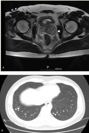
Figure:3

Magnetic resonance imaging (A) at diagnosis showed a 6-cm mass in the left lobe of the prostate, and (B) at recurrence showed multiple nodules in the lungs. Reproduced from: 

[46] Kim H, Kim DY, Seol YM, Ku JY, Choi KU, Choi YJ. Primary malignant peripheral nerve sheath tumor of prostate in a young adult: A case report. Medicine (Baltimore). 2018 Sep;97(39):e12040. doi: 10.1097/MD.0000000000012040. PMID: 30278486; PMCID: PMC6181477. https://www.ncbi.nlm.nih.gov/pmc/articles/PMC6181477/Copyright © 2018 the Author(s). Published by Wolters Kluwer Health, Inc. This is an open access article distributed under the terms of the Creative Commons Attribution-Non-Commercial-No Derivatives License 4.0 (CCBY-NC-ND), where it is permissible to download and share the work provided it is properly cited. The work cannot be changed in any way or used commercially without permission from the journal. http://creativecommons.org/licenses/by-nc-nd/4.0

He had Positron emission tomography-computed tomography (CT) on November 30, 2016 did not demonstrate any evidence of regional lymph node metastasis or distant metastasis. The patient underwent laparoscopic prostatectomy on December 6, 2016. Pathology examination of the prostatectomy specimen showed that the resection margin was positive, for the tumour which indicated R1 resection. The main differential diagnosis was documented by the pathologist to have included MPNST and synovial sarcoma (SS). SS was suspected more on the previous core needle biopsy because the tumour cells had exhibited negative immunohistochemistry staining for S100 protein, but it had exhibited positive staining for pan-cytokeratin (see Figure 4A [4A]). The resected tumour was reported not to have shown the presence of SYT-SSX fusion transcripts upon reverse transcription polymerase chain reaction, however it had exhibited focal immunoreactivity for S100 protein (see Figures 4B and 4C). Based upon the pathology examination features of the tumour, the tumour was reported to have shown features that were compatible with MPNST based on the microscopic findings, and also upon the results of ancillary studies.

Figure:4

Microscopic findings of the prostatectomy specimen. (A) The tumor was composed of fascicles of spindle cells showing alternating hypercellular and hypocellular areas, and heterologous cartilaginous differentiation (center) (H&E, ×40). (B) Malignant spindle cells showed hyperchromatic nuclei and numerous mitoses (H&E, ×200). (C) Immunohistochemistry showed focal staining for S100 protein (×200). Reproduced from: [46] Kim H, Kim DY, Seol YM, Ku JY, Choi KU, Choi YJ. Primary malignant peripheral nerve sheath tumor of prostate in a young adult: A case report. Medicine (Baltimore). 2018 Sep;97(39):e12040. doi: 10.1097/MD.0000000000012040. PMID: 30278486; PMCID: PMC6181477. https://www.ncbi.nlm.nih.gov/pmc/articles/PMC6181477/Copyright © 2018 the Author(s). Published by Wolters Kluwer Health, Inc. This is an open access article distributed under the terms of the Creative Commons Attribution-Non-Commercial-No Derivatives License 4.0 (CCBY-NC-ND), where it is permissible to download and share the work provided it is properly cited. The work cannot be changed in any way or used commercially without permission from the journal. http://creativecommons.org/licenses/by-nc-nd/4.0

He was referred on to the oncology department, and adjuvant radiotherapy was planned. Nevertheless, he had computed tomography (CT) scan on January 1, 2017, preceding his radiotherapy, which demonstrated multiple pulmonary metastases with local recurrence and peritoneal seeding (see figure 3B [3B]). He received a chemotherapy regimen of doxorubicin and ifosfamide and his disease had shown partial response to 3 cycles of the regimen. Nevertheless, he did complain about having severe general weakness and poor oral intake, and therefore, was switched on to doxorubicin monotherapy. He received 3 cycles of doxorubicin monotherapy. His disease was reported to be stable during the administration of his chemotherapy; nevertheless, his disease had progressed 2 weeks after the last dose of his doxorubicin. He received second-line pazopanib chemotherapy treatment, but his tumour did not show any response, and third-line gemcitabine and docetaxel were also associated with no response. The patient further administered everolimus and olaratumab with doxorubicin, but his tumour continued to progress. He died 9 months pursuant to his surgery on September 4, 2017.

Kim et al. [46] made the following summating discussions: 

  • It has been iterated that prostate cancer is the third leading type of cancer death in American men, who are aged 60 to 79 years and the second in those older than 80 years. [51] 
  • With regard to Korean men who are older than 65 years, prostate cancer was documented to be the fourth commonest cancer in 2014. [52] Nevertheless, the majority of prostate cancer is known to be adenocarcinoma, and sarcoma is rare. MPNST is extremely rare within the prostate gland.
  • At the time of the report of their case, four cases of prostate MPNST had been reported globally since 1999. The first case of MPNST, in 1999, was reported in a 21-year-old African-American man with neurofibromatosis. [40] 
  • The man had undergone radical cystoprostatectomy with ileal conduit urinary diversion and he demonstrated no recurrent disease 1 year postoperatively. The second case of MPNST was reported in Poland in 2015. [53] 
  • The third and fourth cases of MPNST were reported in 2016 in Greece and Taiwan, respectively [54] and [1]. See table 1 which Kim et al. [46] had reproduced to summarize these 4 cases.

Reproduced from: [46] Kim H, Kim DY, Seol YM, Ku JY, Choi KU, Choi YJ. Primary malignant peripheral nerve sheath tumor of prostate in a young adult: A case report. Medicine (Baltimore). 2018 Sep;97(39):e12040. doi: 10.1097/MD.0000000000012040. PMID: 30278486; PMCID: PMC6181477. https://www.ncbi.nlm.nih.gov/pmc/articles/PMC6181477/Copyright © 2018 the Author(s). Published by Wolters Kluwer Health, Inc. This is an open access article distributed under the terms of the Creative Commons Attribution-Non-Commercial-No Derivatives License 4.0 (CCBY-NC-ND), where it is permissible to download and share the work provided it is properly cited. The work cannot be changed in any way or used commercially without permission from the journal. http://creativecommons.org/licenses/by-nc-nd/4.0

  • It has been documented that malignant peripheral nerve sheath tumour is a rare disease, and about half of the cases have tended to be sporadic cases, and the other half have tended to be found with type 1 neurofibromatosis (NF1)—which is an autosomal dominant genetic disease. [44]
  • It has been iterated that NF1 is not a prognostic factor for survival in cases of MPNST [44] [55]
  • Like was reported in other soft-tissue sarcomas, manifestation with recurrent disease, large tumour size, and tumour within the trunk (versus the extremities) had been reported as the strongest independent predictors of shorter survival. [55] 
  • Zou et al [56] had analysed 140 MPNST patients and they had reported that size larger than 10 cm and p53 immunohistochemistry expression by the tumour cells were independent poor prognostic factors for MPNST. 
  • The undertaking of complete surgical resection of the tumour with negative margins does represent the mainstay of treatment. 
  • MPNST has been considered to be relatively chemo-insensitive. [57] 
  • Adjuvant radiotherapy could improve the local control of the MPNST tumour and it might benefit survival of patients undergoing treatment for MPNST. Nevertheless, solid evidence based upon randomized controlled trials for the efficacy of any adjuvant treatment has been limited in view of the rarity of MPNST. Despite aggressive surgery and adjuvant treatment, the prognosis of MPNST prognosis is so poor that the 5-year survival rate was reported to be about 34% to 44% [44] [58]
  • They could not be sure that their patient would have had a better disease course if he could have received adjuvant radiotherapy. Despite the fact that he had been administered several lines of chemotherapy for metastatic MPNST, only a doxorubicin-based regimen provided a brief response. They had assumed that the aggressive biology behaviour of the tumour and its poor response to treatment, contributed to the poor prognosis of MPNST. 
  • They would advise physicians to be alert to the symptoms of this rare cancer so that an early diagnosis could be provided, in addition to developing superior treatment modalities.


Ferakis et al. [54] stated that malignant Peripheral Nerve Sheath Tumour (MPNST) is a extremely rare soft tissue sarcoma as well as that sarcomas of the prostate gland are very rare. Ferakis et al. [54] iterated that they were reporting the first case of simultaneous prostatic adenocarcinoma and MPNST in the English literature. Ferakis et al. [54] reported that in December 2015, a 60-year-old man had visited the outpatient clinic of their department for routine urology examination. His past medical history had included hypertension, diabetes mellitus, acute myocardial infarction three years earlier, and ischemic cerebrovascular accident thirteen years earlier. He had digital rectal examination, which demonstrated that his prostate gland was painless, asymmetrically sizable, and tough, with no other clinical findings. The results of the patient’s full blood count and blood chemistry levels were reported to be within the normal range. His serum PSA level was 1 ng/mL He had transabdominal ultrasound scan which showed a solid, large mass, that was in contact with his urinary bladder. The volume of the prostate gland volume was 31 mL and his postvoid residual of urine volume on scanning was 62 mL. He had abdominal/pelvic computed tomography (CT) scan, which showed the presence of a solid, lobed, well-circumscribed mass, which had arisen from the left peripheral zone of his prostate gland and a possible infiltration of his left seminal vesicle. The wall of his urinary bladder was noted to be normal and the distal part of his left ureter had crossed between the mass and the urinary bladder wall. He had trans-rectal ultrasound-guided biopsy of his prostate gland and pathology examination of the biopsy specimen demonstrated a smooth muscle tumour with uncertain malignant potential (STUMP). He had magnetic resonance imaging (MRI) scan which demonstrated a fusiform, lobed, well-circumscribed, 6.5 cm × 6.5 cm × 6.0 cm in size mass, that had regular borders, which had arisen from the left peripheral zone of his prostate gland. The mass was found to have compressed the rectum, the posterior wall of the urinary bladder, and the left seminal vesicle/vas deferens. The lesion was reported to have a hyperintense signal upon T2-weighted sequences and hypointense signal upon T1-weighted sequences and it had contained thin septa. The adipose tissue encompassing the lesion was not infiltrated. There was heterogeneous enhancement of the lesion’s lobules following intravenous administration of paramagnetic contrast agent. He had Computed tomography (CT) scan of his thorax and isotope bone scan which were negative. The patient underwent 18-fluorodeoxyglucose-positron emission tomography (18-FDG-PET-CT) from the base of the skull to the mid-thigh, which demonstrated abnormal uptake within the prostatic mass. He had MRI scan of abdomen and CT scan of his thorax which were normal three months postoperatively and the patient had received adjuvant radiotherapy. His serum PSA level was 0 ng/mL. The patient was reported to have no signs of relapse and he was is in good general condition 6 months pursuant to his operation.


Ferakis et al. [54] made the ensuing summating discussions: 


  • It has been iterated that MPNSTs doe arise from a peripheral nerve or from a pre-existing benign nerve sheath tumour or they do tend to demonstrate Schwann cell differentiation on histology [59] [60] [61] 
  • Additionally, it has been pointed out that any malignant spindle cell tumour in a patient who has neurofibromatosis-1 (NF-1) is considered MPNST, unless proven otherwise [59] 
  • It has been documented that the terminology MPNST has replaced a number of previously used names including malignant schwannoma, neurofibrosarcoma, and neurogenic sarcoma. [60] [61] 
  • It has been iterated that MPNSTs do constitute 5% to 10% of all soft tissue sarcoma [59] [60] 
  • It has been documented that sarcomas of the prostate gland do account for 0.7% of all malignant tumours of the prostate gland. [49] 
  • It has been iterated that forty percent of MPNSTs tend to be sporadic tumours and the incidence in the general population is 0.001% [59] [61] 
  • The median age for sporadic MPNST has been documented to be between 30 and 60 years, with no gender predilection [3]. [61]
  • It has been pointed out that half of MPNSTs do occur in patients who have NF-1. [59] [60] [61]
  • MPNSTs could occur anywhere along the course of myelinated nerves, however, they commonly appear within or near a nerve of the trunk or the limbs. [59] [62] 
  • It has been pointed out that MPNSTs of the pelvis mostly do originate from the sacral or the hypogastric plexus of nerves. [59] [60] [63]
  • It has been pointed out that patients who have MPNST do manifest with an enlarging mass which may cause compression, displacement, or invasion of adjacent structure. [59] [60] [61] [63] 
  • It has been iterated that in most circumstances, the size of the tumour mass has tended to be greater than 5 cm at manifestation and up to 50% of the patients do tend to present with metastases, usually within the lung. [59] [60] [61]
  • With regard to their case, the patient was 60 years old, and he did not have a history of NF-1. He was asymptomatic, even though the size of his tumour mass was greater than 5 cm, and there was no evidence of metastases.
  • The differential diagnoses of MPNST had been stated to include the following: benign neurofibroma, fibrosarcoma, liposarcoma, ganglioneuroma, hydatid cyst, hematoma, and connective tissue diseases. [63] 
  • Pre-operative radiology examinations do play a pivotal role with regard to the diagnosis of a MPNST and in surgical planning. 
  • It has been pointed out that ultrasound scan could discriminate solid tumours from cystic masses. [64] 
  • It has been documented that computed tomography (CT) scan in MPNST does show well-defined, low, or mixed attenuation masses with cystic necrotic central areas [64] 
  • It has been pointed out that haemorrhage, calcification, and hyalinization might be present, in cases of MPNST, however, all these changes are not specific and the main utilization of CT is for the identification of metastases. [59] [60] [63] [64] 
  • It has been iterated that magnetic resonance imaging (MRI) scan is the modality of choice for the characterization of the anatomical extent of the tumour for the planning of surgery planning and it does help with regard to the differentiation of MPNSTs from benign plexiform neurofibromas. [59] [60] [61]
  • It has been pointed out that the lesion usually tends to be fusiform with tapered ends and it tends to be oriented longitudinally along the direction of a peripheral nerve. [59] [60] 
  • It has been stated that fat suppression sequences might allow better visualization of the nerve(s) that are involved in the tumour. [59] 
  • 18-FDG-PET-CT scan does help with regard to the differentiation of MPNST from benign neurofibroma in NF-1 patients and in the detection of malignant transformation of benign plexiform neurofibromas. [59] [61]
  • It has been documented that radiology imaging criteria are generally considered to be unreliable in differentiating MPNST from a benign schwannoma. Big irregular lesion, with rapid growth on interval imaging, heterogeneity, invasion of fat planes, and oedema encompassing the lesion do favour the diagnosis of MPNST. [60] 
  • It has been pointed out that upon macroscopy examination, MPNSTs are found to be globoid or fusiform with regard to shape, fleshy, and firm to hard with regard to consistency and their colour typically tends to be tan-grey upon cut section. [59] 
  • It has also been documented that upon macroscopy examination of specimens of MPNST, areas of necrosis or cyst formation tend to be found to be commonly present and the lesions might be covered by a fibrous pseudo-capsule and they tend to be found to invade encompassing soft tissue, as in their reported case. [59] [64] 
  • It has been pointed out that microscopy examination of specimens of MPNST, does show the tumour to be typified by hypercellular fascicles of spindle cells that are interrupted by hypocellular myxoid areas, and often with hypercellular areas that are localized in close proximity to blood vessels. [59] [61] [63] [64] [65] 
  • It has additionally been stated that upon microscopy examination of MPNST specimens, the spindle cells that are found tend to be relatively large, with long, hyperchromatic, wavy, or “serpentine” nuclei. [59] [65]
  • It has also been iterated that upon microscopy examination of MPNST, malignancy tends to be usually suggested if high mitotic activity, increased cellularity, pleomorphism, nuclear atypia, blood vessel infiltration, and tumour necrosis are demonstrated histologically. [59] [60] [61] [63]
  • Heterologous elements, such as skeletal muscle, bone, and cartilage, are present in about 15% of tumours and could portend an even poorer prognosis. [61] [65] 
  • It has been stated that there is no pathognomonic immunohistochemical study for MPNST. [59] [61]
  •  S-100, which is traditionally regarded as the best marker for MPNST, was positive in their case. Nevertheless, it does have limited diagnostic utility and it has been pointed out that it tends to be positive in about 50% to 90% of the tumours. [60] ] [65].
  • It has been iterated that Leu-7 and myelin basic protein are noted in 50% and 40% of cases, respectively. [59] [60] 
  • In general, a combination of antigens is used to help exclude other spindle cell lesions and confirm the diagnosis of MPNST. [60] 
  • In their case, vimentin, CD34, Bcl-2, and CD56 were only partially positively stained upon immunohistochemistry staining studies of the tumours.
  • It has been iterated that complete surgical extirpation of the tumour with the attainment of clear tumour-free margins is the treatment of choice for MPNST [59] [60] [62] [63] [64] [65]
  • It has been stated that it might be necessary to sacrifice adjacent tissue and viscera in the surgical treatment of MPNST [64] 
  • It has additionally been documented that, in case of malignancy, the local recurrence rate after marginal excision is 72% in comparison with 11.7

Conclusions

  • Malignant peripheral nerve sheath tumour (MPNST) of the prostate gland is a very rare aggressive malignant tumour of the prostate gland. 
  • MPNST refers to malignant neoplasm which does arise from peripheral nerve 
  • MPNST could arise from a pre-existing nerve sheath tumour in neurofibromatosis type 1 (NF1) or in the setting of prior radiotherapy 
  • The absence of an association with NF1 or radiotherapy, the diagnosis of MPNST tends to be very challenging and diagnosis of MPNST of the prostate gland tends to be based upon histology and immunohistochemical staining features of biopsy specimens of the prostate gland and specimens of the prostate tumour in radical prostatectomy specimens that suggest Schwannian differentiation
  • MPNST of the prostate gland could be diagnosed incidentally, or they could be based upon investigations of non-specific specific symptoms of prostatism including lower urinary tract symptoms, haematuria, symptoms related to ureteric obstruction, metastases, and urinary retention. 
  • Considering that MPNST of the prostate gland could also occur in younger men, men who had previously undergone radiotherapy, and individuals who have schwannoma as well as the serum PSA levels of MPNST of the prostate gland tends to be low, a high index of suspicion should be exercised so as to quickly and thoroughly investigate individuals who are young as well as individuals who have predisposing factors in order to establish early diagnosis in order to provide aggressive treatment of curative intent. 
  • There is no consensus opinion; nevertheless, radical prostatectomy with complete excision of the tumour with treatment curative intent should be provided for all patients who have localized MPNST, additionally adjuvant radiotherapy plus adjuvant combination chemotherapy should be provided with an aim to prevent the development of local recurrence, lymph node metastases or distant metastases. 
  • With regard to patients who have locally advanced MPNST, advanced MPNST, and distant metastases, the patients should be treated with utilization of radiotherapy and combination chemotherapy, supportive palliative care and those who have ureteric obstruction should be offered nephrostomy insertion, antegrade ureteric stent insertion, or retrograde ureteric stent insertion in order to avoid the development of renal failure as well as hyperkalaemia. 
  • There is need for Urologists, oncologists, and pharmacology and pharmacy research workers to establish global combination chemotherapy options study in order to find treatment options that would effectively destroy MPNSTs of the prostate gland
  • Individuals who treat MPNSTs of the prostate gland should be encouraged to report their cases in the literature in order that the biological behaviour of the tumour as well as the best treatment options can be established by consensus opinion. 
  • Academic Urologists, oncologists, and pharmacotherapy individuals globally should undertake research studies to ascertain any genomic changes that may be associated with their cases of primary MPNST of the prostate gland and they should undertake studies that would help in the development of novel treatment options that would utilize knowledge of the genomic changes to provide more effective treatment for MPNST of the prostate gland.     

Conflict of interest

None

Acknowledgements

[1] Case Reports in Urology and Hindawi Publishing Group and the authors of articles in the journal for granting permission for reproduction of figures and contents of their journal article under the Creative Commons Attribution License, which permits unrestricted use, distribution, and reproduction in any medium, provided the original work is properly cited. 

[2] Medicine (Baltimore). For granting permission for reproduction of contents of their journal article as well as figures and tables under Copyright © 2018 the Author(s). Published by Wolters Kluwer Health, Inc. This is an open access article distributed under the terms of the Creative Commons Attribution-Non-Commercial-No Derivatives License 4.0 (CCBY-NC-ND), where it is permissible to download and share the work provided it is properly cited. The work cannot be changed in any way or used commercially without permission from the journal. http://creativecommons.org/licenses/by-nc-nd/4.0

References

Clearly Auctoresonline and particularly Psychology and Mental Health Care Journal is dedicated to improving health care services for individuals and populations. The editorial boards' ability to efficiently recognize and share the global importance of health literacy with a variety of stakeholders. Auctoresonline publishing platform can be used to facilitate of optimal client-based services and should be added to health care professionals' repertoire of evidence-based health care resources.

img

Virginia E. Koenig

Journal of Clinical Cardiology and Cardiovascular Intervention The submission and review process was adequate. However I think that the publication total value should have been enlightened in early fases. Thank you for all.

img

Delcio G Silva Junior

Journal of Women Health Care and Issues By the present mail, I want to say thank to you and tour colleagues for facilitating my published article. Specially thank you for the peer review process, support from the editorial office. I appreciate positively the quality of your journal.

img

Ziemlé Clément Méda

Journal of Clinical Research and Reports I would be very delighted to submit my testimonial regarding the reviewer board and the editorial office. The reviewer board were accurate and helpful regarding any modifications for my manuscript. And the editorial office were very helpful and supportive in contacting and monitoring with any update and offering help. It was my pleasure to contribute with your promising Journal and I am looking forward for more collaboration.

img

Mina Sherif Soliman Georgy

We would like to thank the Journal of Thoracic Disease and Cardiothoracic Surgery because of the services they provided us for our articles. The peer-review process was done in a very excellent time manner, and the opinions of the reviewers helped us to improve our manuscript further. The editorial office had an outstanding correspondence with us and guided us in many ways. During a hard time of the pandemic that is affecting every one of us tremendously, the editorial office helped us make everything easier for publishing scientific work. Hope for a more scientific relationship with your Journal.

img

Layla Shojaie

The peer-review process which consisted high quality queries on the paper. I did answer six reviewers’ questions and comments before the paper was accepted. The support from the editorial office is excellent.

img

Sing-yung Wu

Journal of Neuroscience and Neurological Surgery. I had the experience of publishing a research article recently. The whole process was simple from submission to publication. The reviewers made specific and valuable recommendations and corrections that improved the quality of my publication. I strongly recommend this Journal.

img

Orlando Villarreal

Dr. Katarzyna Byczkowska My testimonial covering: "The peer review process is quick and effective. The support from the editorial office is very professional and friendly. Quality of the Clinical Cardiology and Cardiovascular Interventions is scientific and publishes ground-breaking research on cardiology that is useful for other professionals in the field.

img

Katarzyna Byczkowska

Thank you most sincerely, with regard to the support you have given in relation to the reviewing process and the processing of my article entitled "Large Cell Neuroendocrine Carcinoma of The Prostate Gland: A Review and Update" for publication in your esteemed Journal, Journal of Cancer Research and Cellular Therapeutics". The editorial team has been very supportive.

img

Anthony Kodzo-Grey Venyo

Testimony of Journal of Clinical Otorhinolaryngology: work with your Reviews has been a educational and constructive experience. The editorial office were very helpful and supportive. It was a pleasure to contribute to your Journal.

img

Pedro Marques Gomes

Dr. Bernard Terkimbi Utoo, I am happy to publish my scientific work in Journal of Women Health Care and Issues (JWHCI). The manuscript submission was seamless and peer review process was top notch. I was amazed that 4 reviewers worked on the manuscript which made it a highly technical, standard and excellent quality paper. I appreciate the format and consideration for the APC as well as the speed of publication. It is my pleasure to continue with this scientific relationship with the esteem JWHCI.

img

Bernard Terkimbi Utoo

This is an acknowledgment for peer reviewers, editorial board of Journal of Clinical Research and Reports. They show a lot of consideration for us as publishers for our research article “Evaluation of the different factors associated with side effects of COVID-19 vaccination on medical students, Mutah university, Al-Karak, Jordan”, in a very professional and easy way. This journal is one of outstanding medical journal.

img

Prof Sherif W Mansour

Dear Hao Jiang, to Journal of Nutrition and Food Processing We greatly appreciate the efficient, professional and rapid processing of our paper by your team. If there is anything else we should do, please do not hesitate to let us know. On behalf of my co-authors, we would like to express our great appreciation to editor and reviewers.

img

Hao Jiang

As an author who has recently published in the journal "Brain and Neurological Disorders". I am delighted to provide a testimonial on the peer review process, editorial office support, and the overall quality of the journal. The peer review process at Brain and Neurological Disorders is rigorous and meticulous, ensuring that only high-quality, evidence-based research is published. The reviewers are experts in their fields, and their comments and suggestions were constructive and helped improve the quality of my manuscript. The review process was timely and efficient, with clear communication from the editorial office at each stage. The support from the editorial office was exceptional throughout the entire process. The editorial staff was responsive, professional, and always willing to help. They provided valuable guidance on formatting, structure, and ethical considerations, making the submission process seamless. Moreover, they kept me informed about the status of my manuscript and provided timely updates, which made the process less stressful. The journal Brain and Neurological Disorders is of the highest quality, with a strong focus on publishing cutting-edge research in the field of neurology. The articles published in this journal are well-researched, rigorously peer-reviewed, and written by experts in the field. The journal maintains high standards, ensuring that readers are provided with the most up-to-date and reliable information on brain and neurological disorders. In conclusion, I had a wonderful experience publishing in Brain and Neurological Disorders. The peer review process was thorough, the editorial office provided exceptional support, and the journal's quality is second to none. I would highly recommend this journal to any researcher working in the field of neurology and brain disorders.

img

Dr Shiming Tang

Dear Agrippa Hilda, Journal of Neuroscience and Neurological Surgery, Editorial Coordinator, I trust this message finds you well. I want to extend my appreciation for considering my article for publication in your esteemed journal. I am pleased to provide a testimonial regarding the peer review process and the support received from your editorial office. The peer review process for my paper was carried out in a highly professional and thorough manner. The feedback and comments provided by the authors were constructive and very useful in improving the quality of the manuscript. This rigorous assessment process undoubtedly contributes to the high standards maintained by your journal.

img

Raed Mualem

International Journal of Clinical Case Reports and Reviews. I strongly recommend to consider submitting your work to this high-quality journal. The support and availability of the Editorial staff is outstanding and the review process was both efficient and rigorous.

img

Andreas Filippaios

Thank you very much for publishing my Research Article titled “Comparing Treatment Outcome Of Allergic Rhinitis Patients After Using Fluticasone Nasal Spray And Nasal Douching" in the Journal of Clinical Otorhinolaryngology. As Medical Professionals we are immensely benefited from study of various informative Articles and Papers published in this high quality Journal. I look forward to enriching my knowledge by regular study of the Journal and contribute my future work in the field of ENT through the Journal for use by the medical fraternity. The support from the Editorial office was excellent and very prompt. I also welcome the comments received from the readers of my Research Article.

img

Dr Suramya Dhamija

Dear Erica Kelsey, Editorial Coordinator of Cancer Research and Cellular Therapeutics Our team is very satisfied with the processing of our paper by your journal. That was fast, efficient, rigorous, but without unnecessary complications. We appreciated the very short time between the submission of the paper and its publication on line on your site.

img

Bruno Chauffert

I am very glad to say that the peer review process is very successful and fast and support from the Editorial Office. Therefore, I would like to continue our scientific relationship for a long time. And I especially thank you for your kindly attention towards my article. Have a good day!

img

Baheci Selen

"We recently published an article entitled “Influence of beta-Cyclodextrins upon the Degradation of Carbofuran Derivatives under Alkaline Conditions" in the Journal of “Pesticides and Biofertilizers” to show that the cyclodextrins protect the carbamates increasing their half-life time in the presence of basic conditions This will be very helpful to understand carbofuran behaviour in the analytical, agro-environmental and food areas. We greatly appreciated the interaction with the editor and the editorial team; we were particularly well accompanied during the course of the revision process, since all various steps towards publication were short and without delay".

img

Jesus Simal-Gandara

I would like to express my gratitude towards you process of article review and submission. I found this to be very fair and expedient. Your follow up has been excellent. I have many publications in national and international journal and your process has been one of the best so far. Keep up the great work.

img

Douglas Miyazaki

We are grateful for this opportunity to provide a glowing recommendation to the Journal of Psychiatry and Psychotherapy. We found that the editorial team were very supportive, helpful, kept us abreast of timelines and over all very professional in nature. The peer review process was rigorous, efficient and constructive that really enhanced our article submission. The experience with this journal remains one of our best ever and we look forward to providing future submissions in the near future.

img

Dr Griffith

I am very pleased to serve as EBM of the journal, I hope many years of my experience in stem cells can help the journal from one way or another. As we know, stem cells hold great potential for regenerative medicine, which are mostly used to promote the repair response of diseased, dysfunctional or injured tissue using stem cells or their derivatives. I think Stem Cell Research and Therapeutics International is a great platform to publish and share the understanding towards the biology and translational or clinical application of stem cells.

img

Dr Tong Ming Liu

I would like to give my testimony in the support I have got by the peer review process and to support the editorial office where they were of asset to support young author like me to be encouraged to publish their work in your respected journal and globalize and share knowledge across the globe. I really give my great gratitude to your journal and the peer review including the editorial office.

img

Husain Taha Radhi

I am delighted to publish our manuscript entitled "A Perspective on Cocaine Induced Stroke - Its Mechanisms and Management" in the Journal of Neuroscience and Neurological Surgery. The peer review process, support from the editorial office, and quality of the journal are excellent. The manuscripts published are of high quality and of excellent scientific value. I recommend this journal very much to colleagues.

img

S Munshi

Dr.Tania Muñoz, My experience as researcher and author of a review article in The Journal Clinical Cardiology and Interventions has been very enriching and stimulating. The editorial team is excellent, performs its work with absolute responsibility and delivery. They are proactive, dynamic and receptive to all proposals. Supporting at all times the vast universe of authors who choose them as an option for publication. The team of review specialists, members of the editorial board, are brilliant professionals, with remarkable performance in medical research and scientific methodology. Together they form a frontline team that consolidates the JCCI as a magnificent option for the publication and review of high-level medical articles and broad collective interest. I am honored to be able to share my review article and open to receive all your comments.

img

Tania Munoz

“The peer review process of JPMHC is quick and effective. Authors are benefited by good and professional reviewers with huge experience in the field of psychology and mental health. The support from the editorial office is very professional. People to contact to are friendly and happy to help and assist any query authors might have. Quality of the Journal is scientific and publishes ground-breaking research on mental health that is useful for other professionals in the field”.

img

George Varvatsoulias

Dear editorial department: On behalf of our team, I hereby certify the reliability and superiority of the International Journal of Clinical Case Reports and Reviews in the peer review process, editorial support, and journal quality. Firstly, the peer review process of the International Journal of Clinical Case Reports and Reviews is rigorous, fair, transparent, fast, and of high quality. The editorial department invites experts from relevant fields as anonymous reviewers to review all submitted manuscripts. These experts have rich academic backgrounds and experience, and can accurately evaluate the academic quality, originality, and suitability of manuscripts. The editorial department is committed to ensuring the rigor of the peer review process, while also making every effort to ensure a fast review cycle to meet the needs of authors and the academic community. Secondly, the editorial team of the International Journal of Clinical Case Reports and Reviews is composed of a group of senior scholars and professionals with rich experience and professional knowledge in related fields. The editorial department is committed to assisting authors in improving their manuscripts, ensuring their academic accuracy, clarity, and completeness. Editors actively collaborate with authors, providing useful suggestions and feedback to promote the improvement and development of the manuscript. We believe that the support of the editorial department is one of the key factors in ensuring the quality of the journal. Finally, the International Journal of Clinical Case Reports and Reviews is renowned for its high- quality articles and strict academic standards. The editorial department is committed to publishing innovative and academically valuable research results to promote the development and progress of related fields. The International Journal of Clinical Case Reports and Reviews is reasonably priced and ensures excellent service and quality ratio, allowing authors to obtain high-level academic publishing opportunities in an affordable manner. I hereby solemnly declare that the International Journal of Clinical Case Reports and Reviews has a high level of credibility and superiority in terms of peer review process, editorial support, reasonable fees, and journal quality. Sincerely, Rui Tao.

img

Rui Tao

Clinical Cardiology and Cardiovascular Interventions I testity the covering of the peer review process, support from the editorial office, and quality of the journal.

img

Khurram Arshad