
Research Article | DOI: https://doi.org/10.31579/2766-2314/019
University Clinic for Rheumatology, faculty of medicine, University Clinical Center “Mother Therese”, Skopje, Republic of North Macedonia.
*Corresponding Author: Dejan Spasovski, University Clinic for Rheumatology, faculty of medicine, University Clinical Center “Mother Therese”, Skopje, Republic of North Macedonia.
Citation: Spasovski D., (2020) Lysosomal or brush border enzymuria in kidney enzyme activities in seronegative rheumatoid arthritis. J, Biotechnology and Bioprocessing 1(3); DOI: 10.31579/2766-2314/019
Copyright: © 2020, Dejan Spasovski, This is an open access article distributed under the Creative Commons Attribution License, which permits unrestricted use, distribution, and reproduction in any medium, provided the original work is properly cited.
Received: 04 December 2020 | Accepted: 19 December 2020 | Published: 28 December 2020
Keywords: alanine aminopeptidase; (AAP); gama-glutamyl transferase (gama--GT); β2 microglobulin (β2M); rheumatoid arthritis (RA); rheumatoid factor (RF)
Introduction: To determine the effects of non-treated seronegative rheumatoid arthritis (RA) on proximal renal tubule, sensitivity of Alanine aminopeptidase (AAP), g-glutamyltransferase (g--GT), β2 microglobulin in urine β2M), as well as relation with rheumatoid factor (RF) and C-Reactive protein (CRP), DAS 28 disease activity index.
Methods: RF was determined by agglutination test (Latex RF test, while kinetic methods were used for determination of Alanine aminopeptidase (AAP) and g-glutamyltransferase (g-GT), as well as MEIA (Micro particle Enzyme Immunoassay) to determine β2 microglobulin in urine. Samples (serum and urine) of 70 participants were examined (35 RA not treated, 35 health control group).
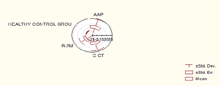
Results: In 35 RF negative RA, AAP enzymuria was present in 12 (34.28%) patients, g-GT was present in 7 patients (20%), while β2 microglobulin was present in 3 patients (8.57%). In the healthy control group, 4 patients showed AAP positivity (11.42%), 2 patients g-GT positivity (5.71%) and 1 patient showed presence of β2 microglobulin in urine (2.85). RF was not present in any patient (0%).
Conclusion: AAP has a higher sensitivity of g-GT and b2 microglobulin in the detection of asymptomatic renal lesions in non treated seronegative RA.
Enzymes in urine can derive from plasma, glands of the urogenital tract, epithelial cells of the urinary tract, leukocytes, and erythrocytes [1] and kidneys. There are about 40 different enzymes [2-6] in the urine that belong to different groups: oxidoreductase, transferase, hydrolase, lyase, while isomerases and ligases are not found in the urine. The occurrence of such large number of enzymes in the urine indicates the dominant role of kidneys in their excretion.
Examination of the cell membranes of the brush epithelium of the proximal tubules confirms the localization of alanine aminopeptidase (AAP) in 90%, alaline phosphatase (AF) in 70% and g-glutamyl transpeptidase (g-GT) in 50% of the total activity of these enzymes in the kidney [7-9].
The aim of this study is to determine the effects of non-treated Rheumatoid arthritis on the tubular function AAP, g-GT and β2M being used as indicators for proximal tubular damage.
In patients included in the study, disease diagnose is based on the revised diagnostic criteria for classification of Rheumatoid arthritis proposed in 1987 by the American Rheumatism Association (ARA) [10-13]. For the classification, i.e. the patients to be included in the RA group it is necessary to satisfy at least 4 of the predicted 7 criteria.
Criteria from 1 to 4 were present at least 6 weeks. The study included 35 patients (age 28, age 7) who were diagnosed with seronegative RA, as well as 35 patients (age 18, age 17) as a healthy control group. The average mean age was 48.5 years (± 4.13) (37-65 years) for the RA group, 36.2 years (±10.78) (29-65) for the healthy group. The average time of onset of disease in months from the beginning was 14.97 (± 15.23), in the interval of (1-14) months. None of the patients in the study had a history of previous or current renal impairment. The others negate use of other drugs before sample were taken. The samples were collected in a period of 1 year.
Including criteria
In the study were included patients with RA at the age of 18-65 years, who were not previously treated with NSAIDs or DMARDs.
Excluding criteria
In the study were excluded patients with symptoms or conditions that can directly or indirectly affect the results, such as:
All participants voluntarily took part in this study, so that the criteria to do it are met.
Clinical assessment of disease activity
Clinical assessment and interpretation was made from the subspecialist in the given area. The disease activity was assessed using DAS 28 index. (Disease Activity Score (DAS 28)) [14]. Indexes use mathematical formula to use the unique composite quantitative score consisting of palatable painfully sensitive joints (maximum number 28) and swollen joints (maximum number 28), global assessment for disease activity (0 – 100 mm Visual Analog Scale VAS), as well as morning stiffness (minutes). DAS 28 index ranges from 0 to 10 and score below 3.2 qualifies the disease as low active.
Laboratory assessment
For clinical assessment of disease, it is necessary to consider the following laboratory variables: Complete blood count (CBC) and differential, acute phase reactants, such as C-reactive protein (CRP), Rheumatoid factor (RF), Erythrocyte sedimentation rate (ESR), alkaline phosphatase (AF), aspartate aminotransferase (AST), alanine aminotransferase (ALT), creatine kinase (CK), lactate dehydrogenase (LDH), urea / serum, creatinine / serum.
Urine samples were taken not only for rutine urinary examination, but also for determination of AAP, g-GT, β2M.
Serum urea is determined by the method of "Kassirer"
Reference values: Serum urea (3-7.8 mmol / L).
Creatine in serum and urine and determined by the method of: "Jaffe"
Reference values: Serum creatine 45 - 109 µmol / L; Creatine in urine 7 - 17 mol / dU.
C-reactive protein (CRP) determined by agglutination test (Latex CRP test)
Reference values: < 6>
Rheumatoid factor (RF) determined by agglutination test (Latex RF test)
Reference values: < 8>
Determination of Alanine Aminopeptidase Activity (AAP): Kinetic Method
Reference values: AAP in urine 0.25-0.75 U / mmol creatinine
Determination of g-glutamyltranspeptidase (g-GT) activity: Ifcc method
Valuable referents
g-GT (urine) 0.84-1.80 u / mmol creatinine
Determination of β2 microglobulin (β2M) concentration in urine by the method “meia” ("micro particle enzyme immunoassay")
Reference values:
β2 microglobulin (urine) = 0.02-0.19 mg / L
Statistical analysis
For testing the significance of the differences between two arithmetic means, i.e. the corresponding proportions, the Student t-test is used, when comparing the mean values of the given number of parameters between two groups, such as Wilcoxon- matched test for independent samples. Sensitivity and productivity for positive and negative tests of the examined markers is determined with tests for sensitivity and specificity. The P value of between 0.05 and 0.1 is considered statistically significant. The data processing is made with the statistical package Statistical 7.0.
In the group of 35 patients with RA, DAS 28 > 3.2 was present in 28 patients (80%).
In these 28 patients DAS 2 > 3.2, AAP positive 10 (35.71%) and their M ± SD (1.25 ± 0.43) range (0.85-2.46), g-GT positive were 5 (17.85%) their M ± SD (2.65 ± 0.46) range (0.95-3.45), while β2M was not present in any patient.
In 7 seronegative RF patients with DAS 28 <3>
In standard medical rheumatology, the greatest emphasis is put on Rheumatoid arthritis as the most exposed disease. Seronegative RA is a rare form, difficult to recognize and most often confused with degenerative rheumatism, probably due to their frequency.
Urinary enzyme activity is normally low in the urine and increases when renal tubular cells are excreted [15]. Urinary enzymes, especially NAG, AAP, and AF are very sensitive indicators of parenchymal renal damage in comparison with functional measurements such as glomerular filtration rate (GFR), creatinine and inulin clearance. The relatively low sensitivity of the GFR can be explained by the large renal functional reserve and its large capacity for compensation [16]. There are indications that elevations in urinary enzyme activity may indicate the location of the primary renal tubular damage due to their localization in the brush border area (microsomal AAP) and tubular lysozyme (NAG). They can be used in early diagnosis of acute renal failure because nephrotoxicity is induced by immunosuppressive drugs, contraceptives, antibiotics and cadmium exposure [17-19].
The sensitivity of AAP is higher in comparison with g-GT and β2M. Other standard routine tests used to assess renal function show low sensitivity: creatine in serum and urine, urea in serum. Seronegativity has an impct on the occurrence of AAP enzymuria. This is also present for seronegative patients with DAS 28 > 3.2 who have a much larger AAP induction than DAS 28 < 3>
Non-treated RA primarily affects tubular brush border area and enzymes that derives from this area have increased sensitivity.
AAP has a higher sensitivity than g-GT and β2M in the detection of asymptomatic renal lesions in the non-treated seronegative RA. AAP and g-GT can be used in the everyday clinical practice to diagnose early, asymptomatic renal lesions.
Dear Editorial Team, Clinical Medical Reviews and Reports. My experience with the journal was highly positive. The peer-review process was rigorous, constructive, and completed in a timely manner. The reviewers provided valuable comments that helped improve the quality and clarity of our manuscript. The editorial office was professional, responsive, and supportive throughout all stages of the publication process. Communication was clear and efficient, and any questions were addressed promptly. Overall, I found the journal to maintain high scientific standards and an excellent publication workflow. I would be pleased to consider submitting future work to this journal. Best wishes from, Elena Popa.

It was my pleasure to submit my testimonial concerning the Reviewer Board of our Scientific Journal “Brain and Neurological Disorders”. The Reviewers focused on some modifications and their contribution was helpful. The ladies of our Editorial Office were also supported my efforts. It was my honor to have such a co-operation and I am looking forward for more collaboration.

Dear Grace Pierce, Editorial Coordinator of Journal of Clinical Research and Reports, Thank you for the speedy and efficient peer review process. I appreciate the fact that your peer reviewers do not take months to respond like with some other journals. I would also like to thank the editorial office for responding quickly to my questions. It is an excellent journal. I plan to submit more manuscripts in the future. Best wishes from, Robert W. McGee

Dear Grace Pierce, Editorial Coordinator of Journal of Clinical Research and Reports, Working with you and your team on our recent publication in JCRR has been a truly wonderful and enjoyable experience. The responses were prompt, and the reviewers were patient, constructive, and highly professional. One reviewer in particular gave me the feeling that a professor was carefully reading and commenting on my coursework, which was deeply touching. The entire process was straightforward and hassle‑free, with no tedious online forms to complete. I highly recommend this journal. Best wishes from, DR Aibing Rao, Head of R&D

I Appreciate the Opportunity to Share my Experience with the Journal of Clinical Research and Reports. The peer review process was timely and constructive, and the feedback provided helped improve the quality of our manuscript. The editorial office was professional, responsive, and supportive throughout the process, ensuring smooth communication and efficient handling of the submission. Overall, it was a positive experience collaborating with your team.

Dear Mercy Grace, Editorial Coordinator of Obstetrics Gynecology and Reproductive Sciences, We would like to express our gratitude for your help at all stages of publishing and editing the article. The editors of the magazine answer all the necessary questions and help at every stage. We will definitely continue to cooperate and publish other works in the Obstetrics Gynecology and Reproductive Sciences! Best wishes from, Alla Konstantinovna Politova,
