
Case Report | DOI: https://doi.org/10.31579/2690-4861/147
*Corresponding Author: Reem Hamdy A Mohammed, Professor of Rheumatology and Rehabilitation, Department of Rheumatology and Rehabilitation- School of Medicine- Cairo University.
Citation: RHA Mohammed, Raghda M Farghaly, Andrea D Matteo. (2021) Longstanding Complicated Wrist Swelling: A Typical Presentation of Primary Tuberculosis a case report and mini review. International Journal of Clinical Case Reports and Reviews. 8(2); DOI:10.31579/2690-4861/147
Copyright: © 2021 Reem Hamdy A Mohammed, This is an open-access article distributed under the terms of the Creative Commons Attribution License, which permits unrestricted use, distribution, and reproduction in any medium, provided the original author and source are credited.
Received: 17 June 2021 | Accepted: 09 August 2021 | Published: 18 August 2021
Keywords: wrist swelling; primary tuberculosis; scaphoid bone
Tuberculosis of the wrist joints is an uncommon clinical entity that most frequently presents with insidious pain and swelling. Isolated carpal bones involvement due to tuberculosis is a relatively uncommon event. In this report, the authors describe the case of a 36-year-old female patient presenting with fracture of the scaphoid bone. The diagnosis of tuberculosis was suspected based on the ultrasound and magnetic resonance imaging findings of the wrist joint, and confirmed by joint fluid culture. Introducing anti-tuberculous drugs under appropriate supervision resulted in clinical improvement and optimal regain of function. No reactivation of the disease was noted after 2 years of follow‑up. This case report describes a rare presentation of wrist joint tuberculosis.
Primary (isolated) tuberculosis (TB) of the hand and wrist has been rarely reported in the literature, representing <1>
Symptoms vary depending on the specific wrist or hand joint which is affected by TB. The involvement of the wrist typically begins in the scapholunate joint. The diagnosis of wrist tuberculosis is often late; when discovered at an early stage, a well-followed medical treatment is usually enough to provide full healing.
The case presented is an Egyptian illiterate 36-year-old housewife patient from a low income family, married with no off springs and has no special habits of medical importance was referred to the outpatient clinic of rheumatology and rehabilitation complaining of painless, swelling of the left wrist of 1 year and 6 months duration.
Previous medical history: The condition started with a painless of the left wrist, of gradual onset and progressive course, not associated with morning stiffness or local redness. The patient sought medical advice at orthopedic clinic at Al Kasr Alainy Hospital, where aspiration of one of the swellings and investigations were done.
The patient was referred to rheumatology and rehabilitation outpatient clinic. On examination there was a boggy synovial thickening with multilocular swelling on the dorsal aspect of the left wrist, with limited dorsiflexion and extension, no local redness or hotness. No evidence of arthritis or limited range of motion in any of the other joints of the upper or lower limbs. Examination of other body systems revealed no abnormality detected. Review of the past history for the present illness, the patient gave history of loss of weight despite of good appetite, vague history of unmeasured fever not associated with skin rash, rigors or excessive night sweats and not associated with an evident source of infection. The patient did not report any chest symptoms. There was no history of subcutaneous nodules, no other joint or musculoskeletal complaints of significance. There was no history of alopecia, oral ulcers, genital ulcers, malar rash, photosensitivity or Raynaud’s phenomenon, no history of DVT, TIAs, stroke or any vascular event, no history of cough, hemoptysis, or dyspnea. No history of tingling, hypothesia or muscle weakness, no history of chronic diarrhea or change of bowel habits or GIT bleeding. No history of bleeding from body orifices.
Laboratory investigations revealed normal complete blood count, normal chemistry and with normal ranges for inflammatory biomarkers (erythrocyte sedimentation rate and C reactive protein), tuberculin test was weak positive.
Plain x ray of the wrist revealed an old fracture of Lt distal forearm with avascular necrosis of the scaphoid bone. Figure 1 (a&b).
Musculoskeletal US revealed significant villous like synovial thickening with power Doppler signal indicating increased vascularity. Scaphoidectomy, wire fixation and synovectomy of extensor compartment were done at the orthopedic clinic. Figure 2.
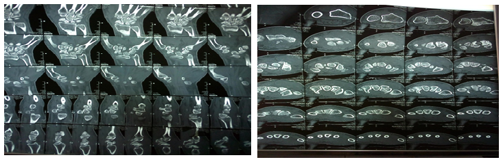
Computerized tomography scan (CT scan) of the Lt wrist revealed an old non-united fracture of the left scaphoid bone waist with gapped sclerosed fracture ends and adjacent tiny bone chips. Scapholunate dissociation with widening intervening spaces. Anterior tilt of the lunate bone denoting ventral intercalated segmental instability. Secondary osteo-arthritic changes of radio-carpal joint with marginal osteophytosis of articular surface. Multi-locular cystic lesion is seen along dorsum of Lt wrist measuring 3.2 x 2.5 cm, likely extensor tendon tenosynovitis. Figure 3 (a &b)
Magnetic resonance imaging (MRI) of the left wrist revealed normal appearance of the proximal and distal carpal bones with no evidence of avascular necrosis (AVN). No fluid collection was seen. Figure 4.


Pathology, microscopy and histopathological examination of the excised synovial tissue:
Gross pathological examination revealed multiple irregular fibrofatty tissue pieces, collectively measuring 6x6 cm with rubbery tan cut section. Multiple irregular tan pink tissue pieces, collectively measuring 2x2 cm totally submitted.
Microscopic tissue examination revealed synovial tissue showing multiple scattered granulomatous tubercles formed of epithelioid cells, multinucleated giant cells and lymphocytes. Moderate fibrosing reaction. Minimal caseation. Fibrinous material.Positive culture for tuberculosis (T.B). Findings consistent with tuberculous infection with tuberculous synovitis.
The patient was started on anti-tuberculous therapy together with radical synovectomy showing a good response to treatment.
In the presented case report authors present a rare case of persistent wrist swelling in a middle-aged female that was diagnosed radiologically and histo-pathologically as a case of primary tuberculosis of the wrist joint. Tuberculosis of wrist joint though uncommon should be considered amongst the differentials in any atypical presentation with wrist pain and/or swelling of the wrist joint with or without significant history of concomitant or past tuberculous infection with special consideration of patient related socioeconomic and educational factors.
Dear Editorial Team, Clinical Medical Reviews and Reports. My experience with the journal was highly positive. The peer-review process was rigorous, constructive, and completed in a timely manner. The reviewers provided valuable comments that helped improve the quality and clarity of our manuscript. The editorial office was professional, responsive, and supportive throughout all stages of the publication process. Communication was clear and efficient, and any questions were addressed promptly. Overall, I found the journal to maintain high scientific standards and an excellent publication workflow. I would be pleased to consider submitting future work to this journal. Best wishes from, Elena Popa.

It was my pleasure to submit my testimonial concerning the Reviewer Board of our Scientific Journal “Brain and Neurological Disorders”. The Reviewers focused on some modifications and their contribution was helpful. The ladies of our Editorial Office were also supported my efforts. It was my honor to have such a co-operation and I am looking forward for more collaboration.

Dear Grace Pierce, Editorial Coordinator of Journal of Clinical Research and Reports, Thank you for the speedy and efficient peer review process. I appreciate the fact that your peer reviewers do not take months to respond like with some other journals. I would also like to thank the editorial office for responding quickly to my questions. It is an excellent journal. I plan to submit more manuscripts in the future. Best wishes from, Robert W. McGee

Dear Grace Pierce, Editorial Coordinator of Journal of Clinical Research and Reports, Working with you and your team on our recent publication in JCRR has been a truly wonderful and enjoyable experience. The responses were prompt, and the reviewers were patient, constructive, and highly professional. One reviewer in particular gave me the feeling that a professor was carefully reading and commenting on my coursework, which was deeply touching. The entire process was straightforward and hassle‑free, with no tedious online forms to complete. I highly recommend this journal. Best wishes from, DR Aibing Rao, Head of R&D

I Appreciate the Opportunity to Share my Experience with the Journal of Clinical Research and Reports. The peer review process was timely and constructive, and the feedback provided helped improve the quality of our manuscript. The editorial office was professional, responsive, and supportive throughout the process, ensuring smooth communication and efficient handling of the submission. Overall, it was a positive experience collaborating with your team.

Dear Mercy Grace, Editorial Coordinator of Obstetrics Gynecology and Reproductive Sciences, We would like to express our gratitude for your help at all stages of publishing and editing the article. The editors of the magazine answer all the necessary questions and help at every stage. We will definitely continue to cooperate and publish other works in the Obstetrics Gynecology and Reproductive Sciences! Best wishes from, Alla Konstantinovna Politova,
