
Research Article | DOI: https://doi.org/ 10.31579/2637-8892/031
Department of Psychotherapy, Lamar University, Beaumont, Texas, USA.
*Corresponding Author: Ricardo Eliécer Neftalí Reyes.Department of Psychotherapy, Lamar University, Beaumont, Texas, USA.
Citation: Ricardo Eliécer Neftalí Reyes, LINAC treating more patients having brain arteriovenous malformations with stereotactic radiosurgery technique. J. Psychology and Mental Health Care. 2(3); DOI: 10.31579/2637-8892/031
Copyright: © Ricardo Eliécer Neftalí Reyes. This is an open-access article distributed under the terms of the Creative Commons Attribution License, which permits unrestricted use, distribution, and reproduction in any medium, provided the original author and source are credited.
Received: 16 April 2018 | Accepted: 26 May 2018 | Published: 11 June 2018
Keywords: linear accelerator, stereotactic radiosurgery, brain arteriovenous malformations, embolization
Background: Linear accelerator (LINAC) based radiosurgery for a brain arteriovenous malformation (bAVM) is replacing gamma knife radiosurgery. We present clinical outcome, obliteration rates and predictor factors of treatment success following LINAC radiosurgery for bAVM which is not much addressed subject in Middle East.
Methods: 13 patients who underwent LINAC radiosurgery for brain arteriovenous malformations from November 2008 to November 2011 in our radiation oncology department were retrospectively analyzed. Recollection of demographic data, AVM and treatment characteristics along with clinical and radiographic follow up information was done by reviewing the electronic data base.
Results: All thirteen patients underwent stereotactic radiosurgery by linear accelerator based treatment delivery system (BrainLab) over three years. These included 7 males and 6 females, with median age of 22 years. Intracranial hemorrhage was a presenting feature in 7 (54 %) of patients. Prior embolization was done in 10 (77%) patients with 7 patients having more than once undergone this procedure. The location of AVM was superficial in 9 (70%) and deep in brain in 4 (30%) patients. The mean AVM score was 0.97 with 3 patients having AVM score ≥ 1 with mean Spetzler-Martin grade of 2.7 and 8 (62%) patients having grade 3 or more. Median follow up was 30 months. Mean dose delivered was 21.7 Gy in single fraction. Complete obliteration of AVM nidus was achieved in 9 (70%) patients while 4 patients (30%) had partial obliteration. Six patients (67 %) achieved complete obliteration among 9 who had AVM score of less than 1. Post radiosurgery neurological deficit occurred in only one patient in form of right temporal field loss.
Conclusions: Linear accelerator based radiosurgery is promising treatment option for brain AVMs in majority of cases with reasonable adverse effect profile.
Cerebral arteriovenous malformations (AVMs) are abnormal connections between the arteries and veins, with poorly formed blood vessels that shunt blood directly from the arterial circulation to the venous system bypassing the capillary network. The high pressures and flow rates in AVM vessels combined with poor construction of the abnormal shunting vessel walls make them vulnerable to rupture and intracranial hemorrhage [1]. Brain AVMs are congenital, but symptoms usually do not appear until the second decade of life. They commonly present with brain hemorrhage, but neurological deficits, seizures and headaches may also occur. The gold standard for diagnosing AVM was conventional angiography in the recent past. However, computed tomography and magnetic resonance angiography are now the first-line diagnostic tools for AVMs [2].
AVMs can occur in the entire central nervous system with a predilection of the supratentorial intracranial compartment. Observation, endovascular embolization, surgical excision and radiosurgery are the main options for management. Based on the characteristics of the lesion, each of these can be used individually or combined as multimodal therapy [3].Microsurgical resection of a cerebral AVM allows for an immediate therapeutic cure whereas stereotactic radiosurgery (SRS) provides an alternative for inoperable or high-risk lesions that require treatment [4]. Radiosurgery is thought to reduce the risk of hemorrhage in AVMs over the course of 2 to 3 years by obliterating the nidus of abnormal vasculature. Depending on the lesion volume, dose of radiation and the pattern of vascular supply and drainage, success in treating AVMs is variable [5].
The role of radiosurgery as a treatment for arteriovenous malformations is particularly aimed at reducing intracranial bleeding due to rupture. Predictive factors for radiosurgery's good results and tolerance include size of nidus, anatomical localization of AVM, prior bleeding or embolization and distributed dose distribution [6]. Although most studies present results of gamma-knife treatment dealing with radiosurgery for cerebral arteriovenous malformations, linear accelerated based radiosurgery is becoming increasingly popular. Much uncertainty still exists about the rationale of combined endovascular and radiosurgical treatment [7]. Stereotactic radiosurgery uses a single fraction high dose radiation while stereotactic radiotherapy uses multifractionated lower dose focused radiation. Our lessons from LINAC precision radiation therapy uphold its value as a promising and effective tool in treating a range of nervous system pathologies [8].
The purpose of this study is to describe our experience in the use of linear accelerator based radiosurgery (RS) for patients with brain arteriovenous malformations (AVMs) in our institute.
This retrospective analysis was performed for all patients with brain AVMs treated with SRS in our department. Between November 2008 and December 2011, a total of 13 patients underwent this procedure. All charts were reviewed for collection of demographic data and treatment characteristics. Pretreatment evaluation consisted of complete history and physical including neurological examination, MRI / MRA scan of brain, Angiogram of cerebral vasculature, Complete blood count, Liver function tests and renal profile.
Initial assessment was done for suitability for SRS in relation to location of the nidus, size of the nidus, arterial supply, and medical suitability for DSA. All cases were evaluated by neuroradiologists, neurosurgeons, and radiation oncologists prior to treatment. Patients selected for SRS had AVMs of <3>
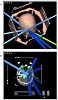
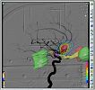
The final plan had to be executed after being finally reviewed and approved by primary radiation oncologist (Figure 1and 3). After radiation dose delivery, stereotactic frame was to be removed and patient to be discharged home on analgesics (as needed).
Stereotactic frame was fixed, and DSA was performed in all 13 patients of AVM planned for SRS. Mean age was 22 years (range 17 - 50 years); these included 7 males and 6 females. Intracranial hemorrhage as shown on CT scan brain was a presenting feature in 7 (54 %) of patients. 3 (23%) patients presented with epilepsy. Prior embolization using Onyx was done in 10 (77%) patients with large AVMs and of whom 7 patients having more than once undergone this procedure. The location of AVM was superficial in 9 (70%) and deep in brain in 4 (30%) patients. The mean AVM score was 0.97 with 3 patients having AVM score ≥ 1 with mean Spetzler-Martin grade of 2.7 and 8 (62%) patients having grade 3 or more. (AVM score= (0.1 Χ AVM volume in ml + 0.02 Χ age in years + 0.3 Χ AVM location); AVM location: 0 = frontal/temporal; 1 = parietal/occipital/intraventricular/ cerebellar/ callosal; 2 = basal ganglia / thalamus / brain stem). Median follow up was 30 months. Mean dose delivered was 21.7 Gy in single fraction. Complete obliteration of AVM nidus was achieved in 9 (70%) patients while 4 patients (30%) had partial obliteration. Six patients (67 %) achieved complete obliteration among 9 who had AVM score of less than 1. Post radiosurgery neurological deficit occurred in only one patient in form of right temporal field loss. The mean number of beams used was 9 (range 7-12). The mean nidus volume was 2.32 cc (range 0.7 - 4 cc). The mean prescribed maximum dose was 21.7 Gy (SD 1.95, range 15-22 Gy).Mean dose to brain stem, optic chiasm and optic nerves was 3.40Gy (range 0.11 – 12.2Gy), 0.55 Gy (range 0.04 – 2.66), and 0.41 Gy (range 0.01-2.66 Gy), respectively. There was no dose attenuation effect by prior Onyx embolization after radiosurgery. No other patients had symptomatic permanent sequelae related to SRS. All patients are leading an active and functional life. Patients and treatment characteristics are summarized in (Table 1).
Variables | N (%) |
Age at presentation | 22 yrs (Range: 17-50) |
Gender |
|
Male | 7 (54%) |
Female | 6 (46%) |
Hemorrhage at presentation |
|
Yes | 7 (54%) |
No | 6 (46%) |
Seizures at Presentation |
|
Yes | 3 (23%) |
No | 10 (77%) |
Times Embolization done |
|
None | 3 (23%) |
Once | 3 (23%) |
Twice | 4 (30%) |
Thrice | 3 (23%) |
Type of imaging done for assessment |
|
Angiogram alone | 3 (23%) |
MRA & Angiogram | 10 (77%) |
Site of AVM |
|
Frontoparietal | 4 (30%) |
Thalamic | 2 (15.4%) |
Temporal | 2 (15.4%) |
Occipital | 1 (7.7%) |
Basal ganglia | 1 (7.7%) |
Corpus callosum | 2 (15.4%) |
Vermian | 1 (7.7%) |
Location of AVM |
|
Superficial | 9 (70%) |
Deep | 4 (30%) |
Mean Spetzler-Martin grade |
|
I | 0 |
II | 5 (38.5%) |
III | 8 (61.5%) |
Treatment Characteristics |
|
No. of beams for SRS | 9 (Range: 7-12) |
Nidus volume | 2.32 cc (Range: 0.7-4) |
Prescribed Dose | 21.7 Gy (Range: 15-22) |
Follow up | 30 months (Range: 13-63) |
Obliteration rate | 9 (70%) |
Complete Partial | 4 (30%) |
Abbreviations:
AVM= Arteriovenous malformations,
cc= cubic centimeters,
Gy= Gray,
SRS= Stereotactic Radiosurgery.
Table 1 : Patients and treatment characteristics.
Cerebral AVMs are developmental malformations of the intracranial arteriovenous system. Usually they are asymptomatic but when symptomatic present mainly with headache and seizures. Because of their fragile walls and tendency for rupture, the major complication is bleeding. The annual rate of hemorrhage from AVMs is 2-4% while the lifetime risk is about 40%. There is a higher risk of subsequent bleeding up to 6
SRS is a viable and safe option in the management of small AVMs. Stringent case selection provides acceptable obliteration rates with minimal long-term toxicity. This study provides a platform for treating more patients having brain arteriovenous malformations with stereotactic radiosurgery technique. The choice of patients is critical depending mainly upon size of AVM and its location within the cranium. Obviously there is a need for collection of large database to fully explore the other prognostic and risk factors.
Dear Editorial Team, Clinical Medical Reviews and Reports. My experience with the journal was highly positive. The peer-review process was rigorous, constructive, and completed in a timely manner. The reviewers provided valuable comments that helped improve the quality and clarity of our manuscript. The editorial office was professional, responsive, and supportive throughout all stages of the publication process. Communication was clear and efficient, and any questions were addressed promptly. Overall, I found the journal to maintain high scientific standards and an excellent publication workflow. I would be pleased to consider submitting future work to this journal. Best wishes from, Elena Popa.

It was my pleasure to submit my testimonial concerning the Reviewer Board of our Scientific Journal “Brain and Neurological Disorders”. The Reviewers focused on some modifications and their contribution was helpful. The ladies of our Editorial Office were also supported my efforts. It was my honor to have such a co-operation and I am looking forward for more collaboration.

Dear Grace Pierce, Editorial Coordinator of Journal of Clinical Research and Reports, Thank you for the speedy and efficient peer review process. I appreciate the fact that your peer reviewers do not take months to respond like with some other journals. I would also like to thank the editorial office for responding quickly to my questions. It is an excellent journal. I plan to submit more manuscripts in the future. Best wishes from, Robert W. McGee

Dear Grace Pierce, Editorial Coordinator of Journal of Clinical Research and Reports, Working with you and your team on our recent publication in JCRR has been a truly wonderful and enjoyable experience. The responses were prompt, and the reviewers were patient, constructive, and highly professional. One reviewer in particular gave me the feeling that a professor was carefully reading and commenting on my coursework, which was deeply touching. The entire process was straightforward and hassle‑free, with no tedious online forms to complete. I highly recommend this journal. Best wishes from, DR Aibing Rao, Head of R&D

I Appreciate the Opportunity to Share my Experience with the Journal of Clinical Research and Reports. The peer review process was timely and constructive, and the feedback provided helped improve the quality of our manuscript. The editorial office was professional, responsive, and supportive throughout the process, ensuring smooth communication and efficient handling of the submission. Overall, it was a positive experience collaborating with your team.

Dear Mercy Grace, Editorial Coordinator of Obstetrics Gynecology and Reproductive Sciences, We would like to express our gratitude for your help at all stages of publishing and editing the article. The editors of the magazine answer all the necessary questions and help at every stage. We will definitely continue to cooperate and publish other works in the Obstetrics Gynecology and Reproductive Sciences! Best wishes from, Alla Konstantinovna Politova,
