
Case Report | DOI: https://doi.org/10.31579/2690-1897/148
1 Department of Maxillofacial Surgery Hospital of Specialities Rabat, Morocco.
2 Department of Otolaryngology and neck surgery Hospital of Specialities Rabat, Morocco.
3 Faculty of Medicine and Pharmacy of Rabat. Mohammed V University in Rabat, Rabat, Morocco.
*Corresponding Author: Rajaa El Azzouzi, Department of Maxillofacial Surgery Hospital of Specialities; CHU Ibn Sina, Av. Abderrahim Bouabid, Rabat-Morocco.
Citation: Rajaa El Azzouzi, Othmane Bouanani, Kawtar Ayyad, Malik Boulaadas, Houssayni leila Essakalli. (2023), The Role of Second Messengers in the Functioning of the Cell, J, Surgical Case Reports and Images 6(3); DOI:10.31579/2690-1897/148
Copyright: : © 2023, Rajaa El Azzouzi. This is a open access article distributed under the Creative Commons Attribution License, which permits unrestricted use, distribution, and reproduction in any medium, provided the original work is properly cited.
Received: 02 March 2023 | Accepted: 14 March 2023 | Published: 22 March 2023
Keywords: case report; histology; kikuchi fujimoto; lymphadenopathy
Kikuchi-Fujimoto's disease is a rare disease, little known by clinicians. It is a necrotizing lymphadenitis, affecting young subjects, with a female predominance. Lymphadenitis are localized, especially in the cervical region, but sometimes also diffuse. The lymph node biopsy allows the diagnosis by finding a necrotizing lymphadenitis with the presence of plasmacytoid monocytes and numerous cells in apoptosis (CD8+ T lymphocytes). The course is usually spontaneously favorable within a few weeks to a few months, only occasionally requiring brief corticosteroid therapy. We report the case of a young patient, who consulted for cervical lymphadenitis in whom the diagnosis of Kikuchi Fujimoto was retained.
Kikuchi's disease may reveal or evolve towards an autoimmune disease, in particular lupus, requiring a long-term clinicobiological follow-up.
Kikuchi-Fujimoto's disease or necrotizing histiocytic lymphadenitis is a benign pathology affecting essentially young women, of unknown etiology. Its diagnosis is primarily histological. It is an often unrecognized cause of cervical adenopathy associated with various clinical symptoms. Kikuchi's disease may reveal or evolve towards an autoimmune disease, in particular lupus, requiring a long-term clinical and biological follow-up.
Kikuchi-Fujimoto's disease or necrotizing histiocytic lymphadenitis is a benign pathology affecting essentially young women, of unknown etiology. Its diagnosis is primarily histological. It is an often unrecognized cause of cervical adenopathy associated with various clinical symptoms. Kikuchi's disease may reveal or evolve towards an autoimmune disease, in particular lupus, requiring a long-term clinical and biological follow-up.
We report the case of a 29-year-old female patient, without any notion of tuberculosis contagion or any past medical history of note, who consulted for the appearance of painful cervical lymphadenitis progressively increasing in size without any other associated signs in a context of fever and conservation of the general state.
Blood tests including immunological tests, chest x-ray and tuberculin test were all normal.
Ultrasound revealed bilateral necrotic lymphadenitis, the cavum was free at nasal endoscopy, a cervicotomy was performed and the histolopathologic evaluation completed by an immunohistochemical study allowed the diagnosis of nonsuppurative histiocytic and necrotizing lymphadenitis refering to Kikuchi Fujimoto disease.
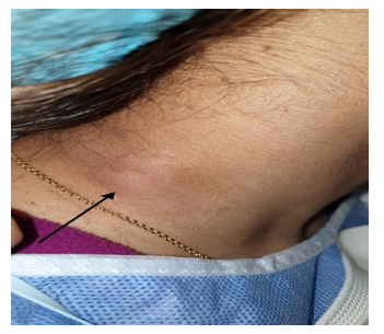
Figure 1: Image of the patient showing the cervical lymphadenitis.
Kikuchi-Fujimoto disease (KF) was first described in 1972. It is generally admitted to be a non-neoplastic disease of lymph node tropism, reactive lymphoid hyperplasia type. KF has been found mainly in young people with a very low recurrence rate (about 3%) [1]. May affect both sexes with female predominance [2]. Mostly observed in East Asia, sporadic cases have been found outside.
A viral or autoimmune cause of KF has been suggested. Some initial reports hinted at Yersinia enterocolitica and Toxoplasma gondii as possible causative agents of KF, mainly on the basis of positive serologic test results. But subsequent studies failed to support these hypotheses. In addition with these microorganisms, the histologic features of lymphadenitis associated clearly differ from those of KF [3,4,5].
To diagnose the disease quickly and accurately, clinical radiological, biological and histological analysis is essential. The clinical manifestations of KF are mainly fever, varying between 38 and 41 °C, lasting about 4 to 6 weeks, superficial lymphadenitis mainly in the neck, 0.5 to 3 cm in diameter, congestive maculo-papular rash, usually on the
trunk, limbs and cheeks, sometimes may be associated with mild hepatosplenomegaly with liver enlargement of about 0.5 to 2 cm. Because of the non-specificity of the symptomatology, other more specific analyses must be performed to avoid misdiagnosis and mistreatment.
An anatomo-pathological analysis of the lymphadenopathy biopsy should be performed. Showing extensive coagulative necrosis and histiocytosis. The histological features, such as clusters of plasma-like mononuclear cells with nuclear debris and crescent-shaped tissue cells, are indistinguishable from those of lymphoma, hence the value of complementing with an immunohistochemical study that can reveal MPO-positive and CD68-positive cells [6], such a pathological tool would be useful for diagnosis and rule out the differential diagnosis.
KF is a subacute disease, usually lasting 1-3 months, but can persist for up to 1 year [2]. There is no universally accepted treatment plan, as each case may be different. The primary treatment for KFD is to manage the disease by supporting the patient mentally and physically to accelerate relief of symptoms. Antibiotics are not effective, but their use in this case [7] prevents potential bacterial infections.
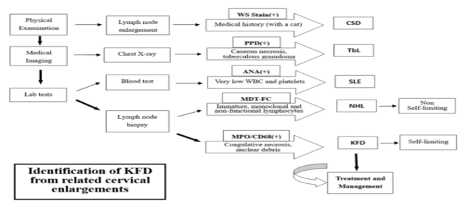
Figure 2: Decision tree of Kikuchi-Fujimoto disease [8].
Cervical lymphadenitis are a very frequent reason for consultation, requiring a wide range of paraclinical examinations in search of the etiology. Kikuchi-Fujimoto's disease is a rare cause and of elimination,
confirmed by histology, an autoimmune work-up is initiated and a long- term clinical-biological monitoring is imposed because of the risk of secondary appearance of autoimmune disease, especially lupus.
Free and informed consent has been given by the patient.
The authors declare no conflicts of interest.
Dear Editorial Team, Clinical Medical Reviews and Reports. My experience with the journal was highly positive. The peer-review process was rigorous, constructive, and completed in a timely manner. The reviewers provided valuable comments that helped improve the quality and clarity of our manuscript. The editorial office was professional, responsive, and supportive throughout all stages of the publication process. Communication was clear and efficient, and any questions were addressed promptly. Overall, I found the journal to maintain high scientific standards and an excellent publication workflow. I would be pleased to consider submitting future work to this journal. Best wishes from, Elena Popa.

It was my pleasure to submit my testimonial concerning the Reviewer Board of our Scientific Journal “Brain and Neurological Disorders”. The Reviewers focused on some modifications and their contribution was helpful. The ladies of our Editorial Office were also supported my efforts. It was my honor to have such a co-operation and I am looking forward for more collaboration.

Dear Grace Pierce, Editorial Coordinator of Journal of Clinical Research and Reports, Thank you for the speedy and efficient peer review process. I appreciate the fact that your peer reviewers do not take months to respond like with some other journals. I would also like to thank the editorial office for responding quickly to my questions. It is an excellent journal. I plan to submit more manuscripts in the future. Best wishes from, Robert W. McGee

Dear Grace Pierce, Editorial Coordinator of Journal of Clinical Research and Reports, Working with you and your team on our recent publication in JCRR has been a truly wonderful and enjoyable experience. The responses were prompt, and the reviewers were patient, constructive, and highly professional. One reviewer in particular gave me the feeling that a professor was carefully reading and commenting on my coursework, which was deeply touching. The entire process was straightforward and hassle‑free, with no tedious online forms to complete. I highly recommend this journal. Best wishes from, DR Aibing Rao, Head of R&D

I Appreciate the Opportunity to Share my Experience with the Journal of Clinical Research and Reports. The peer review process was timely and constructive, and the feedback provided helped improve the quality of our manuscript. The editorial office was professional, responsive, and supportive throughout the process, ensuring smooth communication and efficient handling of the submission. Overall, it was a positive experience collaborating with your team.

Dear Mercy Grace, Editorial Coordinator of Obstetrics Gynecology and Reproductive Sciences, We would like to express our gratitude for your help at all stages of publishing and editing the article. The editors of the magazine answer all the necessary questions and help at every stage. We will definitely continue to cooperate and publish other works in the Obstetrics Gynecology and Reproductive Sciences! Best wishes from, Alla Konstantinovna Politova,
