
Case report | DOI: https://doi.org/10.31579/2690-1919/478
1Division of Infectious Disease, The University of Tennessee Medical Center, Knoxville, TN USA.
2Sri Guru Ram Das Institution of Medical Sciences, Amritsar India.
3Division of Infectious Diseases, The Cleveland Clinic Foundation, Cleveland, OH USA.
*Corresponding Author: Supriya Singh MD, Department of Infectious Diseases, University of Tennessee Medical Center, 1932 Alcoa Highway; Building C, Suite 580; Knoxville, TN 37920 USA.
Citation: Supriya Singh, Vikrant Singh and Jona Banzon, (2025), Isolated Pulmonary Mucormycosis as a Complication of SARS CoV-2 Pneumonia-Case Report and Review of Literature., J Clinical Research and Reports, 18(2); DOI: 10.31579/2690-1919/478
Copyright: © 2025, Supriya Singh. This is an open access article distributed under the Creative Commons Attribution License, which permits unrestricted use, distribution, and reproduction in any medium, provided the original work is properly cited.
Received: 15 January 2025 | Accepted: 22 January 2025 | Published: 29 January 2025
Keywords: sars-cov-2; mucormycosis; steroids: mucor; pneumonia; amphotericin b; remdesivir
SARS-CoV-2 pneumonia is associated with multiple complications. Mucormycosis secondary to SARS-Cov-2 is however a less common entity and isolated lung involvement of mucor is even rarer. We present a case of SARS-CoV-2 infection in a pregnant young female with complicated pulmonary mucormycosis that lead to significant deterioration. We also describe series of similar reported cases after literature review, most of which had poor outcome despite effective treatment. We highlight the importance of timely imaging and diagnostic tests including bronchoscopy to diagnose and treat pulmonary mucor.
Recent advances in prevention and treatment of SARS-CoV-2 pneumonia have significantly decreased the morbidity and mortality associated with this disease. There are still certain conditions associated with SARS-CoV-2 like long covid syndrome [1] and post SARS-CoV-2 fungal pneumonias that significantly complicate the overall clinical outcome of the disease. There have been multiple cases and studies showing post SARS-CoV-2 Aspergillosis and associated increased mortality, but post SARS-CoV-2 associated isolated pulmonary mucormycosis is relatively rare. It is however associated with significantly poorer outcomes. Mucormycosis is a life-threatening disease that has high morbidity and mortality. It requires a high index of suspicion to diagnose secondary pulmonary mucormycosis in patients with SARS-CoV-2 pneumonia. In addition to aggressive reimaging, bronchoscopy and diagnostic tests are performed on the Bronchoalveolar Lavage (BAL) samples. Early diagnosis and timely management can improve the outcome in these patients. We present a case of an extremely complicated pulmonary mucormycosis after a recent SARS-CoV-2 infection and literature review of similarly reported scenarios.
A 31-year-old woman G4P2204 was admitted with respiratory failure. Her laboratory work showed that her hemoglobin was 9.7 g/dl, D-dimer 1349 ng/mL fibrinogen equivalent units procalcitonin 0.44 ng/mL, C reactive Protein 91.4 mg/L, normal white count and her nasal reverse transcription-polymerase chain reaction was positive for SARS-CoV-2. Her Chest x-ray showed bilateral patchy airspace opacities. She underwent emergent cesarean section, was intubated and placed on mechanical ventilation. She was treated with Remdesivir, dexamethasone, vancomycin and piperacillin-tazobactam. On day 11 she developed bilateral pneumothorces which required placement of emergent chest tubes. There was a new elevation in white blood count (16,000/ul) and inflammatory markers. She reached threshold of mechanical ventilatory support so additional support with extracorporeal membrane oxygenation (ECMO) was initiated. On day 15 her Computed Tomography (CT) chest showed a large multiloculated cavity (Figure 1). She underwent Bronchoscopy with BAL and the cultures were noted to be positive for Mucorales (Lichtheimia species). A diagnosis of Pulmonary Mucormycosis was made. There was no evidence of invasive disease on CT abdomen-pelvis, CT head and nasal endoscopy. No tissue pathology was obtained. She was started on treatment with amphotericin B and posaconazole. On day 21 she underwent tracheostomy and successful ECMO weaning in addition to the removal of bilateral chest tubes. Amphotericin B was discontinued, and she was discharged on supplemental oxygen through nasal cannula and oral posaconazole. She continued to show clinical and radiological improvement on outpatient follow-up in clinic visits.
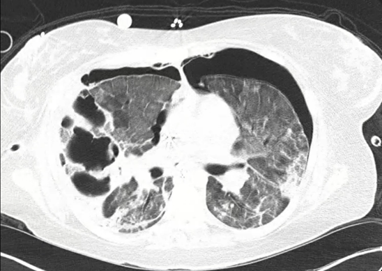
Figure 1: CT chest noted bilateral diffuse interstitial opacities with predominantly right sided large multiloculated cavitary lesion.
Discussion- There have been 10 other cases reported in literature with isolated pulmonary mucormycosis associated with SARS-CoV-2. All cases [2–10]. (Table 1) had significant clinical deterioration despite adequate antimicrobial treatment. Despite the recent advances in Covid 19 treatment and antifungal treatment, this cohort of patients shows that mucor remains a predictor of poor outcome in patients with recent SARS Cov-2 pneumonia. Whether it is the SARS Cov-2 infection itself that alters the immune system [11], or the immunomodulators like Glucocorticoids, Interleukin (IL)-6 inhibitors and monoclonal antibodies that are the risk factors for these secondary fatal infections remains unclear[12]. Another significant predisposing risk factor for infection noted in most of the case reports and case series is presence of underlying diabetes and malignancy.
| Location | Age/Gender | Timeline | Steroid Use | Other Medications | Complications | Microbiology | Diabetes | Medical Treatment | Surgery | Outcome |
| Arizona, USA | 49M | Post Cov (within a month) | YES-IV | Remdesivir, Tocilizumab | Bronchopleural fistula | Rhizopus (BAL Cultures) | NO | Amphotericin B | Yes - fistula repair, pleurodesis | Death |
| Italy | 66M | Post Cov (within a month) | NO | Hydroxychloroquine, Lopinavir-ritonavir | Respiratory failure, new cavitary lesions of lungs | Rhizopus (BAL Cultures) | NO | Amphotericin B, Isavuconazole | No | Death |
| California, USA | 79M | Post Cov (within a month) | YES-IV | Remdesivir | Respiratory failure, new cavitary lesions of lungs | Rhizopus arrhizus and Aspergillus fumigatus (BAL Cultures) | YES | Voriconazole followed by Amphotericin B | No | Vent dependent |
| Delaware, USA | 56M | Post Cov (within a month) | YES-ORAL | Convalescent plasma, Tocilizumab | Respiratory failure, loculated pleural effusions | Rhizopus azygosporus (Pleural fluid cultures and histopath) | NO | Amphotericin B | Yes - VATS | Death |
| Austria | 53M | Co-infection | YES-IV | Tocilizumab | Respiratory failure | Rhizopus microsporus(Tissue Bx) | NO (underlying Hematological malignancy | None | No | Death |
| India | 55M | Post Cov (within a month) | YES-IV | Remdesivir | New cavitary lesion | Rhizopus microspores (Sputum Cultures) | YES | Amphotericin B | No | Survived |
| India | 36F | Post Cov (within a month) | YES | Oxygen therapy, Corticosteroids | Multiple thin- to thick-walled cavities, air-fluid levels, loculated right hydropneumothorax, left pleural effusion | Aseptate, ribbon-like fungal hyphae (KOH wet mount) | NO | Amphotericin B | No | Survived |
| India | 45M | Post Cov (within a month) | NO | None | Patchy GGOs, consolidation with septal thickening, cavitary lesion, mild pneumothorax (resolved) | Aseptate, ribbon-like broad hyphae (KOH mount) | NO | Amphotericin B | No | Survived |
| France | 55M | Post Cov (within a month) | Unknown | Unknown | Respiratory failure, cavitary lesions | Aspergillus fumigatus and Rhizopus microsporus | unknown (Follicular Lymphoma post auto HCT) | Amphotericin B, Voriconazole | No | Death |
| Texas, USA | 44F | Post Cov(within a month | YES | Remdesivir | multiple cavitary lesions | Grocott’s methenamine silver stain- pauciseptated hyphae (zygomycetes) | Yes | Amphotericin B, Voriconazole, Micafungin | No | Death |
Table: 1
This review of literature shows that most of the patients who had pulmonary mucormycosis were males whereas our patient was female and rather younger without any significant comorbidity. The noted time frame for all cases to have superimposed mucormycosis was within a month of initial diagnosis of SARS-CoV-2 pneumonia. New radiological changes were usually seen 2 to 3 weeks from original diagnosis of viral infection. Most of the patients (seven) were given steroids, including our patient as a part of treatment for SARS-CoV-2 pneumonia. Three patients were given Tocilizumab[3,5,6]. Three patients had Type 2 diabetes, which in multiple studies, is associated with invasive fungal infections. Two of the patients had other immunocompromised state due to underlying hematological malignancy[6,7]. The above review showed that two patients who had diabetes got better with antifungal treatment, so it is unclear if diagnosis of diabetes overall impacts the outcome of the disease. Most of the patients had significant complications including bronchopulmonary fistula, pleural effusions and lung cavities which further complicated respiratory failure. Most of the mucor species were identified as Rhizopus although our patient had Lichtheimia species. Two patients had a co-infection with Aspergillus and Rhizopus. Despite treatment with antifungal agents, mortality rates were noted to be relatively high. Six out of these 10 patients expired within same hospitalization.
Mucormycosis is a rare complication and SARS-CoV-2 pneumonia with cases fewer than 1% of the patients infected with SARS-CoV 2 who end up with invasive mucormycosis. Isolated pulmonary mucormycosis remains extremely rare with just 9% of overall cases of invasive mucormycosis secondary to SARS-CoV-2 complication[13,14]. Some studies elaborate multiorgan involvement with mucormycosis (8) whereas our case noted an isolated pulmonary involvement of mucor which is a more unique as opposed to diffuse multiorgan involvement. The commonly isolated species that caused pulmonary mucormycosis belong to genera Rhizopus, Mucor or Lichtheimia. The primary source of infection could be inhalation of the fungal spores, ingestion or direct inoculation. Immunocompromised state like diabetes mellitus,
malignancies organ transplantation, use of immunomodulators are common risk factors for mucormycosis. Acute hyperglycemia has been noted in 50% of SARS-CoV-2 patients [15,16]. Viral pneumonia itself can induce acute diabetes and ketoacidosis by damaging pancreatic islet cells and increased resistance to insulin due to profound inflammatory reaction. In addition, use of glucocorticoids also increases blood glucose levels, insulin resistance and affects immune cells. This can cause an antagonism of macrophage differentiation; decrease in interleukin-1, interleukin-6 as well as tumor necrosis factor; pro inflammatory prostaglandins and leukotrienes production by macrophages. This subsequently can alter the activity of macrophages, suppress neutrophil adhesion to endothelial cells, impair lysosomal enzyme release, alter respiratory burst and chemotaxis, and increase the risk of infection. Iron overload and deferoxamine therapy are also notable risk factors for invasive Mucor infection because free iron incites the growth of Mucor spores. Vascular endothelial injury caused by the community-acquired viruses including SARS-CoV-2 also leads to an increased risk of superimposed fungal infection in this patient population. Hyperglycemia triggers overexpression of glucose regulated protein GRP 78 in the lumen of endoplasmic reticulum [17]. The CotH protein-kinase belonging to the spore coating protein family in Rhizopus acts as a ligand for GRP 78 helping the fungus to adhere and invade the endothelial and epithelial cells of respiratory tract [18]. Sabrili et al demonstrated significantly higher serum GRP78 levels in SARS-CoV-2 patients compared to a SARS-CoV-2 negative control group, hence suggesting pathogenic role of GRP 78 in pulmonary mucormycosis in SARS-CoV-2 patients[19].
Clinically Pulmonary mucor can present as a sudden worsening of respiratory status in a patient diagnosed with SARS-CoV-2 pneumonia with imaging consistent with consolidation, nodules, cavities, necrotizing opacities, recent sign with positive stains and cultures on BAL samples. The fewer number of isolated pulmonary mucormycosis might be secondary to delay in diagnosis including advanced imaging like CT scans as well as invasive procedures for diagnoses including bronchoscopies. This remains a limitation to rule out COVID associated pulmonary mucormycosis. Treatment for pulmonary mucormycosis includes liposomal amphotericin B as well as azoles including posaconazole and isavuconazole[20].
The diagnosis of isolated pulmonary mucormycosis is challenging given the smaller number of invasive diagnostic tests like bronchoscopies performed lack of rapid diagnostic testing and fewer autopsies. Liposomal amphotericin B, posaconazole and isavuconazole remain the main options for treatment along with surgical resection. Pathophysiology of invasive fungal infections including mucormycosis involves impaired T-cell function, impaired phagocytosis, abnormally enhances availability in iron due to displacement of protons by transferrin in diabetic ketoacidosis, increased availability of fungal heme oxygenase which facilitates iron uptake for its metabolism, use of glucocorticoids, IL–6 inhibitors and monoclonal antibodies. Multiple studies have shown that certain patient populations have an increased mortality not only with SARS Cov 2 virus but also with other community acquired viruses [21], and are associated with invasive fungal infections, fungal pneumonias or mycetoma especially those with hematological malignancies or transplant organs[22].
Dear Editorial Team, Clinical Medical Reviews and Reports. My experience with the journal was highly positive. The peer-review process was rigorous, constructive, and completed in a timely manner. The reviewers provided valuable comments that helped improve the quality and clarity of our manuscript. The editorial office was professional, responsive, and supportive throughout all stages of the publication process. Communication was clear and efficient, and any questions were addressed promptly. Overall, I found the journal to maintain high scientific standards and an excellent publication workflow. I would be pleased to consider submitting future work to this journal. Best wishes from, Elena Popa.

It was my pleasure to submit my testimonial concerning the Reviewer Board of our Scientific Journal “Brain and Neurological Disorders”. The Reviewers focused on some modifications and their contribution was helpful. The ladies of our Editorial Office were also supported my efforts. It was my honor to have such a co-operation and I am looking forward for more collaboration.

Dear Grace Pierce, Editorial Coordinator of Journal of Clinical Research and Reports, Thank you for the speedy and efficient peer review process. I appreciate the fact that your peer reviewers do not take months to respond like with some other journals. I would also like to thank the editorial office for responding quickly to my questions. It is an excellent journal. I plan to submit more manuscripts in the future. Best wishes from, Robert W. McGee

Dear Grace Pierce, Editorial Coordinator of Journal of Clinical Research and Reports, Working with you and your team on our recent publication in JCRR has been a truly wonderful and enjoyable experience. The responses were prompt, and the reviewers were patient, constructive, and highly professional. One reviewer in particular gave me the feeling that a professor was carefully reading and commenting on my coursework, which was deeply touching. The entire process was straightforward and hassle‑free, with no tedious online forms to complete. I highly recommend this journal. Best wishes from, DR Aibing Rao, Head of R&D

I Appreciate the Opportunity to Share my Experience with the Journal of Clinical Research and Reports. The peer review process was timely and constructive, and the feedback provided helped improve the quality of our manuscript. The editorial office was professional, responsive, and supportive throughout the process, ensuring smooth communication and efficient handling of the submission. Overall, it was a positive experience collaborating with your team.

Dear Mercy Grace, Editorial Coordinator of Obstetrics Gynecology and Reproductive Sciences, We would like to express our gratitude for your help at all stages of publishing and editing the article. The editors of the magazine answer all the necessary questions and help at every stage. We will definitely continue to cooperate and publish other works in the Obstetrics Gynecology and Reproductive Sciences! Best wishes from, Alla Konstantinovna Politova,
