
Case Report | DOI: https://doi.org/10.31579/2692-9759/079
1 Cardiology Service of the Regional University Hospital of Malaga. Spain.
2 Department of Pediatric Cardiology of the Regional University Hospital of Malaga. Spain.
*Corresponding Author: Patricia Ruiz Martín, Cardiology Service of the Regional University Hospital of Malaga. Spain.
Citation: Patricia R. Martín, Almudena O. Garrido, Juan I. Z. Argüelle, (2023), Isolated Left Ventricular Apical Hypoplasia: A Case Report, Cardiology Research and Reports. 5(4); DOI:10.31579/2692-9759/079
Copyright: © 2023, Patricia Ruiz Martín. This is an open-access article distributed under the terms of the Creative Commons Attribution License, which permits unrestricted use, distribution, and reproduction in any medium, provided the original author and source are credited.
Received: 08 November 2022 | Accepted: 26 June 2023 | Published: 10 July 2023
Keywords: left ventricle; right ventricle; chest x-ray; cardiac magnetic resonance; patients
We show the images of two cases of this rare pathology from our congenital heart disease unit.
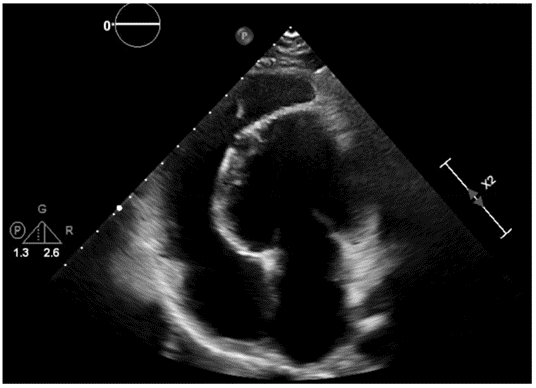
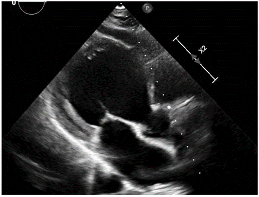
The first patient is a 14-year-old, asmathic male, whose only family history is an ostium secundum atrial septal defect in his mother. During echocardiographic screening performed during pregnancy, a spheroidal left ventricle (LV) and an elongated right ventricle (RV) were observed, confirming these findings at birth.
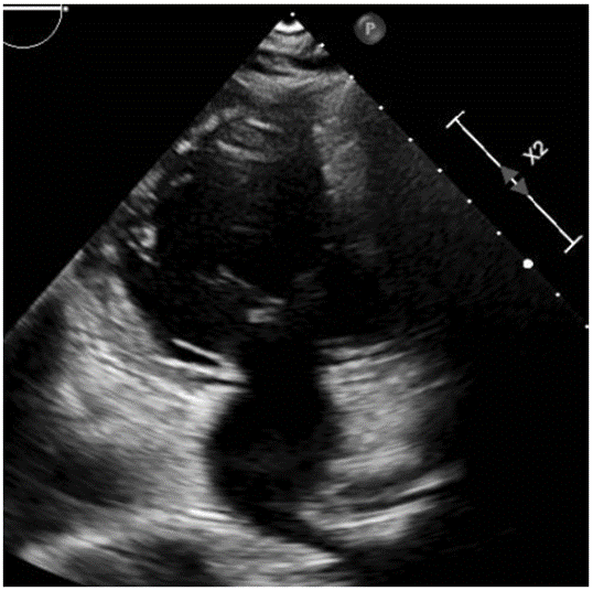
The second patient is an 8-year-old male, whose diagnostic suspicion also began at fetal screening and was confirmed at birth.
Both remain asymptomatic during follow-up, presenting with a normal physical examination, electrocardiogram, chest x-ray and Holter monitoring, so they do not receive any treatment. In addition, a cardiac magnetic resonance imaging (CMR) was performed that confirmed the suspected diagnosis.
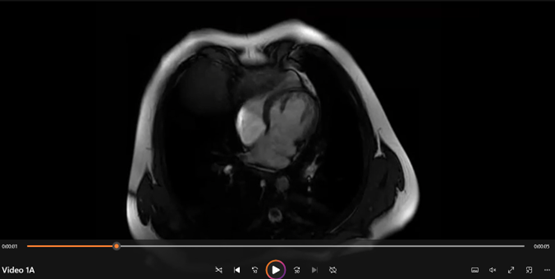
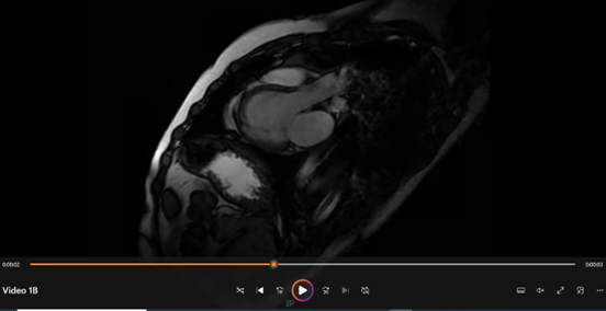
Isolated LV hypoplasia is a rare congenital pathology, characterized by the absence of the LV apex, resulting in a spheroidal ventricle, surrounded by the RV that forms the cardiac apex. It can be associated with an abnormal origin of the papillary muscles and the replacement of the apical myocardium by adipose tissue.
The clinic varies from the absence of symptoms to heart failure, arrhythmias and even sudden death. The differential diagnosis should be made with aneurysms, ventricular diverticulum and hypoplasic LV syndrome. Treatment consists of treat the signs and symptoms of heart failure and arrhythmias if they occur. Its etiology, treatment and prognosis are uncertain since there are few cases published in the literature.
Dear Editorial Team, Clinical Medical Reviews and Reports. My experience with the journal was highly positive. The peer-review process was rigorous, constructive, and completed in a timely manner. The reviewers provided valuable comments that helped improve the quality and clarity of our manuscript. The editorial office was professional, responsive, and supportive throughout all stages of the publication process. Communication was clear and efficient, and any questions were addressed promptly. Overall, I found the journal to maintain high scientific standards and an excellent publication workflow. I would be pleased to consider submitting future work to this journal. Best wishes from, Elena Popa.

It was my pleasure to submit my testimonial concerning the Reviewer Board of our Scientific Journal “Brain and Neurological Disorders”. The Reviewers focused on some modifications and their contribution was helpful. The ladies of our Editorial Office were also supported my efforts. It was my honor to have such a co-operation and I am looking forward for more collaboration.

Dear Grace Pierce, Editorial Coordinator of Journal of Clinical Research and Reports, Thank you for the speedy and efficient peer review process. I appreciate the fact that your peer reviewers do not take months to respond like with some other journals. I would also like to thank the editorial office for responding quickly to my questions. It is an excellent journal. I plan to submit more manuscripts in the future. Best wishes from, Robert W. McGee

Dear Grace Pierce, Editorial Coordinator of Journal of Clinical Research and Reports, Working with you and your team on our recent publication in JCRR has been a truly wonderful and enjoyable experience. The responses were prompt, and the reviewers were patient, constructive, and highly professional. One reviewer in particular gave me the feeling that a professor was carefully reading and commenting on my coursework, which was deeply touching. The entire process was straightforward and hassle‑free, with no tedious online forms to complete. I highly recommend this journal. Best wishes from, DR Aibing Rao, Head of R&D

I Appreciate the Opportunity to Share my Experience with the Journal of Clinical Research and Reports. The peer review process was timely and constructive, and the feedback provided helped improve the quality of our manuscript. The editorial office was professional, responsive, and supportive throughout the process, ensuring smooth communication and efficient handling of the submission. Overall, it was a positive experience collaborating with your team.

Dear Mercy Grace, Editorial Coordinator of Obstetrics Gynecology and Reproductive Sciences, We would like to express our gratitude for your help at all stages of publishing and editing the article. The editors of the magazine answer all the necessary questions and help at every stage. We will definitely continue to cooperate and publish other works in the Obstetrics Gynecology and Reproductive Sciences! Best wishes from, Alla Konstantinovna Politova,
