
Research | DOI: https://doi.org/10.31579/2640-1053/121
1 Professor of Cytogenetic. Head of Cytogenetic Lab at Radiation Biology Department, National Centre for the Radiation Research and Technology. Egyptian Atomic Energy Authority. Cairo, Egypt.
2 Assistant Professor of Physiology at Radiation Biology Department, National Centre for the Radiation Research and Technology. Egyptian Atomic Energy Authority. Cairo, Egypt.
*Corresponding Author: Sherien Abdelwhab Montaser, Assistant Professor of Physiology at Radiation Biology Department, National Centre for the Radiation Research and Technology. Egyptian Atomic Energy Authority. Cairo, Egypt.
Citation: Mahmoud Mohamed Ahmed and Sherien Abdelwhab Montaser. (2022). Irradiation Consequences from Chromosome Damages to Triggering of Diverse Immunological Responses. Cancer Research and Cellular Therapeutics. 6(4); Doi:10.31579/2640-1053/121
Copyright: © 2022 Sherien Abdelwhab Montaser, this is an open-access article distributed under the terms of the Creative Commons Attribution License, which permits unrestricted use, distribution, and reproduction in any medium, provided the original author and source are credited.
Received: 18 May 2022 | Accepted: 04 June 2022 | Published: 15 June 2022
Keywords: micronucleus; CBMN; innate immune response; interferon and TNF-α
Present work was designed to investigate DNA damages post irradiation via cytokinesis-block micronucleus (CBMN) test and its corresponding immunological response. Determination of interferon- α, β for innate, interferon-γ for acquired response, TNF- α and immunoglobulin concentration IgG & IgM.
Six human blood samples were divided into 4 groups (control & 3irradiated) which exposed to doses (0.5 - 2 and 4 Gy). Triplet blood samples (control and irradiated groups) were cultured for 72 hours after 1 hour of irradiation.
γ - Irradiation induced significant increase of IFN-α (innate immunology hallmark) in all experimental doses (0.5 -2.0 and 4.0 Gy). IFN-β also recorded significant increase with control at dose 4.0 Gy. The results showed significant increase in IFN-γ representing acquired immune response at 2.0 and 4.0 Gy. These results confirmed by exhibits increase in the level of IgG and IgM production. TNF-α late immune response started to give significant increase at 2.0 and 4.0 Gy.
TNF-α and IFN-β recorded significant difference when compared with control at 1.0 and 4Gy exposure also 4Gy group recorded significant increase compared with 1.0 Gy exposure.
INF-α recorded significant increase at all doses when compared with control and each other. IFN-γ recorded significant increase in 1.0 and 4.0 Gy when compared with control with no significant difference between them.
We conclude that immune system can sense when cells damaged. Mni which come from a variety of sources such as irradiation can lead to an immune response similar to that observed during viral infection.
Normal cells involvement several of DNA lesions per day over usual cellular metabolism, exposure to both radiotherapy or chemotherapy depend on DNA impairment to destroy tumour cells. In reaction to such injuries, the DNA damage response triggers cell-cycle checkpoints, initiates DNA healing mechanisms, or may be helps the clearance of irreparable cells. Work over the past decade has revealed broader influences of the DNA damage response, involving inflammatory gene expression following unresolved DNA damage, and immune surveillance of damaged or mutated cells [1].
There is fact has given rise to the term immunogenic death mediated by radiation, So, radiotherapy remains a basis of oncological treatment for many types of tumors. It has been demonstrated that ionizing radiation may exert interesting effects over the tumor microenvironment and the effectiveness of patients’ antitumor immune responses in the clinical setting even at distant sites [2].
Polly Matzinger in 1994 postulated that not only microenvironment but also the release an effective immune response is the induction of a threatening “danger signal,” related to the stress signs generated by the damaged tissue [3]. The most potent subcellular mediatoris called micronuclei (Mni), containing broken fragments of DNA or whole chromosomes that have been isolated away from the rest of the genome. Mni can initiate pro-inflammatory signalling cascades, or massively degrade to invoke distinct forms of genomic instability. It is also considered as a reservoir of immune-stimulatory nucleic acids [4].
IFN-α and -β (called type I interferon) were produced by many cell types following viral infection. Dendritic cells (DCs) have been identified as being the most potent producers of type I IFNs in response to antigen and have thus been called natural IFN-producing cells. On the other hand IFN-γ (the sole member of type II IFNs) is produced by activated natural killer (NK) cells and effector T cells, and thus a marker for induction of the acquired immune responses. In addition to their antiviral activities, IFN-α and IFN-β induce increased MHC class I expression on most uninfected cells, thus enhancing their resistance to NK cells as well as making newly infected cells more susceptible to killing by CD8+cytotoxic T cells. Finally, they activate NK cells, which contribute to early host responses to viral infections [5].
TNF-α produced by activated macrophages and mast cells, involved in inflammatory responses affecting damaging cells to activates endothelial cells and other cells of immune and normal cells [5].
MNi generation which followed the DNA damage, represented a mediator between the DNA damage response and immune recognition. So, several mechanisms through which DNA damage can be reach immune system crosstalk [4].
Five different types of biological responses have been induced after irradiation which known as 5Rs of radiobiology: faster repopulation, healing of sublethal damage, reoxygenation, rearrangement in the cell cycle and intrinsic radio sensitivity. Most cells which still survive for a limited period of time after irradiation and, through this period, they produce molecular signals that enhance the overexpression of specific genes that control the expression of cytokines, cell surface receptors, growth factors, and chemokine [6].
Whereas cell survival depends mainly on the previous responses and its ability to repair damaged DNA, being these phenomena of main importance in radiation treatments, as they may regulate the final effects over the surrounding microenvironment [7].
Additionally, it has been defined that the radiobiological response causes the stimulation of different T-cell lines, which generate the “switch-on” of the adaptive immune response [8]. These outcomes have led the scientific communities to discover the relation between the immunotherapy and the radiotherapy effects and be applied as synergic tools in cancer treatment protocols [9].
IgG is the most commonly found immunoglobulin in our body. It constitutes around 80% of the total antibody concentration in blood serum. Our body produces them in the later stages of the infection. And thus, it is a part of the secondary or delayed immune response [5]. The B-cells also manufacture IgG. Thus, at the time of their production, B-cells have to undergo class switching. For this reason, it takes time for B-cells to generate IgG.
While IgM is the most successful immunoglobulin, it is playing an important role in fixing the complement system. It’s the first antibody that our body produces after encountering the pathogen. Thus, it’s an essential part of the primary immune response. While diagnosis, they are the best indicators to determine whether a person is suffering from a disease or not. They fight with the foreign antigen until our body generates a more specific immune response in the form of IgG. They also in carrying out the immune reaction like agglutination and neutralisation [5].
In this work, we aim to study and verify the immunological consequences after irradiation which induced from chromosomal damages (MNi formations) leading to triggering of varied innate or acquired immunological responses.
Chemicals
Chemicals of the blood cultures were purchased from GIBCO-BRL, USA. FA, RBMI media, cytochalasin-B, heat-inactivated foetal calf serum (FCS), solvents and other chemicals were purchased from Sigma/ Aldrich Chemical Company, St. Louis (USA).
Blood sampling
To overcome possible inter-individual variability in response to treatments, blood sample was obtained from matching three healthy females (average age 35 years and non-smokers) who gave an informed consent for participation in the study. The donors were selected according to current International Programme on Chemical Safety rules for the observing of genotoxic effects of carcinogens in humans [10]. Venous blood was collected under sterile conditions in heparinised vacationer tubes (V= 5 ml, Becton Dickinson, USA) containing lithium heparin as anticoagulant.
Experimental design
Blood were divided into 4 groups; in each group, 3 samples were processed (n=3). Group 1: control blood (not exposed to irradiation), Group 2: blood irradiated with 0.5Gy. Group 3: blood irradiated with 2Gy. Group 4: blood irradiated with 4Gy. After irradiation blood samples were added to the culture CBMN and immunological parameters were measured in plasma and culture (72h).
Blood culture
To 0.5 ml of the whole blood, 5ml culture medium (RPMI-1640) supplemented with 20 FCS, 200 m M l-glutamine, penicillin 100 units/ ml and streptomycin 100 μg/ ml were added in 15ml conical tubes. Phyto-haemagglutinin-M (PHA-M); 0.2 ml was added to the culture to initiate cell division. Then, cells were incubated at 37°C.
Irradiation source
137Cs γ-rays Canadian source unit was belonging to the National Centre of Radiation Research and Technology, Egyptian Atomic Energy authority. The dose rate was 3.16 Gy/ min. The samples were kept at 37°C after irradiation immediately till the treatment started.
Cytokinesis-blocked micronucleus assay (CBMN):
The presence of MN in a BNC was assayed by blocking the cell at the cytokinesis stage by the method of Fenech [11] and Fenech et al. [12]. Blood culture was set as described previously. Cytochalasin- B (3μg/5ml culture) was added to the culture at 48h after the initiation. The cells were further incubated at 37°C for another 24 h.
At the harvesting time, after centrifugation at 1500 round per minute (rpm) for 5min, cell pellets treated with 5ml of mild hypotonic solution (0.1 M KCl) was used for 3min, and a further 10 min of centrifugation at 800 rpm. Next to another centrifugation, cells were washed carefully once with the fixative solution consist of [3:1 methanol: acetic acid (v/v)]. Then fixed cells were dropped very gently on clean labelled microscope slides then keep for 5 min to air- dried. After dried it collected in staining jar and stained with fresh prepared 10% Giemsa stain and keep in for 8min. then washed carefully with distilled water and started for investigation.
In each group a total of 1500 BNC (500 from each experiment) were scored and the frequency of cells with one (MN1) and two (MN2) micronuclei were recorded.
Biochemical investigations
-Determination of IFNs:
Assay of Interferon- α, β and –γ were performed by Myobiosource, USA. ELISA plate.
-Determination of TNF-α:
Human TNF-α ELISA Kit was purchased from ALPCO with Catalog Number: 45-TNFHU-E01.
-Determination of immunoglobulin’s:
IgG and IgM were estimated in culture supernatant by Radial Immune-diffusion method (RID), according to Lentner [13]. Kits was purchased from BINDRID which is a trademark of the binding site group Ltd. Birmingham, UK.
Radical Immune-diffusion
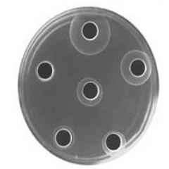
RID is a variation of the agar coated plastic plates. Precipitation technique is used in clinical immunology for the detection and quantitation of all classes of immunoglobulin’s complement, ceroplastic transferring and other serum components.
Data are presented as distribution analysis, means± S. E. and analysed using two ways analysis of variance “F” test according to Abramowitz and Stegun [14]. The level for statistical significance was P< 0>
As shown in Table (1), there are non-significant differences between the measured cytogenetic parameters of the 0.5Gy group when compared with the control group. The first scoring of 2 MNi and the significantly expression of the genetic damage (Mni) is at dose of 2 Gy. Moreover, the recorded MNi were directly proportional to the exposed radiation doses (0.5, 2, 4 Gy). The genetic damages were represented by Mono-MN, Bi-MN, and total MN.
It’s noticeable that in the 4 Gy group the total number of mononucleated cells recorded the lowest value, and on the contrary, the total number of binucleated recorded the highest value when compared with the other groups.
Table (2) showed that all immunological parameters recorded non-significant difference when 0.5 Gy group compared with control group except for IFN-α and IgG. It is well noticed that, there are extremely significant increase of all parameters in group 4 Gy except IgG& IgM.
TNF-α concentration recorded significantly increased by (7folds) at 4Gy when compared with control and (3folds) when compared with 2Gy.
About INF-α, it recorded significant increase at 0.5, 2 and 4 Gy γ-irradiationapplied when compared with control and each other while IFN-β levels started to be significant at 2 and 4 Gy. IFN-γ recorded significant increase in 2 and 4 Gy when compared with control group with no significant difference between them.
The responses of IgG& IgM post different gamma-irradiation doses showed fluctuation manner. For IgG gradually significant decreases at 0.5,2 and 4 Gy while IgM recorded spontaneous significant increase at 0.5 Gy then decreased significantly at both 2 and 4 Gy irradiation.
Table (3) showed that by using statistical correlation method, results showed positive correlation between micronucleus formation and both IFN-α, IFN-β and TNF-α. On the other hands there was negative correlation between micronucleus formation and IFN-γ, IgG and IgM.
Genomic instabilities and damages can be sensed by immune system via MNi which following aneuploidy and chromosome segregation [15] then it has been directly up-regulate the expression of natural killer cell receptors. In addition, ligands and the senescence-associated secretory phenotype (SASP) recruiting innate immune cells [16]. The SASP, usually defined as a pro-inflammatory secrete sand it is one of the comprehensively described processes of irradiation-induced cytokine secretions [17]. Large subset of SASP cytokines are reliant on DNA damage response which signalling for its production. Since of this, the SASP can considered an extracellular allowance of the DNA damage response that influences the microenvironment through paracrine signalling [18].
In the present work, Table (3) revealed positive correlation between MN formation and innate immune system response (IFN-α –β and TNF-α) post γ -irradiation,
Irradiated and extensively damaged cancer cells also suffering obvious DNA damage responses and initiating the pro-inflammatory SASP which can employee immune cells reaction for tumor dealing [19].
The role of the DNA damage response in transforming all previous extracellular communications may be uncover possible therapeutic targets helping anti-tumor immunity, and lead to a better approval for their role in carcinogenesis [18].
Interferon also up regulate the expression of major histocompatibility complex (MHC) class I and II molecules and are major activators of natural killer cells [20] which mainly produced from macrophages and neutrophils. In addition, IFN-α/β has recently been reported to be of importance in the amplification of dendritic cell responses [21] and in stimulating the persistence of activated lymphocytes [22] altogether associated with innate immune responses.
On contrary, IFN-γ exerts stimulatory effects on macrophage function and regulates the balance of cytokine production during immune responses [23]. Cellular sources of IFNs vary, with IFN-α being produced by cells of the lymphoid lineage, IFN-β being produced by epithelial and fibroblast cells [24], and IFN-γ being produced by T cells and large granular lymphocytes but also by macrophages and B cells [25].
Automatic or semi-automatic images analysis system can also performed using MN assay [26], and the scoring of many thousands of cells in short time may be possible. For many years, dicentrics and MNi measurements have been used for bio-dosimetry.
In the present study the frequency of MN formation was associated with immune-stimulating factors. MN recorded at different three doses (0.5 – 2 and 4 Gy) of ionizing irradiation. In conclusion, there is correlation between innate immunological response consequences compared with acquired immune response after irradiation due to formation of micronuclei in cells especially for white blood cells cultures studies which seen as abnormal or foreign cells. In addition, the potential fate for cells surviving checkpoints adaptation with unresolved DNA damage is the formation of MNi.
At mitosis, the acentric fragments or whole sheathing chromosomes are left behind at the metaphase plate as the rest of the genome separates, and they are not included within the newly forming nuclear envelopes at mitotic outlet [27]. As a substitute, they are confiscated into MNi, a fragment of double-stranded DNA covered in a version of a nuclear envelope, existed in in the cytosol at interphase and separate from the main nucleus [28]. Mni have developed as important features of and functional entities in cells that have experienced DNA damage, in at least two major ways: directly, when micronucleus envelope rupture exposes double-stranded DNA to the cytosol, where it is recognized by viral pattern recognition receptors and raised an inflammatory signalling program [29]; or/and indirectly, as micronucleation which initiating events in a flow of accumulating genomic instability, leading to the creation of neoantigens implicated in cancer immune editing. Mni generation following unresolved DNA damage therefore acts as a intermediary between the DNA damage response and immune detection [30].
Every diploid cell in the human body contains the same genes as every other cell. The only exceptions are lymphocytes, which differ from other cells and each other in the actual content of genes coding for their antigen-specific receptors.
Cells within an individual differ from one another because they transcribe and translate different genes. So the expression of specific pattern of genes determines the cell’s function. As all cells contain Ig genes but only B lymphocytes (and their differentiated form, plasma cells) express Ig genes and synthesize Ig molecules [5].
Chromatin bridge breakage is example for nuclear envelope rupture which considered another mechanism for double strands DNA exposure to the cytosol [30]. While double strands breaks can accumulate in the primary nucleus if these ruptures are not immediately repaired, which primes micronuclei formation in subsequent mitoses. Understanding the specific influences that result in death over damage repair in both the primary nucleus and mitochondria has long been an area of active research [31].
The UNSCEAR 2006 report [32] published considerable data on the effect of γ-irradiation on the immune system including both high and low dose effects and concentrated on the complex functional changes within the immune system in response to radiation. This was the first report released by an international organisation investigating radiation health effects which deserted the “classical” model that ionizing irradiation is purely immune suppressive [33]. Actually, this report suggested that ionizing irradiation is an immunomodulatory agent due to the massive amount and sometimes opposing pathways it can influence the immune system depending on various parameters such as dose, dose rate, health status, comorbidities, lifestyle, genetic background, age, and environmental co-stressors.
Also, pathway of immune recognition after MNi formation starts when MN envelopes are structurally rupture in interphase due to its fragility unalike to normal cells envelope and exposing ds DNA to the cytosol [34].
It is clear that noticeable gaps remain in our thoughtful of DNA damage and immune communications, and their role at both systemic and local levels. Future work is needed to fully interpreted the immune-mediated interaction to DNA damages, and whether this is useful to recognize in a clinical setting when treating a single lesion in metastatic disease and highlighting the specific cell-intrinsic systems that join DNA damages to immune action, which have the potential to be applied for clinical use.
Dear Editorial Team, Clinical Medical Reviews and Reports. My experience with the journal was highly positive. The peer-review process was rigorous, constructive, and completed in a timely manner. The reviewers provided valuable comments that helped improve the quality and clarity of our manuscript. The editorial office was professional, responsive, and supportive throughout all stages of the publication process. Communication was clear and efficient, and any questions were addressed promptly. Overall, I found the journal to maintain high scientific standards and an excellent publication workflow. I would be pleased to consider submitting future work to this journal. Best wishes from, Elena Popa.

It was my pleasure to submit my testimonial concerning the Reviewer Board of our Scientific Journal “Brain and Neurological Disorders”. The Reviewers focused on some modifications and their contribution was helpful. The ladies of our Editorial Office were also supported my efforts. It was my honor to have such a co-operation and I am looking forward for more collaboration.

Dear Grace Pierce, Editorial Coordinator of Journal of Clinical Research and Reports, Thank you for the speedy and efficient peer review process. I appreciate the fact that your peer reviewers do not take months to respond like with some other journals. I would also like to thank the editorial office for responding quickly to my questions. It is an excellent journal. I plan to submit more manuscripts in the future. Best wishes from, Robert W. McGee

Dear Grace Pierce, Editorial Coordinator of Journal of Clinical Research and Reports, Working with you and your team on our recent publication in JCRR has been a truly wonderful and enjoyable experience. The responses were prompt, and the reviewers were patient, constructive, and highly professional. One reviewer in particular gave me the feeling that a professor was carefully reading and commenting on my coursework, which was deeply touching. The entire process was straightforward and hassle‑free, with no tedious online forms to complete. I highly recommend this journal. Best wishes from, DR Aibing Rao, Head of R&D

I Appreciate the Opportunity to Share my Experience with the Journal of Clinical Research and Reports. The peer review process was timely and constructive, and the feedback provided helped improve the quality of our manuscript. The editorial office was professional, responsive, and supportive throughout the process, ensuring smooth communication and efficient handling of the submission. Overall, it was a positive experience collaborating with your team.

Dear Mercy Grace, Editorial Coordinator of Obstetrics Gynecology and Reproductive Sciences, We would like to express our gratitude for your help at all stages of publishing and editing the article. The editors of the magazine answer all the necessary questions and help at every stage. We will definitely continue to cooperate and publish other works in the Obstetrics Gynecology and Reproductive Sciences! Best wishes from, Alla Konstantinovna Politova,
