AUCTORES
Globalize your Research

Research Article | DOI: https://doi.org/10.31579/2641-5194/058
1Department of Health Sciences, Division of Science, Technology, Engineering, and Math, Friends University, Wichita, KS 67213, USA.
*Corresponding Author: Prince N. Agbedanu, Department of Health Sciences, Division of Science, Technology, Engineering, and Math, Friends University, Wichita, KS 67213, USA.
Citation: Troy B. Puga, Schafer J., Harris P., Branum G., Strasser N., Prince N. Agbedanu (2022), Investigation of Reactive Oxygen Species production in Human Hepatocytes. J. Gastroenterology Pancreatology and Hepatobilary Disorders. 6(2) DOI: 10.31579/2641-5194/058
Copyright: © 2022, Prince N. Agbedanu, This is an open access article distributed under the Creative Commons Attribution License, which permits unrestricted use, distribution, and reproduction in any medium, provided the original work is properly cited.
Received: 17 December 2021 | Accepted: 28 December 2021 | Published: 12 January 2022
Keywords: jaundice; reactive oxygen species; furosemide; morphine; bilirubin; hepatocyte; 2',7'-dichlorodihydrofluorescein diacetate; dichlorodihy drofluorescein diacetate; dimethyl sulfoxide; tert-butyl hydro peroxide
Abstract:
1. Aim/Background: Reactive oxygen species (ROS) have been identified as compounds responsible for producing cellular damage. The purpose of this research is to examine if there is production of reactive oxygen species through free radical intermediates within human hepatocytes treated with morphine, bilirubin, or furosemide. The investigation examines the early stages of biotransformation by measuring the levels of reactive oxygen species produced inside of the treated hepatocytes within the first and second hours of treatment. The experiment was designed upon a case of a jaundiced (elevated bilirubin) infant who received morphine and furosemide and later died through unknown mechanisms. The experiment looks to examine if these drug compounds could contribute to cellular damage. This can help to further understand the potential interactions and complications of free radical intermediates produced during the phases of biotransformation.
2. Method: Previously cultured human hepatocytes were washed by centrifugation and re-suspended in 1x supplemental buffer to a concentration of 1x10⁶ cells/mL and seeded in a dark clear bottom 96-well microplate at 100,000 stained cells/well. The cells were treated with either furosemide, morphine, bilirubin, a Tert-Butyl hydro peroxide (TBHP) positive control, or left as a background. Reactive oxygen generated in the presence of these agents were quantified by fluorescence excitation/emission measurement at 495nm/529nm. Fluorescence was measured at one and two hours. ROS generated convert 2',7'-dichlorodihydrofluorescein diacetate to 2',7'-dichlorodihydrofluorescein within the cells, which fluoresces. The fluorescence intensity detected is equivalent to the level of ROS generated. Wells that were untreated were used as blanks and subtracted from background and TBPH.
3. Results: Furosemide and Morphine did not produce statistically significant levels of ROS (p >0.05) above the background in both hours 1 and 2 of biotransformation and ROS measurement (Figure 1). Although Bilirubin did not produce statistically significant (p >0.05) levels of ROS above the background (Figure 2) during the first hour, it did produce statistically significant levels in the second hour of biotransformation. Each compound’s level of ROS was reduced during the second hour, signaling the removal of intermediate ROS metabolites (Figure 2). The production of ROS in each compound signifies that there is biotransformation to an intermediate that produces ROS.
4. Conclusion: The production of ROS above the background by each of the compounds shows there is an intermediate free radical compound that is produced during the biotransformation of each compound [21]. In this study, although furosemide and morphine did not produce statistically significant levels of ROS in both hours of biotransformation, bilirubin did produce significant levels of ROS in the second hour of biotransformation. This finding is in line with previous studies that shows morphine to offer protective effects against ROS production [16, 17]; and bilirubin demonstrating deleterious production of ROS at high doses [18]. Further work must be done to examine the correlation between the levels of ROS and extent of hepatocellular damage.
H2DCFDA: 2',7'-dichlorodihydrofluorescein diacetate
DFCDA: dichlorodihydrofluorescein diacetate
TBHP: Tert-Butyl Hydroperoxide
DMSO: dimethyl sulfoxide
PBS: Phosphate buffered saline
This investigation is based upon the story of a jaundiced infant who was administered furosemide and morphine (while on supplemental oxygen), and later died due to unknown mechanisms. The infant presented with high levels of bilirubin shortly after birth, noted through jaundice (yellowing of the skin and sclera). Jaundice occurs due to a build-up of bilirubin that is not metabolized through conjugation and excretion in the urine (urobilinogen) or stool (stercobilinogen) [1]. Bilirubin is conjugated by the enzyme UDP-Glucuronosyltransferase within hepatocytes [1]. It is common for neonates to have elevated levels of bilirubin through physiologic neonatal jaundice, due to low levels of the enzyme UDP-Glucuronosyltransferase at the beginning of life [2]. As the patient’s condition became severe, it became necessary under the Physician’s determination to provide supplemental oxygen, furosemide, and morphine.
Many xenobiotics have been shown to undergo biotransformation in the liver through a two-phase transformation. Phase 1 typically involves the processing of the xenobiotic through the CYP450 family of enzymes. Phase 2 typically involves the conjugation, sulfonation, or acetylation of the xenobiotic intermediate [3] although, these phases can occur out of order. The purpose of biotransformation is to deactivate potentially toxic substances within the body [3]. Biotransformation can also transform compounds into a toxic form [3]. Production of a toxic intermediate can affect the body and is dependent on several factors, such as how quickly it can be metabolized to a less toxic compound [3]. The free radical intermediates may produce Reactive Oxygen Species (ROS). ROS are unstable molecules composed of oxygen free radicals that can produce numerous random reactions [4]. ROS is formed in mitochondria as a side-product of the electron transport chain [5]. At low, biological levels, ROS is involved in immune protection and cell signalling [6]. Many life forms, including mitochondria, protect themselves from the toxic effects of ROS by utilizing antioxidants [7]. When ROS production in cells exceeds biological limits, it is capable of damaging macromolecules and causing cell death [8]. This experiment investigates the levels of ROS produced in the presence of furosemide, morphine, and bilirubin. The study will examine the biotransformation of each compound to determine if there is production of free radical intermediates that may produce cellular damage.
Buffers
Fetal Bovine Serum (2ml) was added to 18mL of 1x PBS buffer to create a supplemental buffer. A 20 micromolar solution of 2',7'-dichlorodihydrofluorescein diacetate (H2DCFDA) was prepared by adding 2.5 microliters of 20mM of dichlorodihydrofluorescein diacetate (DFCDA) in dimethyl sulfoxide (DMSO) to 2.5mL solution of a previously prepared 1x PBS buffer [10]. In addition, a tert-Butyl hydroperoxide (TBHP) solution was prepared as a positive control, by adding 4.5 microliters of 55mM TBHP to 2.5mL of 1x supplemental buffer, yielding a 100 micromolar solution [10].
ROS Detection Assay
ROS production was detected using the DCFDA/H2DCFDA - Cellular ROS Assay Kit (ab113851) by abcam [10]. Human Suspension Hepatocytes (Gibco™ Human Suspension Hepatocytes, Metabolism Qualified) were maintained in William’s Medium E (Gibco, A1217601) in the presence of 4% CO2 and media changed at 48-hour intervals. The media was supplemented with the Gibco Primary Hepatocyte Maintenance Supplements (Gibco CM4000). The cultured cell suspensions were washed by centrifugation in phosphate buffer solution [10]. The cells were stained with the DCFDA solution at a concentration of 1x10⁶ cells/mL and incubated at 37 degrees Celsius for 30 minutes in the dark. The cells were washed by centrifugation with 1x buffer to remove excess dye. The washed cells were re-suspended in 1x supplemental buffer to a concentration of 1x10⁶ cells/mL and seeded in a dark clear bottom 96-well microplate at 100,000 stained cells/well. The cells were treated with furosemide, morphine, bilirubin, a TBPH positive control, or left as a background. Reactive oxygen generated in the presence of these agents were quantified by fluorescence excitation/emission measurement at 495nm/529nm. ROS generated convert 2',7'-dichlorodihydrofluorescein diacetate to 2',7'-dichlorodihydrofluorescein within the cells which fluoresces. The fluorescence intensity detected is equivalent to the level of ROS generated. Fluorescence was measured at one and two hours. Wells untreated were measured as blanks and subtracted from background and TBPH [10].
Data analysis
An unpaired t-test (Suppl. Table 1.) was performed to compare the means of each of the compounds to the background to test for significance at hours 1 and 2.
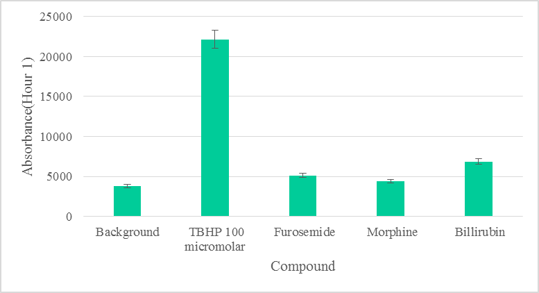
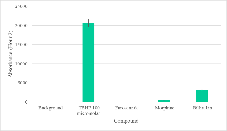
Furosemide, Morphine, or Bilirubin did not produce statistically significant levels of ROS (p >0.05) above the background within the first hour of biotransformation (Figure 1). ROS due to Furosemide was completely depleted in the second hour. Morphine did not produce statistically significant (p >0.05) levels of ROS during the second hour above the background (Figure 2). ROS levels from Bilirubin in the second hour, however, were found to be statistically significant. ROS produced by each compound was depleted to various degrees in the second hour, suggesting the removal of intermediate ROS metabolites (Figure 2). The production of ROS in each compound also signifies that there was biotransformation to an intermediate that produces ROS [21].
Furosemide is a diuretic use to treat hypertension, with a unique sparing effect on the clearance of potassium [11], by blocking Na+, K+, 2Cl− cotransporter (NKCC2) in the thick ascending limb (TAL) of the loop of Henle [12,13]. Despite its sparing effects on potassium, furosemide is known to induce a compartmentalized oxidative stress in hepatic mitochondria [14]. In this study, we have used whole liver cells without isolating mitochondria. We aim to mimic physiological conditions as much as possible, although there is still a limitation to the use of liver cells in assays, such as this type. The low levels of ROS detected due to furosemide may be attributable to the cellular environmental differences compared to organelle compartments (e.g., the mitochondria). Previous studies have shown that mitochondria is a key source of ROS production [19], hence, ROS levels in the mitochondria and its surroundings may be higher compared to remote areas in whole cells [22]. However, given our goal to mimic physiological conditions of the subject in question, we have used whole liver cells, as opposed to isolated mitochondria.
At low concentration, morphine is found to have promoted cell proliferation and suppressed nicotine-induced cell death in PC12 cells [15, 16]. It was also demonstrated to have neuroprotective roles in Parkinson’s disease models by the improvement of mitochondrial function via the inhibition of ROS production [16, 17]. Although furosemide has the potential to induce ROS, morphine has been shown to inhibit oxidative stress via the improvement of mitochondrial function. This antagonistic effect of morphine on ROS generation [16, 17] explain the very low and insignificant levels of ROS detected compared to background (Figure 1).
The detection of ROS above background levels in our experiments suggests the production of an intermediate compound—endogenous highly reactive oxygen-bearing molecule [20] during biotransformation. However, the insignificant levels of free radical intermediate due to furosemide, and the possible antagonistic effect of morphine on ROS production [16, 17] by furosemide, may not have allowed high enough levels of ROS in the subject to induce cellular damage.
At low doses, bilirubin in its unconjugated form has antioxidant effects, but at high doses produces deleterious neurological damage and death due to oxidative stress [18]. Although ROS detected due to bilirubin was also insignificant in the first hour, ROS levels due to bilirubin were statistically significant in the second hour (Figure 1, Suppl. Table 1) and clinically may be implicated in cell death [23]. Based on our data, we can safely rule out significant ROS production due to furosemide and morphine (Figure 1).
The biologically tolerable levels of bilirubin have been documented to be as high as 12.9 mg/dL among newborns [22]. Our results suggest that 100 micromolar (1.131 mg/dL) of bilirubin could induce the production of significant levels of ROS within two hours in hepatocytes, in vitro. This result from a direct cellular exposure to biliubin in vitro, an outcome which may be different from whole body exposure and that explains why the body tolerates as high as 12.9mg/dL [22]. Nonetheless, the damaging effect of jaundice are known to be cumulative [24]. Although the body’s homeostasis may allow it to tolerate such levels, the cumulative effects may not be immediately obvious.
We have not performed cell death assays to determine the possibility of hepatocellular damage due to these compounds in isolation. We aimed to determine the production of ROS in the presence of furosemide, morphine and bilirubin. We found the levels of ROS produced due to furosemide and morphine in both hours of biotransformation were statistically insignificant compared to background levels of ROS. We found the level of ROS produced due to bilirubin in the second hour of biotransformation to be statistically significant. Based on the level of ROS production, we conclude that cell death may be likely due to bilirubin and less likely due to furosemide or morphine. The minimum level of free radical accumulation needed to cause cellular damage due to bilirubin, especially, must be further investigated along with cell death assays.
The experiments were performed in vitro rather than in vivo. There could be potential differences in the effects of the compounds in vivo. Further investigation could not be performed at this time due to budget constraints. We encourage other researchers to continue investigations into these compounds in vivo to explore these possibilities.
Further work must be done to examine the correlation between the levels of ROS and the extent of cell damage.
We thank the Friends University VPAA office, and the division Chair, Dr. Nora Strasser, for the provision of starter funds to support research involving undergraduates. We thank Ms. Amy Morgan for the procurement of equipment and materials needed to execute all projects
G.B. conceived the project. P.N.A, T.P, J.S. and P.H. designed the experiments. T.P., J.S., and P.H. performed all experiments. T.P. and N.S. analyzed the data. T.P. wrote the manuscript. P.N.A. and G.B. edited and proofread the manuscript. P.N.A. procured funding.
The authors of this article do not have any conflicts of interest regarding this research. The authors did not discriminate any participants or aspects of research based on race, gender, or religion.
Clearly Auctoresonline and particularly Psychology and Mental Health Care Journal is dedicated to improving health care services for individuals and populations. The editorial boards' ability to efficiently recognize and share the global importance of health literacy with a variety of stakeholders. Auctoresonline publishing platform can be used to facilitate of optimal client-based services and should be added to health care professionals' repertoire of evidence-based health care resources.

Journal of Clinical Cardiology and Cardiovascular Intervention The submission and review process was adequate. However I think that the publication total value should have been enlightened in early fases. Thank you for all.

Journal of Women Health Care and Issues By the present mail, I want to say thank to you and tour colleagues for facilitating my published article. Specially thank you for the peer review process, support from the editorial office. I appreciate positively the quality of your journal.

Journal of Clinical Research and Reports I would be very delighted to submit my testimonial regarding the reviewer board and the editorial office. The reviewer board were accurate and helpful regarding any modifications for my manuscript. And the editorial office were very helpful and supportive in contacting and monitoring with any update and offering help. It was my pleasure to contribute with your promising Journal and I am looking forward for more collaboration.

We would like to thank the Journal of Thoracic Disease and Cardiothoracic Surgery because of the services they provided us for our articles. The peer-review process was done in a very excellent time manner, and the opinions of the reviewers helped us to improve our manuscript further. The editorial office had an outstanding correspondence with us and guided us in many ways. During a hard time of the pandemic that is affecting every one of us tremendously, the editorial office helped us make everything easier for publishing scientific work. Hope for a more scientific relationship with your Journal.

The peer-review process which consisted high quality queries on the paper. I did answer six reviewers’ questions and comments before the paper was accepted. The support from the editorial office is excellent.

Journal of Neuroscience and Neurological Surgery. I had the experience of publishing a research article recently. The whole process was simple from submission to publication. The reviewers made specific and valuable recommendations and corrections that improved the quality of my publication. I strongly recommend this Journal.

Dr. Katarzyna Byczkowska My testimonial covering: "The peer review process is quick and effective. The support from the editorial office is very professional and friendly. Quality of the Clinical Cardiology and Cardiovascular Interventions is scientific and publishes ground-breaking research on cardiology that is useful for other professionals in the field.

Thank you most sincerely, with regard to the support you have given in relation to the reviewing process and the processing of my article entitled "Large Cell Neuroendocrine Carcinoma of The Prostate Gland: A Review and Update" for publication in your esteemed Journal, Journal of Cancer Research and Cellular Therapeutics". The editorial team has been very supportive.

Testimony of Journal of Clinical Otorhinolaryngology: work with your Reviews has been a educational and constructive experience. The editorial office were very helpful and supportive. It was a pleasure to contribute to your Journal.

Dr. Bernard Terkimbi Utoo, I am happy to publish my scientific work in Journal of Women Health Care and Issues (JWHCI). The manuscript submission was seamless and peer review process was top notch. I was amazed that 4 reviewers worked on the manuscript which made it a highly technical, standard and excellent quality paper. I appreciate the format and consideration for the APC as well as the speed of publication. It is my pleasure to continue with this scientific relationship with the esteem JWHCI.

This is an acknowledgment for peer reviewers, editorial board of Journal of Clinical Research and Reports. They show a lot of consideration for us as publishers for our research article “Evaluation of the different factors associated with side effects of COVID-19 vaccination on medical students, Mutah university, Al-Karak, Jordan”, in a very professional and easy way. This journal is one of outstanding medical journal.

Dear Hao Jiang, to Journal of Nutrition and Food Processing We greatly appreciate the efficient, professional and rapid processing of our paper by your team. If there is anything else we should do, please do not hesitate to let us know. On behalf of my co-authors, we would like to express our great appreciation to editor and reviewers.

As an author who has recently published in the journal "Brain and Neurological Disorders". I am delighted to provide a testimonial on the peer review process, editorial office support, and the overall quality of the journal. The peer review process at Brain and Neurological Disorders is rigorous and meticulous, ensuring that only high-quality, evidence-based research is published. The reviewers are experts in their fields, and their comments and suggestions were constructive and helped improve the quality of my manuscript. The review process was timely and efficient, with clear communication from the editorial office at each stage. The support from the editorial office was exceptional throughout the entire process. The editorial staff was responsive, professional, and always willing to help. They provided valuable guidance on formatting, structure, and ethical considerations, making the submission process seamless. Moreover, they kept me informed about the status of my manuscript and provided timely updates, which made the process less stressful. The journal Brain and Neurological Disorders is of the highest quality, with a strong focus on publishing cutting-edge research in the field of neurology. The articles published in this journal are well-researched, rigorously peer-reviewed, and written by experts in the field. The journal maintains high standards, ensuring that readers are provided with the most up-to-date and reliable information on brain and neurological disorders. In conclusion, I had a wonderful experience publishing in Brain and Neurological Disorders. The peer review process was thorough, the editorial office provided exceptional support, and the journal's quality is second to none. I would highly recommend this journal to any researcher working in the field of neurology and brain disorders.
Dear Agrippa Hilda, Journal of Neuroscience and Neurological Surgery, Editorial Coordinator, I trust this message finds you well. I want to extend my appreciation for considering my article for publication in your esteemed journal. I am pleased to provide a testimonial regarding the peer review process and the support received from your editorial office. The peer review process for my paper was carried out in a highly professional and thorough manner. The feedback and comments provided by the authors were constructive and very useful in improving the quality of the manuscript. This rigorous assessment process undoubtedly contributes to the high standards maintained by your journal.

International Journal of Clinical Case Reports and Reviews. I strongly recommend to consider submitting your work to this high-quality journal. The support and availability of the Editorial staff is outstanding and the review process was both efficient and rigorous.

Thank you very much for publishing my Research Article titled “Comparing Treatment Outcome Of Allergic Rhinitis Patients After Using Fluticasone Nasal Spray And Nasal Douching" in the Journal of Clinical Otorhinolaryngology. As Medical Professionals we are immensely benefited from study of various informative Articles and Papers published in this high quality Journal. I look forward to enriching my knowledge by regular study of the Journal and contribute my future work in the field of ENT through the Journal for use by the medical fraternity. The support from the Editorial office was excellent and very prompt. I also welcome the comments received from the readers of my Research Article.

Dear Erica Kelsey, Editorial Coordinator of Cancer Research and Cellular Therapeutics Our team is very satisfied with the processing of our paper by your journal. That was fast, efficient, rigorous, but without unnecessary complications. We appreciated the very short time between the submission of the paper and its publication on line on your site.

I am very glad to say that the peer review process is very successful and fast and support from the Editorial Office. Therefore, I would like to continue our scientific relationship for a long time. And I especially thank you for your kindly attention towards my article. Have a good day!

"We recently published an article entitled “Influence of beta-Cyclodextrins upon the Degradation of Carbofuran Derivatives under Alkaline Conditions" in the Journal of “Pesticides and Biofertilizers” to show that the cyclodextrins protect the carbamates increasing their half-life time in the presence of basic conditions This will be very helpful to understand carbofuran behaviour in the analytical, agro-environmental and food areas. We greatly appreciated the interaction with the editor and the editorial team; we were particularly well accompanied during the course of the revision process, since all various steps towards publication were short and without delay".

I would like to express my gratitude towards you process of article review and submission. I found this to be very fair and expedient. Your follow up has been excellent. I have many publications in national and international journal and your process has been one of the best so far. Keep up the great work.

We are grateful for this opportunity to provide a glowing recommendation to the Journal of Psychiatry and Psychotherapy. We found that the editorial team were very supportive, helpful, kept us abreast of timelines and over all very professional in nature. The peer review process was rigorous, efficient and constructive that really enhanced our article submission. The experience with this journal remains one of our best ever and we look forward to providing future submissions in the near future.

I am very pleased to serve as EBM of the journal, I hope many years of my experience in stem cells can help the journal from one way or another. As we know, stem cells hold great potential for regenerative medicine, which are mostly used to promote the repair response of diseased, dysfunctional or injured tissue using stem cells or their derivatives. I think Stem Cell Research and Therapeutics International is a great platform to publish and share the understanding towards the biology and translational or clinical application of stem cells.

I would like to give my testimony in the support I have got by the peer review process and to support the editorial office where they were of asset to support young author like me to be encouraged to publish their work in your respected journal and globalize and share knowledge across the globe. I really give my great gratitude to your journal and the peer review including the editorial office.

I am delighted to publish our manuscript entitled "A Perspective on Cocaine Induced Stroke - Its Mechanisms and Management" in the Journal of Neuroscience and Neurological Surgery. The peer review process, support from the editorial office, and quality of the journal are excellent. The manuscripts published are of high quality and of excellent scientific value. I recommend this journal very much to colleagues.

Dr.Tania Muñoz, My experience as researcher and author of a review article in The Journal Clinical Cardiology and Interventions has been very enriching and stimulating. The editorial team is excellent, performs its work with absolute responsibility and delivery. They are proactive, dynamic and receptive to all proposals. Supporting at all times the vast universe of authors who choose them as an option for publication. The team of review specialists, members of the editorial board, are brilliant professionals, with remarkable performance in medical research and scientific methodology. Together they form a frontline team that consolidates the JCCI as a magnificent option for the publication and review of high-level medical articles and broad collective interest. I am honored to be able to share my review article and open to receive all your comments.

“The peer review process of JPMHC is quick and effective. Authors are benefited by good and professional reviewers with huge experience in the field of psychology and mental health. The support from the editorial office is very professional. People to contact to are friendly and happy to help and assist any query authors might have. Quality of the Journal is scientific and publishes ground-breaking research on mental health that is useful for other professionals in the field”.

Dear editorial department: On behalf of our team, I hereby certify the reliability and superiority of the International Journal of Clinical Case Reports and Reviews in the peer review process, editorial support, and journal quality. Firstly, the peer review process of the International Journal of Clinical Case Reports and Reviews is rigorous, fair, transparent, fast, and of high quality. The editorial department invites experts from relevant fields as anonymous reviewers to review all submitted manuscripts. These experts have rich academic backgrounds and experience, and can accurately evaluate the academic quality, originality, and suitability of manuscripts. The editorial department is committed to ensuring the rigor of the peer review process, while also making every effort to ensure a fast review cycle to meet the needs of authors and the academic community. Secondly, the editorial team of the International Journal of Clinical Case Reports and Reviews is composed of a group of senior scholars and professionals with rich experience and professional knowledge in related fields. The editorial department is committed to assisting authors in improving their manuscripts, ensuring their academic accuracy, clarity, and completeness. Editors actively collaborate with authors, providing useful suggestions and feedback to promote the improvement and development of the manuscript. We believe that the support of the editorial department is one of the key factors in ensuring the quality of the journal. Finally, the International Journal of Clinical Case Reports and Reviews is renowned for its high- quality articles and strict academic standards. The editorial department is committed to publishing innovative and academically valuable research results to promote the development and progress of related fields. The International Journal of Clinical Case Reports and Reviews is reasonably priced and ensures excellent service and quality ratio, allowing authors to obtain high-level academic publishing opportunities in an affordable manner. I hereby solemnly declare that the International Journal of Clinical Case Reports and Reviews has a high level of credibility and superiority in terms of peer review process, editorial support, reasonable fees, and journal quality. Sincerely, Rui Tao.

Clinical Cardiology and Cardiovascular Interventions I testity the covering of the peer review process, support from the editorial office, and quality of the journal.

Clinical Cardiology and Cardiovascular Interventions, we deeply appreciate the interest shown in our work and its publication. It has been a true pleasure to collaborate with you. The peer review process, as well as the support provided by the editorial office, have been exceptional, and the quality of the journal is very high, which was a determining factor in our decision to publish with you.

The peer reviewers process is quick and effective, the supports from editorial office is excellent, the quality of journal is high. I would like to collabroate with Internatioanl journal of Clinical Case Reports and Reviews journal clinically in the future time.

Clinical Cardiology and Cardiovascular Interventions, I would like to express my sincerest gratitude for the trust placed in our team for the publication in your journal. It has been a true pleasure to collaborate with you on this project. I am pleased to inform you that both the peer review process and the attention from the editorial coordination have been excellent. Your team has worked with dedication and professionalism to ensure that your publication meets the highest standards of quality. We are confident that this collaboration will result in mutual success, and we are eager to see the fruits of this shared effort.

Dear Dr. Jessica Magne, Editorial Coordinator 0f Clinical Cardiology and Cardiovascular Interventions, I hope this message finds you well. I want to express my utmost gratitude for your excellent work and for the dedication and speed in the publication process of my article titled "Navigating Innovation: Qualitative Insights on Using Technology for Health Education in Acute Coronary Syndrome Patients." I am very satisfied with the peer review process, the support from the editorial office, and the quality of the journal. I hope we can maintain our scientific relationship in the long term.

Dear Monica Gissare, - Editorial Coordinator of Nutrition and Food Processing. ¨My testimony with you is truly professional, with a positive response regarding the follow-up of the article and its review, you took into account my qualities and the importance of the topic¨.

Dear Dr. Jessica Magne, Editorial Coordinator 0f Clinical Cardiology and Cardiovascular Interventions, The review process for the article “The Handling of Anti-aggregants and Anticoagulants in the Oncologic Heart Patient Submitted to Surgery” was extremely rigorous and detailed. From the initial submission to the final acceptance, the editorial team at the “Journal of Clinical Cardiology and Cardiovascular Interventions” demonstrated a high level of professionalism and dedication. The reviewers provided constructive and detailed feedback, which was essential for improving the quality of our work. Communication was always clear and efficient, ensuring that all our questions were promptly addressed. The quality of the “Journal of Clinical Cardiology and Cardiovascular Interventions” is undeniable. It is a peer-reviewed, open-access publication dedicated exclusively to disseminating high-quality research in the field of clinical cardiology and cardiovascular interventions. The journal's impact factor is currently under evaluation, and it is indexed in reputable databases, which further reinforces its credibility and relevance in the scientific field. I highly recommend this journal to researchers looking for a reputable platform to publish their studies.

Dear Editorial Coordinator of the Journal of Nutrition and Food Processing! "I would like to thank the Journal of Nutrition and Food Processing for including and publishing my article. The peer review process was very quick, movement and precise. The Editorial Board has done an extremely conscientious job with much help, valuable comments and advices. I find the journal very valuable from a professional point of view, thank you very much for allowing me to be part of it and I would like to participate in the future!”

Dealing with The Journal of Neurology and Neurological Surgery was very smooth and comprehensive. The office staff took time to address my needs and the response from editors and the office was prompt and fair. I certainly hope to publish with this journal again.Their professionalism is apparent and more than satisfactory. Susan Weiner

My Testimonial Covering as fellowing: Lin-Show Chin. The peer reviewers process is quick and effective, the supports from editorial office is excellent, the quality of journal is high. I would like to collabroate with Internatioanl journal of Clinical Case Reports and Reviews.

My experience publishing in Psychology and Mental Health Care was exceptional. The peer review process was rigorous and constructive, with reviewers providing valuable insights that helped enhance the quality of our work. The editorial team was highly supportive and responsive, making the submission process smooth and efficient. The journal's commitment to high standards and academic rigor makes it a respected platform for quality research. I am grateful for the opportunity to publish in such a reputable journal.

My experience publishing in International Journal of Clinical Case Reports and Reviews was exceptional. I Come forth to Provide a Testimonial Covering the Peer Review Process and the editorial office for the Professional and Impartial Evaluation of the Manuscript.

I would like to offer my testimony in the support. I have received through the peer review process and support the editorial office where they are to support young authors like me, encourage them to publish their work in your esteemed journals, and globalize and share knowledge globally. I really appreciate your journal, peer review, and editorial office.

Dear Agrippa Hilda- Editorial Coordinator of Journal of Neuroscience and Neurological Surgery, "The peer review process was very quick and of high quality, which can also be seen in the articles in the journal. The collaboration with the editorial office was very good."

I would like to express my sincere gratitude for the support and efficiency provided by the editorial office throughout the publication process of my article, “Delayed Vulvar Metastases from Rectal Carcinoma: A Case Report.” I greatly appreciate the assistance and guidance I received from your team, which made the entire process smooth and efficient. The peer review process was thorough and constructive, contributing to the overall quality of the final article. I am very grateful for the high level of professionalism and commitment shown by the editorial staff, and I look forward to maintaining a long-term collaboration with the International Journal of Clinical Case Reports and Reviews.

To Dear Erin Aust, I would like to express my heartfelt appreciation for the opportunity to have my work published in this esteemed journal. The entire publication process was smooth and well-organized, and I am extremely satisfied with the final result. The Editorial Team demonstrated the utmost professionalism, providing prompt and insightful feedback throughout the review process. Their clear communication and constructive suggestions were invaluable in enhancing my manuscript, and their meticulous attention to detail and dedication to quality are truly commendable. Additionally, the support from the Editorial Office was exceptional. From the initial submission to the final publication, I was guided through every step of the process with great care and professionalism. The team's responsiveness and assistance made the entire experience both easy and stress-free. I am also deeply impressed by the quality and reputation of the journal. It is an honor to have my research featured in such a respected publication, and I am confident that it will make a meaningful contribution to the field.

"I am grateful for the opportunity of contributing to [International Journal of Clinical Case Reports and Reviews] and for the rigorous review process that enhances the quality of research published in your esteemed journal. I sincerely appreciate the time and effort of your team who have dedicatedly helped me in improvising changes and modifying my manuscript. The insightful comments and constructive feedback provided have been invaluable in refining and strengthening my work".

I thank the ‘Journal of Clinical Research and Reports’ for accepting this article for publication. This is a rigorously peer reviewed journal which is on all major global scientific data bases. I note the review process was prompt, thorough and professionally critical. It gave us an insight into a number of important scientific/statistical issues. The review prompted us to review the relevant literature again and look at the limitations of the study. The peer reviewers were open, clear in the instructions and the editorial team was very prompt in their communication. This journal certainly publishes quality research articles. I would recommend the journal for any future publications.

Dear Jessica Magne, with gratitude for the joint work. Fast process of receiving and processing the submitted scientific materials in “Clinical Cardiology and Cardiovascular Interventions”. High level of competence of the editors with clear and correct recommendations and ideas for enriching the article.

We found the peer review process quick and positive in its input. The support from the editorial officer has been very agile, always with the intention of improving the article and taking into account our subsequent corrections.

My article, titled 'No Way Out of the Smartphone Epidemic Without Considering the Insights of Brain Research,' has been republished in the International Journal of Clinical Case Reports and Reviews. The review process was seamless and professional, with the editors being both friendly and supportive. I am deeply grateful for their efforts.

To Dear Erin Aust – Editorial Coordinator of Journal of General Medicine and Clinical Practice! I declare that I am absolutely satisfied with your work carried out with great competence in following the manuscript during the various stages from its receipt, during the revision process to the final acceptance for publication. Thank Prof. Elvira Farina

Dear Jessica, and the super professional team of the ‘Clinical Cardiology and Cardiovascular Interventions’ I am sincerely grateful to the coordinated work of the journal team for the no problem with the submission of my manuscript: “Cardiometabolic Disorders in A Pregnant Woman with Severe Preeclampsia on the Background of Morbid Obesity (Case Report).” The review process by 5 experts was fast, and the comments were professional, which made it more specific and academic, and the process of publication and presentation of the article was excellent. I recommend that my colleagues publish articles in this journal, and I am interested in further scientific cooperation. Sincerely and best wishes, Dr. Oleg Golyanovskiy.

Dear Ashley Rosa, Editorial Coordinator of the journal - Psychology and Mental Health Care. " The process of obtaining publication of my article in the Psychology and Mental Health Journal was positive in all areas. The peer review process resulted in a number of valuable comments, the editorial process was collaborative and timely, and the quality of this journal has been quickly noticed, resulting in alternative journals contacting me to publish with them." Warm regards, Susan Anne Smith, PhD. Australian Breastfeeding Association.

Dear Jessica Magne, Editorial Coordinator, Clinical Cardiology and Cardiovascular Interventions, Auctores Publishing LLC. I appreciate the journal (JCCI) editorial office support, the entire team leads were always ready to help, not only on technical front but also on thorough process. Also, I should thank dear reviewers’ attention to detail and creative approach to teach me and bring new insights by their comments. Surely, more discussions and introduction of other hemodynamic devices would provide better prevention and management of shock states. Your efforts and dedication in presenting educational materials in this journal are commendable. Best wishes from, Farahnaz Fallahian.

Dear Maria Emerson, Editorial Coordinator, International Journal of Clinical Case Reports and Reviews, Auctores Publishing LLC. I am delighted to have published our manuscript, "Acute Colonic Pseudo-Obstruction (ACPO): A rare but serious complication following caesarean section." I want to thank the editorial team, especially Maria Emerson, for their prompt review of the manuscript, quick responses to queries, and overall support. Yours sincerely Dr. Victor Olagundoye.

Dear Ashley Rosa, Editorial Coordinator, International Journal of Clinical Case Reports and Reviews. Many thanks for publishing this manuscript after I lost confidence the editors were most helpful, more than other journals Best wishes from, Susan Anne Smith, PhD. Australian Breastfeeding Association.

Dear Agrippa Hilda, Editorial Coordinator, Journal of Neuroscience and Neurological Surgery. The entire process including article submission, review, revision, and publication was extremely easy. The journal editor was prompt and helpful, and the reviewers contributed to the quality of the paper. Thank you so much! Eric Nussbaum, MD

Dr Hala Al Shaikh This is to acknowledge that the peer review process for the article ’ A Novel Gnrh1 Gene Mutation in Four Omani Male Siblings, Presentation and Management ’ sent to the International Journal of Clinical Case Reports and Reviews was quick and smooth. The editorial office was prompt with easy communication.

Dear Erin Aust, Editorial Coordinator, Journal of General Medicine and Clinical Practice. We are pleased to share our experience with the “Journal of General Medicine and Clinical Practice”, following the successful publication of our article. The peer review process was thorough and constructive, helping to improve the clarity and quality of the manuscript. We are especially thankful to Ms. Erin Aust, the Editorial Coordinator, for her prompt communication and continuous support throughout the process. Her professionalism ensured a smooth and efficient publication experience. The journal upholds high editorial standards, and we highly recommend it to fellow researchers seeking a credible platform for their work. Best wishes By, Dr. Rakhi Mishra.

Dear Jessica Magne, Editorial Coordinator, Clinical Cardiology and Cardiovascular Interventions, Auctores Publishing LLC. The peer review process of the journal of Clinical Cardiology and Cardiovascular Interventions was excellent and fast, as was the support of the editorial office and the quality of the journal. Kind regards Walter F. Riesen Prof. Dr. Dr. h.c. Walter F. Riesen.

Dear Ashley Rosa, Editorial Coordinator, International Journal of Clinical Case Reports and Reviews, Auctores Publishing LLC. Thank you for publishing our article, Exploring Clozapine's Efficacy in Managing Aggression: A Multiple Single-Case Study in Forensic Psychiatry in the international journal of clinical case reports and reviews. We found the peer review process very professional and efficient. The comments were constructive, and the whole process was efficient. On behalf of the co-authors, I would like to thank you for publishing this article. With regards, Dr. Jelle R. Lettinga.

Dear Clarissa Eric, Editorial Coordinator, Journal of Clinical Case Reports and Studies, I would like to express my deep admiration for the exceptional professionalism demonstrated by your journal. I am thoroughly impressed by the speed of the editorial process, the substantive and insightful reviews, and the meticulous preparation of the manuscript for publication. Additionally, I greatly appreciate the courteous and immediate responses from your editorial office to all my inquiries. Best Regards, Dariusz Ziora

Dear Chrystine Mejia, Editorial Coordinator, Journal of Neurodegeneration and Neurorehabilitation, Auctores Publishing LLC, We would like to thank the editorial team for the smooth and high-quality communication leading up to the publication of our article in the Journal of Neurodegeneration and Neurorehabilitation. The reviewers have extensive knowledge in the field, and their relevant questions helped to add value to our publication. Kind regards, Dr. Ravi Shrivastava.

Dear Clarissa Eric, Editorial Coordinator, Journal of Clinical Case Reports and Studies, Auctores Publishing LLC, USA Office: +1-(302)-520-2644. I would like to express my sincere appreciation for the efficient and professional handling of my case report by the ‘Journal of Clinical Case Reports and Studies’. The peer review process was not only fast but also highly constructive—the reviewers’ comments were clear, relevant, and greatly helped me improve the quality and clarity of my manuscript. I also received excellent support from the editorial office throughout the process. Communication was smooth and timely, and I felt well guided at every stage, from submission to publication. The overall quality and rigor of the journal are truly commendable. I am pleased to have published my work with Journal of Clinical Case Reports and Studies, and I look forward to future opportunities for collaboration. Sincerely, Aline Tollet, UCLouvain.

Dear Ms. Mayra Duenas, Editorial Coordinator, International Journal of Clinical Case Reports and Reviews. “The International Journal of Clinical Case Reports and Reviews represented the “ideal house” to share with the research community a first experience with the use of the Simeox device for speech rehabilitation. High scientific reputation and attractive website communication were first determinants for the selection of this Journal, and the following submission process exceeded expectations: fast but highly professional peer review, great support by the editorial office, elegant graphic layout. Exactly what a dynamic research team - also composed by allied professionals - needs!" From, Chiara Beccaluva, PT - Italy.

Dear Maria Emerson, Editorial Coordinator, we have deeply appreciated the professionalism demonstrated by the International Journal of Clinical Case Reports and Reviews. The reviewers have extensive knowledge of our field and have been very efficient and fast in supporting the process. I am really looking forward to further collaboration. Thanks. Best regards, Dr. Claudio Ligresti

Dear Chrystine Mejia, Editorial Coordinator, Journal of Neurodegeneration and Neurorehabilitation. “The peer review process was efficient and constructive, and the editorial office provided excellent communication and support throughout. The journal ensures scientific rigor and high editorial standards, while also offering a smooth and timely publication process. We sincerely appreciate the work of the editorial team in facilitating the dissemination of innovative approaches such as the Bonori Method.” Best regards, Dr. Matteo Bonori.

I recommend without hesitation submitting relevant papers on medical decision making to the International Journal of Clinical Case Reports and Reviews. I am very grateful to the editorial staff. Maria Emerson was a pleasure to communicate with. The time from submission to publication was an extremely short 3 weeks. The editorial staff submitted the paper to three reviewers. Two of the reviewers commented positively on the value of publishing the paper. The editorial staff quickly recognized the third reviewer’s comments as an unjust attempt to reject the paper. I revised the paper as recommended by the first two reviewers.

Dear Maria Emerson, Editorial Coordinator, Journal of Clinical Research and Reports. Thank you for publishing our case report: "Clinical Case of Effective Fetal Stem Cells Treatment in a Patient with Autism Spectrum Disorder" within the "Journal of Clinical Research and Reports" being submitted by the team of EmCell doctors from Kyiv, Ukraine. We much appreciate a professional and transparent peer-review process from Auctores. All research Doctors are so grateful to your Editorial Office and Auctores Publishing support! I amiably wish our article publication maintained a top quality of your International Scientific Journal. My best wishes for a prosperity of the Journal of Clinical Research and Reports. Hope our scientific relationship and cooperation will remain long lasting. Thank you very much indeed. Kind regards, Dr. Andriy Sinelnyk Cell Therapy Center EmCell

Dear Editorial Team, Clinical Cardiology and Cardiovascular Interventions. It was truly a rewarding experience to work with the journal “Clinical Cardiology and Cardiovascular Interventions”. The peer review process was insightful and encouraging, helping us refine our work to a higher standard. The editorial office offered exceptional support with prompt and thoughtful communication. I highly value the journal’s role in promoting scientific advancement and am honored to be part of it. Best regards, Meng-Jou Lee, MD, Department of Anesthesiology, National Taiwan University Hospital.
