
Research Article | DOI: https://doi.org/10.31579/2578-8868/169
1Neurology Department, Hospital Central Ignacio Morones Prieto, San Luis Potosí, Mexico.
2Dermatology Department, Hospital Central Ignacio Morones Prieto, San Luis Potosí, Mexico.
3Pathology Department, Hospital Central Ignacio Morones Prieto, San Luis Potosí, Mexico.
4Universidad Autónoma de Baja California, Mexico.
*Corresponding Author: Ildefonso Rodriguez-Leyva, Neurology Department, Hospital Central Ignacio Morones Prieto, San Luis Potosí, Mexico.
Citation: Rubio-Hernandez M., Rodriguez-Leyva I, Castanedo-Cazares JP, Johnson-Ponce O., Fonseca-Leal MDP., et al. (2021) Intraepidermal Nerve fiber Density in healthy Subjects with Diabetic Family History. ¿Is Neuropathy an early Biomarker? A Pilot study. J. Neuroscience and Neurological Surgery. 8(3); DOI:10.31579/2578-8868/169
Copyright: © 2021 Ildefonso Rodriguez Leyva, This is an open-access article distributed under the terms of The Creative Commons Attribution License, which permits unrestricted use, distribution, and reproduction in any medium, provided the original author and source are credited
Received: 06 March 2021 | Accepted: 22 March 2021 | Published: 29 March 2021
Keywords: diabetic neuropathy; symmetrical distal polyneuropathy; diabetes mellitus type 1 (dm1); diabetes mellitus type 2 (dm2); intradermal nerve fibers; intradermal nerve fiber density; glycosylated hemoglobin
Objective: Evaluate the intraepidermal nerve fiber density in healthy subjects with diabetic family history compared with diabetic patients and controls.
Introduction: Neuropathy is the most prevalent chronic complication of diabetes, presenting various symptoms that interfere with daily living activities, psychosocially disability, and reducing life quality. The skin biopsy is recognized as a minimally invasive procedure that allows morphometric quantification of intraepidermal nerve fibers and has made possible the study of peripheral neuropathies involving thin fibers that traditional methods cannot diagnose.
Methods: Analytical cross-sectional observational pilot study with seven patients per group including healthy, diabetic, and healthy with diabetic family history subjects. For the statistical analysis, we used the R package, R software version 3.3.2, with a confidence level of 95%. The research was performed with ANOVA and Kruskal-Wallis test to test the primary objective.
Results: The density of intraepidermal nerve fibers is similar between the group with diabetic family history 6.8 ± 2.1 (3.5 - 10.1) and diabetic patients 6.3 ± 2.9 (3.5 - 7.05) while the control group reported a density in parameters of normality of 10± 1.2 (8.2 - 10.1) with a p= 0.01 between the three groups. The decrease of intraepidermal nerve fibers showed a tendency to decrease with increasing age and BMI with a ratio coefficient for age of r= -0.342, 95% CI (-0.67 - 0.106), p= 0.129; and for BMI of r= -0.36, 95% CI (-0.685 - 0.0847), p= 0.109.
Conclusion: Intraepidermal nerve fiber density is decreased in subjects with a family history of diabetes mellitus type 2 and even more so in diabetics, with no statistical difference.
Diabetic neuropathy (DN) is the most prevalent and painful chronic complication of diabetes [1].
Diabetic distal symmetric sensorimotor polyneuropathy (DSSP) is the most common clinical subtype, which accounts for approximately 75% of cases. Observational cohorts suggest that DSPN appears in at least 20% of people with DM1 when they have 20 years of disease progression and is detected in at least 10-15% of newly diagnosed patients with DM2, increasing in frequency to 50% after ten years of disease duration [1-3].
Symptoms are predominantly sensitive length-dependent, i.e., they affect the feet first and present proximal progression. They can be divided into "positive" (tingling, burning, shooting pain, and other abnormal sensations) or "negative" (sensory loss, weakness, and numbness). Motor symptoms are less common and occur late.
Painful diabetic neuropathy occurs in 10-26% of patients, mainly due to thin fibers' involvement. There is an exaggerated response to painful and inclusive, not usual painful stimuli (hyperalgesia), and pain evoked by contact with, for example, socks, shoes, or bed sheets (allodynia) worsens at night. Neuropathic pain interferes with daily living activities, is psychosocially disabling, and reduces the quality of life. On the contrary, when it affects thick fibers, patients present numbness, tingling without pain, and loss of sensation of protection. They report feelings such as "having their feet trapped in concrete" or "walking on thick shoes," the loss of pain sensation allows the patient to walk on the neuropathic ulcers, promoting chronicity that is frequently associated with infection [1-3].
All sensory modalities may be affected, reduced vibration detected with 128 Hz tuning fork is an early indicator of neuropathy, 1.0-g. Semmes-Weinstein monofilament can be used to detect changes in sensation. In contrast, abnormal sense with 10-g. Monofilament, including assessing the dorsal aspect of the first orthotic of both feet described by Perkins et al., indicates an increased risk of ulceration and amputation [3].
Treatment involves pharmacological and non-pharmacological measures such as lifestyle interventions through diet and exercise that can prevent or reverse polyneuropathy. However, the main recommendation is prevention through strict glycemic control. Therapeutic options for symptomatic management of ND pain include gabapentin, pregabalin, topiramate, and venlafaxine [1,2].
DSSP can affect different nerve fibers and various degrees; this implies difficulties when selecting the screening instrument in the population; this is mainly due to the subjective interpretation that patients have of the sensory stimuli commonly applied, which results in poor reproducibility.
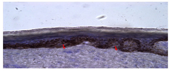
Skin biopsy is a minimally invasive procedure that allows morphometric quantification of intraepidermal nerve fibers (IENF), expressed by section length (IENF/mm). This ratio has been widely used in recent years for the study of peripheral neuropathies involving thin nerve fibers that are not diagnosed by physical (128 Hz tuning fork vibration test, Semmes-Weinstein monofilament test) neurophysiological (nerve conduction studies), or traditional neuropathological (sural nerve biopsy) tests [4-6].
Skin samples can be taken from anybody's site. However, the most common areas are the distal portion of the leg (10 cm above the lateral malleolus) and the thigh (20 cm below the iliac spine). The preferred technique is a 3-mm punch biopsy, as it is minimally invasive and safe under local anesthesia. Quantification of linear INFD density is performed on at least three 50 μm thick sections per biopsy, fixed in 2% periodate-lysine-formaldehyde(PLP) for brightfield microscopy, or Zamboni fixative solution (2% picric acid-formaldehyde) for confocal microscopy, by immunohistochemistry using rabbit polyclonal anti-PGP 9.5.5-8 antibodies.
PGP 9.5 is a carboxyl-terminal ubiquitin hydrolase found mainly in neurons and accompanies the slow axonal transport component. It is widely distributed in the peripheral nervous system and is a non-diffuse pan-axonal marker [5-7].
Brightfield immunohistochemistry and indirect immunofluorescence with confocal microscopy are the two most used immunostaining methods for investigating cutaneous innervation. Based on the results obtained from various studies, typical values for the density of INFD in the distal portion of healthy subjects using brightfield immunohistochemistry are currently available; these values range from 12.4 + 4.6 fibers/mm to 15 + 5/mm.3,4 Furthermore, it has been observed that the density of INFD shows a decreasing gradient from proximal to distal regions of the body, being 60% higher in the thigh region than in the supra malleolar area [8,9].
In patients with ND, low INFD density was first described by Levy et al [10,11], and confirmed by Kennedy et al. [9]. Epidermal denervation must increase with the duration of diabetes, and even nerve fiber reduction has been observed in subjects with prediabetes [7,8] In 2004, Pittenger et al. [8] described decreased mean dendritic length and INFD density in diabetic and non-diabetic patients with neuropathy compared to a healthy control group. The degree of decrease in INFD density was greater in patients with a longer duration of diabetes.
The European Federation of Neurological Societies/Peripheral Nerve Society made a review and update of the guidelines for the use of skin biopsies for the diagnosis of peripheral neuropathy were made in 2010 [12]. These guidelines indicate reference values for INFD density in healthy subjects using the brightfield immunohistochemistry technique; however, not for confocal immunofluorescence. Also, serial skin biopsies could help detect early INFD density changes, which could predict neuropathy progression and assess INFD degeneration and regeneration rates. However, studies are needed to confirm the usefulness of this technique.
Reference values in healthy subjects from the 2010 guidelines range from 9.8 + 3.6 to 13.8 + 6.7 fibers/mm [12]. The largest normative study included 188 healthy subjects stratified by age and gender, establishing values for each decade. Authors observed that the density of INFD in the distal portion of the leg is lower in men than in women and that weight and height do not have a significant impact, but that their values decrease with age.
After the 2010 guidelines, another study was conducted with 550 healthy participants. Patients with symmetric distal polyneuropathy had lower INFD density and dermal and epidermal nerve fiber length with higher axonal swelling rates than healthy controls [13,14]. Regarding the use of INFD as a biomarker of progression in diabetic neuropathy, Narayanaswamy et al [15] observed an average annual rate in INFD loss in patients with ND of 3.76 fibers/mm.
In 2016, Divisova et al [16] observed that the annual rate of INFD loss in patients with DM2 is several times higher than in healthy. Importantly, this study identified that the change in INFD density is not linear and should be expressed as a proportion of the initial INFD density to serve as a marker of the course of ND. The loss of INFD in these patients was less than that described by Narayanaswamy et al in 2012 [15]; however, patients in this first study had a longer duration of diabetes and worse glycemic control, supporting the theory that these factors accelerate the loss of thin fibers.
To fulfill the primary objective, we made an observational, cross-sectional, analytical study was designed. The study's universe was formed by the patients who attended the outpatient services of the endocrinology, dermatology, and neurology departments of the Hospital Central "Dr. Ignacio Morones Prieto." Since there were no similar studies in the literature, a pilot study was carried out, including 21 patients, seven subjects per group [17]. Patients were formed according to the selection criteria, aged 18 to 65 years and of either sex. In group 1, healthy subjects with diabetic family history of first-degree diabetes mellitus type 2 with glycosylated hemoglobin levels of less than 5.7%; in group 2, healthy subjects with no family history of diabetes mellitus type 2 and with glycosylated hemoglobin levels of less than 5.7%; and in group 3, subjects with a diagnosis of type 2 diabetes mellitus with glycosylated hemoglobin levels of more than 6.5%. All agreed to participate and signed a letter of informed consent.
All the patients who had a history of HIV, immune-mediated neuropathies such as systemic lupus erythematosus, Sjögren's syndrome, celiac disease, or sarcoidosis were excluded. Also, hereditary neuropathies such as Fabry disease, Charcot-Marie-Tooth, or Friedreich's ataxia. Besides leprosy, hypothyroidism, use of neurotoxic drugs, Parkinson's disease. We also excluded subjects with non-diabetic neuropathy.
Biopsy technique, tissue handling, and cutting procedures are described in Annex 1. The morphometric evaluation and the quantification of intraepidermal nerve fibers were performed by a certified pathologist who evaluated the slides in a blinded manner. Table 1 describes the variables studied.
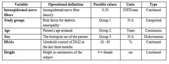
Table 1: Variables included in the study.
STATISTICAL ANALYSIS.
For the statistical analysis, we used the R package of the R software version 3.3.2 [18] with a confidence level of 95%. The normality of the distribution of continuous variables was evaluated with the Shapiro Wilk test. Continuous variables are reported as mean (median) [Q1, Q3] (min-max), discrete variables as frequencies (%). The analysis was performed with ANOVA and Kruskal-Wallis test to test the primary objective.
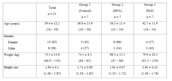
We included twenty-one subjects, 7 in each group, age, sex, weight, and height are described in Table 2.
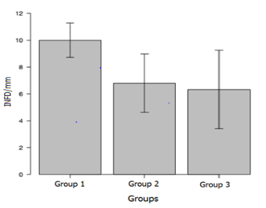
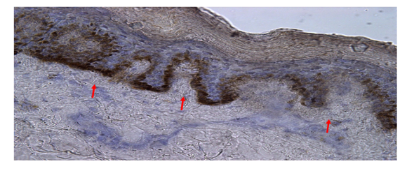
The intraepidermal nerve fibers density (INFD/mm) is similar between the group with family history 6.8 ± 2.1 (3.5 - 10.1) and in the diabetics 6.3 ± 2.9 (3.5 - 7.05) while the control group reported a density of 10± 1.2 (8.2 - 10.1) with a p= 0.01 between the three groups. Figure 1. Figures 2, 3, and 4 show photographs of the epidermis of the three study groups.

Body mass index (BMI) was 28.8 (22.7- 40.2) for patients with DM2 compared to controls 24.8 (23.35 - 26.9) and subjects diabetic family history 24.9 (19.2 - 36.7) (Figure 5).
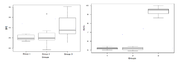
Glycosylated hemoglobin (HbA1c) was 5.2 (5-5.4) for group 1, 5.2 (4.9-5.4) in group 2 and 9.5 (6.8-10) in group 3 with a p=0.001. (Figure 5b).
The decrease in INFD showed a tendency to decrease with increasing age and BMI with a relationship coefficient for age of r= -0.342, 95% CI (-0.67 - 0.106), p= 0.129; and for BMI of r= -0.36, 95% CI (-0.685 - 0.0847), p= 0.109. (Figures 7 and 8).
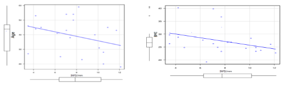
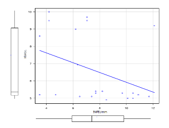
No direct correlation was found between HbA1c levels and INFD/mm with r = -0.375, 95% CI -0.694-0.068, p = 0.0942. (Figure 7).
In the multivariate linear regression analysis to evaluate the factors associated with the variation of INFD/mm, the initial model included the group, HbA1c, and BMI, leaving in the final model only the group, in the relatives of patients with diabetes, the variation was - 2. 7/mm (95% CI -4.8 - -0.58) and in the diabetic group -3.14/mm (95% CI -5.2 - -1.0) concerning healthy patients, this model explains 38.4% of the variation (p = 0.01).
The presence of DM2 in any first-degree relative increases the subject's lifetime risk of presenting DN in 2 to 3 more times. If both parents raise DM2, the risk increases 5 to 6 more times [19,20] The fact that the group with diabetic family history and diabetes present a similar INFD/mm is exciting since it can be inferred that this population is at imminent risk not only of giving DM2; but one of the complications that have the most significant socioeconomic impact on the people.
As described by Gallo et al [21], Mexicans show a greater tendency to develop diabetes; our study is the first to evaluate the INFD/mm in Mexicans, suggesting that this is also a population with a high risk of developing diabetic neuropathy. These results indicate that alterations in INFD/mm begin even before a diagnosis of prediabetes or diabetes is established if the subject already has a genetic load for DM2, being the first study worldwide to describe these findings.
Diabetic neuropathy causes disability due to pain, loss of sensation, gait instability, injuries secondary to falls, ulceration, and amputation of the foot.
Although there are no specific medical treatments and effective glycemic control, overweight control and exercise could change the future lives of those who do not know they have diabetes but have all the potential to be so and certainly to suffer from neuropathy be presented in a previous form.
Skin biopsy for detecting intraepidermal nerve fibers is a minimally invasive procedure that can be of great utility as a screening method in subjects at high risk of developing DM2, perhaps the most pertinent candidates for those subjects with high BMI and family history. As described in the literature, our study shows an inversely proportional relationship between increasing BMI and INFD/mm. HbA1c does not seem to directly connect subjects with a family history of DM2 for the decrease in INFD/mm, so perhaps a prevention strategy in these subjects is an adequate exercise plan.
We could not determine at what age and what other risk factors would be essential to choose the test candidates. Therefore, obtaining a larger sample would enrich the possible parameters to generate a scale or score that would allow us to establish which subjects would be convenient to examine.
Objectively indicating that the subject has morphometric alterations in nerve fibers' density (even without a serological diagnosis of prediabetes or diabetes) can open the door to new strategies. The first contact physician could apply those strategies for the prevention and awareness of the population and then reduce the complications and costs caused by these pathologies.
This study's main limitation is the small sample population; even though the results show a clear tendency to decrease INFD/mm in subjects with diabetic family history it is necessary to have a larger sample to increase the statistical performance of the test.
It is necessary to generate a strategy to establish which subjects are ideal for testing and obtaining data to help the clinician.
The study must continue recruiting participants in the coming years to establish this procedure as one of the screening tools in the arduous work of health education and prevention.
Intraepidermal nerve fiber density is decreased in subjects with a family history of diabetes in comparison with healthy subjects, leading to the possibility to use de skin biopsy as an early biomarker to detect neuropathy. The Mexicans as a population present a higher risk to develop DM2 and diabetic neuropathy, according to the results of this study. Agreeing to previous literature higher BMI and older age are significant risk factors for the decrease of INFD/mm. Glycosylated hemoglobin does not correlate directly with fiber density. Height, weight, and gender in this study were not associated with a decrease in INFD/mm. To our knowledge this is the first study realized in the Mexican population, we propose to continue studying this parameter trying to establish an early biomarker of diabetes o neuropathy.
Dear Editorial Team, Clinical Medical Reviews and Reports. My experience with the journal was highly positive. The peer-review process was rigorous, constructive, and completed in a timely manner. The reviewers provided valuable comments that helped improve the quality and clarity of our manuscript. The editorial office was professional, responsive, and supportive throughout all stages of the publication process. Communication was clear and efficient, and any questions were addressed promptly. Overall, I found the journal to maintain high scientific standards and an excellent publication workflow. I would be pleased to consider submitting future work to this journal. Best wishes from, Elena Popa.

It was my pleasure to submit my testimonial concerning the Reviewer Board of our Scientific Journal “Brain and Neurological Disorders”. The Reviewers focused on some modifications and their contribution was helpful. The ladies of our Editorial Office were also supported my efforts. It was my honor to have such a co-operation and I am looking forward for more collaboration.

Dear Grace Pierce, Editorial Coordinator of Journal of Clinical Research and Reports, Thank you for the speedy and efficient peer review process. I appreciate the fact that your peer reviewers do not take months to respond like with some other journals. I would also like to thank the editorial office for responding quickly to my questions. It is an excellent journal. I plan to submit more manuscripts in the future. Best wishes from, Robert W. McGee

Dear Grace Pierce, Editorial Coordinator of Journal of Clinical Research and Reports, Working with you and your team on our recent publication in JCRR has been a truly wonderful and enjoyable experience. The responses were prompt, and the reviewers were patient, constructive, and highly professional. One reviewer in particular gave me the feeling that a professor was carefully reading and commenting on my coursework, which was deeply touching. The entire process was straightforward and hassle‑free, with no tedious online forms to complete. I highly recommend this journal. Best wishes from, DR Aibing Rao, Head of R&D

I Appreciate the Opportunity to Share my Experience with the Journal of Clinical Research and Reports. The peer review process was timely and constructive, and the feedback provided helped improve the quality of our manuscript. The editorial office was professional, responsive, and supportive throughout the process, ensuring smooth communication and efficient handling of the submission. Overall, it was a positive experience collaborating with your team.

Dear Mercy Grace, Editorial Coordinator of Obstetrics Gynecology and Reproductive Sciences, We would like to express our gratitude for your help at all stages of publishing and editing the article. The editors of the magazine answer all the necessary questions and help at every stage. We will definitely continue to cooperate and publish other works in the Obstetrics Gynecology and Reproductive Sciences! Best wishes from, Alla Konstantinovna Politova,
