
Research | DOI: https://doi.org/10.31579/2693-4779/067
Voltammetry-NIRS Lab, Medicine Center, Verona, Italy.
*Corresponding Author: Francesco Crespi, Voltammetry - NIRS Lab., Medicine Center, Verona, Italy.
Citation: Francesco Crespi. (2021) Influence of nicotine upon human brain metabolism, an in vivo noninvasive Near Infrared Spectroscopy (NIRS) study. Clinical Research and Clinical Trials. 4(4); DOI: 10.31579/2693-4779/067
Copyright: © 2021 Francesco Crespi, This is an open access article distributed under the Creative Commons Attribution License, which permits unrestricted use, distribution, and reproduction in any medium, provided the original work is properly cited.
Received: 11 October 2021 | Accepted: 20 October 2021 | Published: 27 October 2021
Keywords: NIRS; human volunteers; hae¬moglobin; brain metabolism
Nicotine, a natural alkaloid derived from tobacco, is involved in various outcomes ranging from addiction to toxicity and/or neuro-protective actions.
Nevertheless, the literature on the effects of nicotine administration upon the activity of brain regions is mixed; either increased, decreased, or no overall effect was reported when being evaluated by various methodologies such as positron emission tomography (PET), functional Magnetic Resonance Imaging (fMRI).
In this work, Near Infrared Spectroscopy (NIRS) is applied as it allows monitoring oxygen saturation in the living tissue as well as changes in oxygenation of hemoglobin and when applied on brain studies, it gives indications of cerebral haemo-dynamics as well as brain metabolism.
In particular, here NIRS has been applied in human volunteers as this methodology is based upon the use of harmless radiations so that to provide a non-invasive, non-ionizing procedure to monitor 2 main forms of haemoglobin: oxy-haemoglobin (HbO2) and deoxy-haemoglobin (Hb).
The data gathered indicate an overall positive influence of nicotine upon HbO2 levels, as well as total blood volume (V) therefore suggesting an increased brain metabolism.
Finally these data further propose NIRS with its characteristics of noninvasiveness, easy to-use, portable, restraint-free therefore relatively psychologically undemanding, as replicable and ideal methodology for clinical applications and translational approaches.
Various works have analyzed the influence of nicotine upon brain activity. In particular, different studies have shown that nicotine causes a small overall reduction in global cerebral metabolism of glucose in healthy tobacco smoking adult male volunteers as monitored by positron emission tomography (PET) [1, 2].
Furthermore, experimental studies performed in rodents have shown that nicotine and/or electronic cigarette (E-Cig) exposure induce a state of glucose deprivation at the neurovascular unit because of decreases brain glucose uptake under normoxic and ischemic conditions along with down-regulation of GLUT1 and GLUT3 expressions [3].
A different approach to assess central nervous system actions of nicotine is by the use of cerebral blood flow (CBF) measurement i.e. by means of [O-15]-labeled water and PET [4] or using functional Magnetic Resonance Imaging (fMRI) [5].
A variety of effects either increase, decrease or none have been shown (for a review see REF 6).
For istance:
- A significant fall in CBF was observed by transcranial Doppler ultrasound in six volunteers as resulting of the immediate effects of smoking [7]
- In contrast various authors have reported localized increases in CBF after nicotine administration, for example, Nagata et al (1995) using brain imaging showed a significant increase in CBF after cigarette smoking, mainly in the frontal lobes and cerebellum [8].
Stein et al (1998) found fMRI activation in brain areas involved in reinforcement, following i.v. treatment with nicotine [9].
- Other works reported mixed effects: i.e. using PET in habitual smokers receiving nasal nicotine spray it appeared increased blood flow in the right hemisphere thalamus, but decreased blood flow in the left anterior temporal cortex and right hemisphere amygdala [10]. Similar observations were reported in healthy volunteer smokers when exposed to nicotine-containing cigarette as well as receiving intravenous nicotine injections [11, 12].
Finally, no overall effect of nicotine upon CBF was also reported in normal adults with a smoking habit during cigarette smoking [13, 14].
In order to analyze further the influence of nicotine possibly in the attempt to better clarify its effect upon brain metabolism here the methodology of NIRS has been applied in human volunteers.
NIRS is indeed a non-invasive non-ionizing technique that can be used to monitor oxygen saturation in the living tissue as well as changes in oxygenation of hemoglobin [15, 16]. Importantly, the absorption spectra of near-infrared light differ for the oxygenation–deoxygenation states of hemoglobin (oxygenated form HbO2 vs.deoxygenated form Hb, respectively) so that the two compounds can be directly monitored. Such total haemoglobin concentration (HbO2 + Hb) is considered as total blood volume (THC or V) [17]. All together, these measurements are indicative of the state of vascular activity and the state of the metabolism in the tissue analyzed.
The NIRS apparatus used in this work has been described earlier [18], briefly, a schematic configuration of the NIRS system consists of four main blocks: the optical head (optic fibers laser sources and the receiver), the emitter unit, the receiver unit, and the control unit. The optical head is placed onto the surface of the tissue under test in order to monitor non-invasively the oxygenation.
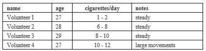
Four healthy tobacco smoking adult male volunteers (aged 27-29) smoking either few or number of cigarettes daily (from 1 up to 12) were prepared for NIRS measurements as described earlier [19], briefly they were wearing a cap in which the optic fibers and the receiver were embedded so that external light cannot influence the measurements (see scheme in figure 1).
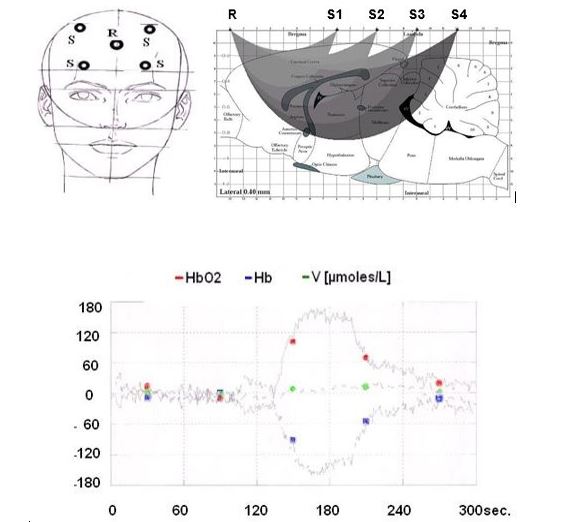
TOP left: Scheme of the positioning of the receiver R and of the four laser sources (S).
R and S are deeply embedded in a cap to avoid influence of external light (not shown).
TOP right : Theoretical brain areas monitored in the brain i.e. computer simulation of photon paths based on photon migration theory [21, 22).
BOTTOM: Response of NIRS parameters to exogenous supply of pure oxygen (1 min from 120 to 180 sec) shown as changes from basal levels that are recorded from zero to 120 sec and that are normalized as = zero μmoles/L. Colored symbols are inserted to facilitate the observation of the continuous recordings. This graphic has been obtained in one volunteer. Similar traces have been observed in all the volunteers.
The optical components were positioned in order to involve the cortical region of the brain accordingly to previous work indicating this area among the most negatively sensitive to nicotine [20].
The protocol of the experiment was as follow: 10min recordings for control baseline of the three parameters monitored i.e. HbO2, Hb, HbT (or V). During this period the volunteers were asked to mimic the smoking act, in order to inspect the influence of forced air inhalation upon NIRS parameters. Successively the volunteers were asked to smoke during 5min. Then measurements were continued other 10min.
Ethical clearance and permission was obtained from the Ethical Review Committee of Public Health and Medical Sciences. Data were collected from the participants after getting informed consents. All the information obtained in due time were kept confidentially.
Statistical analysis have been performed using Statistica 6. Row data were subjected to ANOVA, with comparison between “control period”
and “treatment period” values (one-way ANOVA and Dunnett test).
Results are presented as μmoles/L, mean ± s.e.m., p <0>
In figure 1 the positioning of the Receiver (R) and the four laser Sources (S) is shown as well as the theoretical brain areas of brain that can be monitored when using such positioning accordingly to computer simulation of photon paths based on photon migration theory [21, 22]. Furthermore, the response of NIRS parameters to exogenous supply of pure oxygen (1 min from 120 to 180 sec) is shown as continuous changes from basal levels that are recorded from zero to 120 sec and that are normalized as = zero μmoles/L. This graphic has been obtained in one volunteer. It is evident the large increase of HbO2 to approximately +150µmole/L, the parallel decrease of Hb to approximately -150 µmole/l, while no significant modification of V is observed.
Similar traces have been observed in all the volunteers.
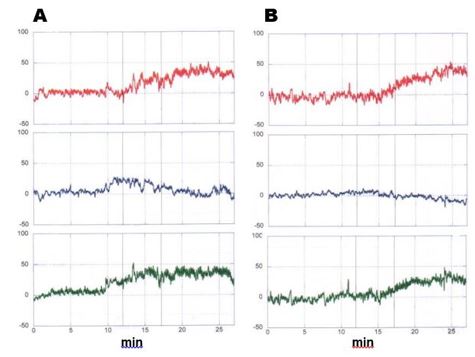
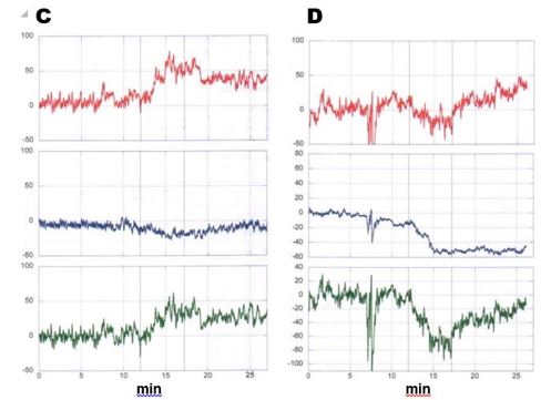
Protocol of the experiment: first 10min recordings for control baseline of the three parameters monitored i.e. HbO2, Hb, HbT (or V) i.e. red, blue or green line respectively. During this period the volunteers were asked to mimic the smoking act, in order to examine the influence of forced air inhalation upon NIRS parameters. Successively the volunteers were asked to smoke during 5min. Then measurements were continued other 10min.
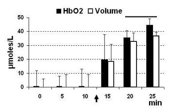
Note that forced air inhalation did not modify significantly any of then3 NIRS parameters i.e. HbO2, Hb, V. oppositsely, smoking was followed by significant modifications of these parameters (see also Figure 3).
It appeared that mimicking the smoking act did not influence NIRS parameters in any one of the volunteers, in contrast HbO2 was significantly increased in all subjects, in particular when taking into account the volunteers #1, 2, 3 that were avoiding large movements (i.e. walking around, gesticulation, volunteer #4) while under recordings (see Table 1).
In figure 3 the mean of the data gathered in the four volunteers is shown. It appeared that both HbO2 and volume V are significantly increased within 20 -25 min following smoking up to approximately 45 and 38 µmole/l, respectively, when considering basal levels as = zero μmoles/L.
The natural alkaloid nicotine, derived from tobacco, is involved in addictive effects [23], toxic effects [24, 25] and neuro-protective actions [26, 5, 19, 32].
Nevertheless and as underlined in the Introduction, the literature on the effects of nicotine administration on the activity of brain regions is mixed; either increased, decreased, or no overall effect was reported in particular when evaluated by CBF analysis.
In this work, NIRS is applied as it allows monitoring oxygen saturation in the living tissue as well as changes in oxygenation of hemoglobin (15, 16). In particular, when applied on brain studies, it gives indications of cerebral haemo-dynamics as well as brain metabolism as already demonstrated [18, 28].
The data gathered indicate an overall positive influence of nicotine upon HbO2 levels, therefore possibly upon brain metabolism. This is further supported by the observation of increased values of the volume (NIRS parameter V) that could be related to a vasodilator influence of nicotine. Indeed, Uchida and Hotta have reported enhanced cerebral flow in the cortex of anesthetized rats treated with nicotine [29]. Such effect has been related to the capability of nicotine to activate the nitric oxide system that is indeed inducing vasodilatation in brain [30] following activation of nicotinic receptors [29, 31].
In a previous work performed in anaesthetised rodents prepared for NIRS analysis, a positive effect of nicotine treatment upon HbO2 levels was detected in the similar progressive manner as obtained here in man with a maximum increase obtained 20-25 min later [32].
Another in vivo non-invasive methodology such as functional magnetic resonance imaging (fMRI) has employed nicotine as a prototypical agent for the analysis of drug-induced changes in the human brain [33, 34].
It resulted increase in brain activity in particular in cortical and sub cortical regions that is also temporally consistent with the NIRS data presented here.
These results, together with our previous data with other drugs of abuse [5, 19, 32], support the applicability of NIRS for direct measurement of turnover of endogenous oxygen that is directly related to neuronal functions coupled with blood flow. In such application NIRS might be complementary to fMRI capabilities, with some advantages versus fMRI that are: no need for ’tracers” , for the subject to be completely immobile, no noise, small dimensions of the instrumentation which is mobile and portable, but most of all direct detection of both HbO2 and Hb; fMRI measures Hb only.
Within the recent decades, more and more studies are using NIRS to analyse the neural mechanisms underlying more and more subtle (i.e. functional, cognitive, perceptual) brain functions in man [35, 36]. In this contest, the NIRS methodology with the characteristics of noninvasiveness, easy to-use, portable, restraint-free therefore relatively psychologically undemanding, and replicable [37] is ideal for clinical applications and translational approaches [38].
Finally, the present results obtained in man together with our recent data achieved with NIRS both in rodents and man [19] support NIRS as a valuable tool for analysis of brain etabolism and its reliable efficacy on direct, rapid translational studies from animals to man.
Acknowledgment to Francesco Congestri for technical support.
Dear Editorial Team, Clinical Medical Reviews and Reports. My experience with the journal was highly positive. The peer-review process was rigorous, constructive, and completed in a timely manner. The reviewers provided valuable comments that helped improve the quality and clarity of our manuscript. The editorial office was professional, responsive, and supportive throughout all stages of the publication process. Communication was clear and efficient, and any questions were addressed promptly. Overall, I found the journal to maintain high scientific standards and an excellent publication workflow. I would be pleased to consider submitting future work to this journal. Best wishes from, Elena Popa.

It was my pleasure to submit my testimonial concerning the Reviewer Board of our Scientific Journal “Brain and Neurological Disorders”. The Reviewers focused on some modifications and their contribution was helpful. The ladies of our Editorial Office were also supported my efforts. It was my honor to have such a co-operation and I am looking forward for more collaboration.

Dear Grace Pierce, Editorial Coordinator of Journal of Clinical Research and Reports, Thank you for the speedy and efficient peer review process. I appreciate the fact that your peer reviewers do not take months to respond like with some other journals. I would also like to thank the editorial office for responding quickly to my questions. It is an excellent journal. I plan to submit more manuscripts in the future. Best wishes from, Robert W. McGee

Dear Grace Pierce, Editorial Coordinator of Journal of Clinical Research and Reports, Working with you and your team on our recent publication in JCRR has been a truly wonderful and enjoyable experience. The responses were prompt, and the reviewers were patient, constructive, and highly professional. One reviewer in particular gave me the feeling that a professor was carefully reading and commenting on my coursework, which was deeply touching. The entire process was straightforward and hassle‑free, with no tedious online forms to complete. I highly recommend this journal. Best wishes from, DR Aibing Rao, Head of R&D

I Appreciate the Opportunity to Share my Experience with the Journal of Clinical Research and Reports. The peer review process was timely and constructive, and the feedback provided helped improve the quality of our manuscript. The editorial office was professional, responsive, and supportive throughout the process, ensuring smooth communication and efficient handling of the submission. Overall, it was a positive experience collaborating with your team.

Dear Mercy Grace, Editorial Coordinator of Obstetrics Gynecology and Reproductive Sciences, We would like to express our gratitude for your help at all stages of publishing and editing the article. The editors of the magazine answer all the necessary questions and help at every stage. We will definitely continue to cooperate and publish other works in the Obstetrics Gynecology and Reproductive Sciences! Best wishes from, Alla Konstantinovna Politova,
