
Research Article | DOI: https://doi.org/10.31579/2642-973X/157
1Department of Neurology, Faculty of Medicine, Mansoura University, Mansoura, Egypt.
2Department of Neurology, Faculty of Medicine, Ain Shams University, Mansoura, Egypt.
3Department of Diagnostic Radiology, Faculty of Medicine, Mansoura University, Egypt.
4Department of Interventional Neuroradiology, Faculty of Medicine, Paris Saclay University, France.
*Corresponding Author: Ahmed Mohamed Hosny Mahmoud Elawady, Department of Neurology, Faculty of Medicine, Mansoura University, Mansoura, Egypt. E-mail: dr_ahmedhosny@mans.edu.eg
Citation: Elawady A, El-Bassiony A, Wessam M, Amer T, Laurent Spelle, Bdeir A, (2022), Influence of Angiographic Features on the Obliteration Rate of Cerebral Arteriovenous Malformations Treated by Endovascular Embolization Using Onyx, J. Brain and Neurological Disorders, 5(5): DOI:10.31579/2642-973X/157
Copyright: © 2022, Ahmed Elawady. This is an open-access article distributed under the terms of The Creative Commons Attribution License, which permits unrestricted use, distribution, and reproduction in any medium, provided the original author and source are credited.
Received: 10 October 2022 | Accepted: 27 December 2022 | Published: 30 October 2022
Keywords: brain avms; onyx embolization; angioarchitecture; obliteration rate; endovascular treatment; cerebral vascular malformation
Background: Brain arteriovenous malformations (AVMs) are complex vascular anomalies with abnormal connections between arteries and veins, posing significant risks of hemorrhage and neurological deficits. Endovascular embolization with Onyx has emerged as a minimally invasive therapeutic option. While prior studies have focused on general treatment outcomes, limited research has assessed the predictive value of specific angiographic characteristics on treatment success.
Objective: To evaluate the influence of AVM angioarchitectural features on the post-embolization obliteration rate and complication profile using Onyx as the embolic agent.
Methods: This retrospective study included patients diagnosed with cerebral AVMs and treated with Onyx embolization. Demographic, clinical, and angiographic data—including nidus size, location, venous drainage pattern, feeder type, and rupture status—were analyzed. The primary outcomes were total or partial obliteration and the incidence of complications. Modified Rankin Scale (mRS) was used to assess clinical outcomes.
Results: Of the 21 patients (mean age 37.3 ± 17.09 years; 52.4% male), 95.2% presented with hemorrhage. Most AVMs (90%) had nidus sizes between 1–3 cm; 76.2% were lobar. Terminal feeders were present in 76.2% of cases. Total obliteration was achieved in 42.9% of patients, partial in 57.1%. AVMs with superficial location, compact nidus, and terminal feeders had significantly higher obliteration rates. Deep-seated lesions and perforator-type feeders were associated with incomplete embolization and complications.
Conclusions: Angiographic features of AVMs are critical determinants of embolization success. Careful pre-procedural assessment of these features can guide treatment planning, improve obliteration rates, and reduce procedural risks.
Cerebral arteriovenous malformations (AVMs) are rare, complex vascular anomalies characterized by direct arteriovenous shunting without an intervening capillary bed. These malformations pose a significant clinical concern due to their potential for life-threatening intracranial hemorrhage, seizures, and progressive neurological deficits. While the annual risk of hemorrhage from AVMs ranges between 2% to 4%, nearly half of the cases present initially with bleeding, often leading to considerable morbidity and mortality [1]. Various treatment modalities have evolved over the past decades, including microsurgical resection, stereotactic radiosurgery, and endovascular embolization. Among these, endovascular embolization using Onyx—a non-adhesive liquid embolic agent—has gained prominence for its ability to achieve deep penetration within the nidus and facilitate preoperative devascularization or even standalone cure in selected cases [2, 3].
Despite advancements in embolization techniques and embolic materials, predicting treatment outcomes remains a challenge. Most existing studies focus on patient demographics, AVM size, and volume as predictors of treatment success [4]. However, there is a relative paucity of research evaluating the influence of detailed angiographic features—such as nidus compactness, feeder type (e.g., terminal vs. en-passage), venous drainage pattern, and location—on obliteration rates and procedural complications. This represents a critical knowledge gap in optimizing treatment strategies and tailoring patient selection [5].
The novelty of the current study lies in its specific focus on the angioarchitectural parameters of cerebral AVMs and their correlation with embolization outcomes using Onyx. By systematically analyzing these vascular characteristics, this study provides a nuanced understanding of which anatomical configurations are most amenable to successful obliteration and which may predispose patients to procedural risks or incomplete treatment [6].
This study was performed to address this gap and assist neurointerventionists in risk stratification, procedural planning, and counseling of patients undergoing endovascular treatment. Better understanding of these variables is essential for improving clinical outcomes and minimizing treatment-related morbidity.
This study aims to evaluate the influence of specific angiographic features on the obliteration rate and complication profile of cerebral AVMs treated by endovascular embolization using Onyx. It seeks to identify which angioarchitectural patterns are predictive of treatment success or failure, thereby refining selection criteria and enhancing therapeutic outcomes.
After obtaining approval from the Institutional Research Ethics Committee and informed consent from all participating patients or their guardians, this retrospective observational study was conducted at the Interventional Neurology Unit, Mansoura University Hospital. The study included patients with cerebral arteriovenous malformations (AVMs) who underwent endovascular embolization using Onyx between July 2015 and October 2020. All procedures were conducted in accordance with the principles of the Declaration of Helsinki.
A total of 146 cases were initially screened. Based on inclusion and exclusion criteria, 21 patients were selected and enrolled in the study.
Inclusion and Exclusion Criteria:
Patients were included if they had a confirmed diagnosis of cerebral AVM based on digital subtraction angiography (DSA), and were treated with endovascular embolization using Onyx.
Exclusion criteria included:
• Prior surgical resection or radiosurgical treatment of the AVM
• Incomplete angiographic data
• Associated vascular pathologies that may interfere with outcome assessment
• Lost follow-up or non-consent to participation.
Study Procedures: All participants were submitted to the following:
All patients enrolled in this study underwent a standardized and systematic evaluation protocol comprising clinical assessment, radiological investigation, and interventional management. The following steps outline the study procedures in detail:
1. Initial Clinical Evaluation
Each patient was subjected to a thorough review of medical records and clinical history. Baseline demographic data—including age, sex, presenting symptoms (e.g., hemorrhage, seizures, focal neurological deficits), and prior interventions—were recorded. Neurological examination findings were documented, and functional status was assessed using the modified Rankin Scale (mRS) before and after embolization.
2. Diagnostic Imaging and Angiographic Assessment
All patients underwent Digital Subtraction Angiography (DSA) as the gold standard for cerebral AVM diagnosis and characterization. The angiographic study included:
• Assessment of AVM nidus size, measured in centimeters and categorized into micro (<1>6 cm).
• Determination of location, classified as lobar, deep (basal ganglia/thalamus), cerebellar, brainstem, or intraventricular.
• Identification of arterial feeders, distinguishing between terminal, en-passage, or perforator types.
• Analysis of venous drainage pattern as superficial or deep.
• Evaluation for associated aneurysms, both intranidal and flow-related.
• Assessment of rupture status, based on clinical and radiological evidence.
This pre-procedural angiographic evaluation was used to determine the complexity of the lesion and guide therapeutic planning.
3. Endovascular Embolization Procedure
All embolization procedures were performed in a dedicated neuro-interventional suite under general anesthesia by experienced interventional neuroradiologists. The following standardized steps were applied:
•Vascular access was obtained via the common femoral artery using the Seldinger technique.
•A guiding catheter was navigated into the appropriate parent artery supplying the AVM nidus.
•Superselective microcatheterization of the feeder vessel was performed to achieve precise localization.
•Onyx™ (ethylene-vinyl alcohol copolymer) was used as the embolic agent. The Onyx was slowly injected under continuous fluoroscopic control using the "plug and push" technique to achieve deep nidus penetration and flow arrest.
•Embolization continued until complete occlusion of the nidus or until safe limits were reached (to avoid reflux or non-target embolization).
•Post-procedural angiography was performed to assess the degree of nidus obliteration.
Multiple sessions were performed if required in cases of high-flow or complex AVMs.
4. Post-Procedural Evaluation and Follow-Up
Immediately following the procedure, all patients were monitored in a neurosurgical intensive care unit. Neurological assessments were repeated, and early post-embolization complications such as hemorrhage, ischemia, or neurological deterioration were recorded.
Follow-up imaging (DSA or MRI) was conducted at 3 to 6 months post-embolization to evaluate:
• Obliteration status (complete or partial)
• Residual nidus
• New-onset complications or recurrence
Modified Rankin Scale (mRS) was re-evaluated at follow-up to document changes in functional status.
Outcome measures:
Primary Outcome Measures
1. Degree of AVM Obliteration
The primary endpoint was the angiographic assessment of the extent of AVM obliteration, categorized as:
o Complete Obliteration
o Partial Obliteration
Obliteration was assessed immediately post-procedure and confirmed during follow-up imaging at 3 to 6 months’ post-treatment.
2. Treatment-Related Complications
Documented intra-procedural or post-procedural complications included:
o Hemorrhage (intranidal or perinidal rupture)
o Ischemic stroke or infarction due to unintended embolization
o Neurological deterioration
o Non-target embolization or catheter entrapment
Secondary Outcome Measures
1. Functional Outcome
Assessed using the Modified Rankin Scale (mRS) before embolization and at follow-up. Changes in mRS were used to evaluate the impact of treatment on neurological function and quality of life.
2. Correlation Analysis
Associations were analyzed between angiographic features (e.g., AVM size, location, feeder type, venous drainage pattern) and:
o Degree of obliteration
o Risk of procedural complications
o Post-treatment mRS scores
3. Rebleeding and Recurrence (if applicable)
Documented during clinical follow-up if patients presented with new symptoms or imaging suggested recurrence of the lesion.
Statistical analysis: Data entry and analysis was done by statistical package of social sciences “spss” version 23. Qualitative variables were summarized in number & percent. Chi square test is used for comparison of qualitative variables between groups. Fisher exact test was used when 50% of cell value was less than 5. Non normally distributed data are summarized in median minimum and maximum. Wilcoxon signed rank test was used for comparison of non-parametric continuous variables. Level of significance less than 0.05 is considered statistically significant.
Demographic data:
From July 2015 to October 2020, 146 cases of cerebral AVM were studied, of them, 125 patients were ruled out by exclusion criteria as shown in figure 1:
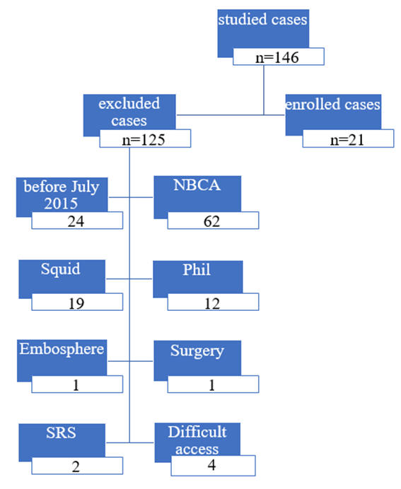
Figure 1: total number of studied and excluded patients
21 patients were recruited in the study including 11 males (52.4%) and 10 females (47.6%), with mean age 37.3 ± 17.09 years. Table (1) illustrates the demographics and clinical features of patients included in the study.
Noteworthy, the initial presentation varied among the study population. variable symotoms were observed as disturbed conscious level, focal deficit, seizures and manifestations of increased ICT, the incidence of these presentations is illustrated in table (1).
Study subjects n=21 | |
| 37.3 (17.09)12-68 | Age: mean (s) Min-max |
11(52.4) 10(47.6) | Sex Male Female |
| 3(14.3) | Hyperlipidemia |
| 4(19) | Hypertension |
| 4(19) | Smokers |
9(42.9) 2(9.5) 1(4.8) 1(4.8) 8(38.1) | Clinical presentation ICT Focal deficit Seizure Disturbed consciousness Multiple symptoms |
Table 1: Patients' demographic baseline characteristics
Angiographic features of the AVMs in the studied population
16 AVMs (76.2%) were located in the cerebral lobes, two (9.5%) were seated deeply in the basal ganglia and thalamus, two in the brainstem and cerebellum (9.5%) and one AVM (4.8%) located lobar and intraventricular. (Table 2)
19 AVMs nidi (90%) sizes ranged from one to three cm, with one micro nidus (< 1cm) and one nidus (3-6 cm). (Table 2)
Most of the AVM (95.2%) presented with hemorrhge due to rupture, while only one AVM didn’t show rupture which presented with resistant symptomatic epilepsy. (Table 2)
Regarding the arterial feeders, 16 AVMs (76.2%) had terminal feeders, three with en-passage type and two had perforator feeders.
Aneurysms were observed in nine AVMs (42.9%), of them; seven were intranidal (77.8%) and the other two were extranidal (22.2%). (Table 2)
16 AVMs (76.2%) had distinct borders (compact) while the other five AVMs (23.8%) were diffuse. The plexiform type of communication between the arterial and venous side was observed in 20 patients (95.2%), and one patient (4.8%) with mixed plexiform – fistulous type. (Table 2)
Nine AVMs (42.9%) had single venous drainage and six patients (28.6%) had several venous draining channels. Ten of them (47.6%) had superficial venous drainage, while six of them (28.6%) had deep drainage and five patients (23.8%) had mixed drainage. Venous ectasia presented in 11 patients (52.4%). (Table 2).
According to Spetzler – Martin grading scale, grade I was observed in eight patients (38.1%), seven AVMs (33.3%) of grade II and the remaining six AVMs (28.6%) were of grade III. (Table 2)
Study subjects n=21 | Variables |
16(76.2) 2(9.5) 2(9.5) 1(4.8) | Location Lobar Brain stem& cerebellar Basal ganglia & thalamus Lobar & Choroidal |
1(4.8) 19(90.5) 1(4.8) | Size <1> 1-3 cm 3-6 cm |
10(47.6) 6(28.6) 5(23.8) | Draining veins Super Deep Mixed |
9(42.9) 6(28.6) 6(28.6) | No of draining veins 1 2 >2 |
16(76.2) 3(14.3) 2(9.5) | Feeder terminal en passage perforator |
| 9(42.9) | Aneurysms |
7(77.8) 2(22.2) | Aneurysms Location Intranidal Extranidal |
20(95.2) 1(4.8) | Shape Plexiform Mixed |
16(76.2) 5(23.8) | Borders Compact Diffuse |
| 11(52.4) | Venous Ectasia |
| 20(95.2) | Rupture |
8(38.1) 7(33.3) 6(28.6) | Spetzler and Martin scale 1 2 3 |
Table 2: AVM characteristics:
Treatment modalities and outcome:
18 patients (85.7%) were treated via the transarterial route, two patients (9.5%) treated through the venous side and one patient (4.8%) treated via both routes. 14 patients (66.7%) underwent one session of embolization, while six of them (28.6%) had two sessions and one patient (4.8%) underwent four embolization sessions (3 arterial and one venous). (Table 5.3)
The results of nidi obliteration were as follows; 18 patients (85.7%) showed complete obliteration (with 5 of them had more than one session to show complete obliteration), one patient (4.8%) had subtotal obliteration and two patients (9.5%) had partial obliteration. (Table 5.3)
Complications were observed among 4 patients (19%); of them three had postoperative ischemic symptoms and one patient had intracerebral with intraventricular haemorrhge. (Table 3)
Table 3 summarizes the treatment modalities adopted in the study population.
| Variables | Study subjects n=21 |
Approach Transvenous Trans arterial Both |
2(9.5) 18(85.7) 1(4.8) |
Number of embolization sessions 1 2 4 |
14(66.7) 6(28.6) 1(4.8) |
Nidus obliteration Complete Subtotal Partial |
18(85.7) 1(9.5) 2(4.8) |
| Procedure related complications | 4(19) |
Table 3: Treatment characterization
The number of patients with mRS score of zero increased from one patient before the treatment to become 15 patients postoperative during follow up, which is statistically significant (P < 0.00001), as shown in Table (4).
| mRS score | preoperative | Postoperative | P value |
| 0 | 1 | 15 | * < 0.00001 |
| > 0 | 20 | 6 |
Table 4: mRS score before and after treatment
Angiographic features predicting obliteration of AVMs
None of the angiographic features was found to be statistically significant in relation to obliteration rate as size, eloquent location, type of arterial feeders, presence of aneurysms, number of draining veins, venous ectasia or stenosis, nidi shape and borders (Table 5).
The rupture state of the AVM and the route of treatment weren’t found to be of significant value on the obliteration rate (Table 5).
Complete n=18 | Partial or subtotal n=3 | P value | |
Size < 3cm> > 3cm |
17(85) 1(100) |
3(15) 0(0) | 1.00 |
Location Eloquent non eloquent |
4(66.7) 14(93.3) |
2(33.3) 1(0.7) | 0.18 |
No of draining veins 1 2 > 2 |
8(88.9) 6(100) 4(66.7) |
1(11.1) 0(0) 2(33.3) | 0.2 |
Feeders Terminal En passage Perforators |
14(87.5) 3(100) 1(50) |
2(12.5) 0(0) 1(50) | 0.2 |
| Aneurysm | 8(88.9) | 1(11.1) | 1.00 |
Shape Plexiform Mixed |
17(85) 1(100) |
3(15) 0(0) | 1.00 |
Borders Compact Diffuse |
14(87.5) 4(80) |
2(12.5) 1(20) | 1.00 |
| Venous Ectasia | 9(81.8) | 2(18.2) | 1.00 |
| Rupture | 18(90) | 2(10) | 0.14 |
Spetzler and Martin 1 2 3 |
8(100) 6(85.7) 4(66.7) |
0(0) 1(14.3) 2(33.3) |
0.08 |
Approach Transvenous Trans arterial Both Row percent is considered |
2(100) 15(83.3) 1(100) |
0(0) 3(16.7) 0(0) |
0.4 |
Table 5: Relation between AVM characteristics and Obliteration
There was no angiographic feature of statistical significance in relation to the complications related to the treatment of the AVMs (Table 6).The way of treatment was of no significant value too.
| Complications | P value | ||
Yes n=4 | No n=17 | ||
Size < 3cm> > 3cm |
3(15) 1(100) |
17(85) 0(0) | 0.19 |
Location Eloquent Non eloquent |
1(16.7) 3(20) |
5(83.3) 12(80) | 1.00 |
No of draining veins 1 2 > 2 |
1(11.1) 1(16.7) 2(33.3) |
8(88.9) 5(83.3) 4(66.7) | 0.5 |
Feeder Terminal En passage Perforator |
3(18.8) 0(100) 1(50) |
13(81.3) 3(100) 1(50) | 0.3 |
| Aneurysms | 3(33.3) | 6(66.7) | 0.2 |
Shape Plexiform Mixed |
4(20) 0(0) |
16(80) 1(100) | 1.00 |
Borders Compact Diffuse |
3(18.8) 1(20) |
13(81.3) 4(80) | 1.00 |
| Ectasia | 3(27.3) | 8(72.7) | 0.5 |
| Rupture | 4(20) | 16(80) | 1.00 |
Spetzler and Martin 1 2 3 |
0(100) 2(28.6) 2(33.3) |
8(100) 5(71.4) 4(66.7) |
0.2 |
Approach Transvenous Trans arterial Both |
0(0) 3(16.7) 1(100) |
2(100) 15(83.3) 0(0) |
0.1 |
Table 6: Relation between AVM characteristics and presence of complications
Brain arteriovenous malformations (bAVMs) are rare lesions characterized by the presence of a nidus containing abnormal tortuous vascular channels between feeding arteries and draining veins without an intervening capillary network [7], this abnormal connection of cerebral arteries and vein leads to the brain hemodynamics disorder, thus causing clinical symptoms such as epileptic seizure, intracranial hemorrhage, or progressive deficit. Brain malformation mass rupture hemorrhage is a severest symptom with high mortality [8].
Traditionally, parenchymal AVMs have been considered congenital lesions. However, several reports of de novo AVM formation and the observation that parenchymal brain AVMs (unlike vein of Galen malformations) are never seen on prenatal ultrasound, have challenged this dogma. Concordant with the increasingly pervasive use of noninvasive neuroimaging, incidental AVMs are being detected with greater frequencies [9].
The overall hemorrhage risk of an untreated, unruptured AVM is estimated to be 1% – 3% per year. This risk is higher among ruptured versus unruptured AVMs, especially within the first year after initial hemorrhage. Prior hemorrhage is the most consistent predictor of subsequent hemorrhage. AVM angioarchitectural features, including venous drainage pattern, fewer draining veins, nidus location, nidus size, presence of associated arterial aneurysms or venous varices, are other potential risk factors. It is unclear if patient demographics, such as age and sex, influence an AVM’s hemorrhage risk [10].
Angiography is the gold standard for the diagnosis and treatment planning for AVMs. Angiograms show the location, size, and number of arterial feeders and draining veins and locate weak points such as intranidal aneurysms, flow-related aneurysms, venous drainage stenosis, ectasia, or aneurysm of the draining veins [11].
Grading scales have been developed to predict outcomes after AVM intervention to stratify patients by operative risk. The Spetzler-Martin (SM) grading scale is the most commonly used classification system. The SM grade comprises 5 tiers, with points allocated for size, venous drainage pattern, and location [12].
With increasingly pragmatic classification to assess the threat and risk of hemorrhage and outcome with or without treatment, controversies about the optimal treatment paradigms are ongoing, especially as treatment modalities become more diverse and multimodal. The target of brain AVM treatment is to eliminate the risk of bleeding. This target could be achieved by complete occlusion of AVM. Unless the nidus is occluded, recruitment of deep perforators and secondary arterialization occurs, consequently aggravating the situation and further restricting therapeutic options for a cure [13].
Various options are available for AVM treatment (medical management, embolization, microsurgery, and radiosurgery) either alone or in combination. ARUBA, largest multicenter randomized trial to date, showed that medical management alone is superior to interventional therapy for the prevention of death and stroke in patients with unruptured AVMs. Nevertheless, ruptured AVMs, unruptured AVMs with significant risk factors, and some symptomatic AVMs in young patients must be treated [14].
The stepwise goals of the microsurgery are wide exposure of the relevant anatomy, occlusion of the feeding arteries while preserving en-passage vessels, circumferential dissection of the lesion, disconnection of the draining veins, and finally en-bloc extirpation of the nidus [10]. The advantages of microsurgery, compared to alternate AVM interventions, are a high rate of complete obliteration, immediate elimination of hemorrhage risk, and long-term durability. The disadvantages of AVM resection are that it requires an open craniotomy, longer hospital stay, longer recovery, and the risk of perioperative neurological and systemic morbidity [15].
SRS is a definitive therapy for AVMs initially reserved for surgically high-risk lesions. SRS is best suited for small- or medium-sized AVMs (volume < 12>< 3cm>
Endovascular treatment of cerebral AVMs can be used before open surgery, before radiosurgery, for cure as a stand-alone treatment, to target the weak angioarchitectural points, or for palliative purposes. The advantages of endovascular treatment include minimally invasiveness, immediate angiographic evaluation during and after the treatment, and immediate occlusive effect [17].
Presurgical embolization is the most common application of endovascular AVM intervention, and the goals are to reduce intraoperative bleeding and facilitate safer dissection of the nidus, thereby decreasing surgical complications. Embolization can be performed in a single stage or in multiple stages [18].
Currently, pre-SRS embolization is primarily employed to selectively target high-risk angiographic features (e.g., intranidal or prenidal arterial aneurysms, intranidal arteriovenous fistulas) that predispose the AVM to rupture during the latency period between SRS and obliteration [15].
In recent years, treatment of brain AVMs in many centers has changed their goal of embolization from adjunctive therapy before surgery or radiosurgery to primary curative embolization. Although higher rates can be achieved among angioarchitecturally simple AVMs, complete obliteration rates with AVM embolization alone have been reported in up to 51%. Either curative or adjuvant embolization can result in postprocedural hemorrhage and ischemia [19]. Although the vast majority of AVM embolizations are performed from a transarterial approach, transvenous embolization has recently emerged as a potentially curative technique for appropriately selected lesions [20].
Certain angiographic features of brain AVMs such as a well-defined nidus and undilated feeder arteries contribute to AVM occlusion [21]. Improvements in microcatheter technology and embolic agents, especially n-BCA and EVOH, have led to an increased curative rate of brain AVMs [22]. Since Onyx has been approved and widely implemented for the endovascular treatment of cerebral AVMs. The implementation of this agent has been almost universally recognized as a step forward in the treatment of these complex vascular lesions. There have, however, recently been suggestions that the use of Onyx for the embolization of AVMs does not improve overall outcomes, and its use may even be associated with worse overall outcomes when compared with n-butyl cyanoacrylate (n-BCA). It has been hypothesized that this may be a result of increased complications with Onyx embolization or increased surgical aggressiveness in patients treated with Onyx [23].
The rapid evolution of endovascular techniques and devices presents a constant challenge to the practicing physician. Endovascular devices and techniques have seen substantial changes over the past 10 to 20 years. This becomes more apparent, and problematic, when assessing the outcomes of patients treated with endovascular methods. Certainly long-term follow-up is necessary to accurately determine the risks and benefits of a procedure [23].
Recent studies reported morbidity ranging from 3–11%, and mortality from 0– 4%, with associations to angioarchitecture, embolic agents, microcatheters, and the manipulator’s skills [22]. Complications of embolization can be categorized into technical- related and non–technical-related. Technical-related complications included vascular perforation, normal branch occlusion, and bleeding caused by catheter removal. Recent improvement in flow-directed microcatheter and nonadhesive embolic agents has reduced the risk of microcatheter delivery and removal. Non–technical-related complications are mainly associated with angioarchitectural characteristics of brain AVMs. Many studies correlated venous outflow obstruction with hemorrhagic presentation after embolization. Furthermore, by progressively blocking draining veins of the AVM, as venous stenosis or occlusion would result in redistribution of blood flow in the nidus and cause intranidal hypertension. Nonetheless, not all venous occlusions would result in hemorrhage; the extent and severity of outflow obstruction matter as well, that is why AVM should be completely or nearly completely embolized as soon as possible when venous outflow obstruction is identified [24].
The main objective of this study was thus to establish the angioarchitectural parameters of AVMs affecting the obliteration and complication rates as a result of embolization using Onyx.
In our study, we found that among the recruited patients, male: female ratio was (1.09:1) which shows insignificant sex difference, which is in harmony with most of the studies as that conducted by Hetts et al., [25], Wahood et al., [26] and Reitz et al., [13].
Complete occlusion was defined as demonstration by arteriography of the total absence of the AVM nidus and AVM venous drainage, with recovery of normal venous brain circulation. In the current study, complete occlusion was achieved in 18 patients (85.7%). The BRAVO trial was a European prospective multicenter trial to assess the safety and efficacy of Onyx, It showed 23.5% complete occlusion rate using onyx [27], Meanwhile, the range of complete occlusion rates in published case series was 8.3–53.9% (10). In the study carried out by Elewa [19], the complete occlusion was achieved in 42.9%, while de Castro-Afonso et al. [28] cure rate was 91.3%. This high cure rate could be explained by the prior intention to completely occlude the lesion whenever possible, the use of Oynx with detachable tip catheters which permit longer injection time, and the use of staged approach.
Meanwhile, obliteration rates in patients treated by SRS alone ranges from 60 to 70% for margin doses of 15–16 Gy to 90% or more for margin doses of 20–25 Gy, and it occurs between 1 and 5 years after treatment. Technical improvements (e.g., in radiosurgical approaches [volume staged radiosurgical downgrading]) for high-grade AVMs might in the future represent an additional option in AVM treatment [29]. In the study done by Paúl et al. [21], The obliteration rate after a single RS was 69.3%; after multiple RS, it was 75%.
We reported a procedural related complications rate of 19% (n=4) in our treated patients, of them three were ischemic which caused by Onyx reflux into arterial territory supplying normal brain area, and one patient with hemorrhagic insult which was attributed to migration of Onyx into the draining veins causing obstruction with subsequent increasing of the intranidal pressure which led to rupture. In concordance with these findings, de Castro-Afonso et al. [28] reported a complication rate of 13% without significant morbidity or mortality. Similar complications results of 13.1% were obtained by Sato et al. [24]. It seems that the results of different studies in this area should not be generalized, as it depends on operator experience which is variable, and cautious slow controlled injections that avoid the draining veins can minimize the procedural risk even in eloquent locations with longer injection time especially with the use of detachable tip microcatheters.
On the other hand, complications in patients treated with SRS ranges from 5% to 13% most likely because of recurrent hemorrhages of incompletely treated AVM or adverse radiation effects. Patients with AVM in the thalamus and brainstem are at the highest risk for symptomatic imaging changes. The fact that patients remain at risk for bleeding until obliteration occurs is the primary drawback of radiosurgery when compared to other modalities. Moreover, Advances in neuroimaging and dose-planning software have reduced the incidence of early adverse radiation effects. Volume-staged procedures can be safely performed for large-volume AVM that were previously considered too large for Gamma knife [29].
Over the years, pre-GKRS embolization has been used to reduce the AVM volume, thereby theoretically reducing the chance of adverse radiation effects. However, most studies have found pre-GKRS to be a negative predictor of obliteration for several reasons. First, nidus recanalization has been reported after both glue and Onyx embolization, which places the patient at risk for future ICH. Second, pre-GKRS embolization can make it more difficult to accurately visualize the nidus or, if the AVM has been divided into multiple pieces, the overall volume that needs to be covered may be no different. As an alternative to pre-GKRS, numerous centers have managed patients with large AVM using volume-staged SRS (VS-SRS). Volume staging of large AVM consists of treating separate components of the nidus in multiple procedures to allow the entire nidus at a therapeutic radiation dose while reducing the radiation exposure to the adjacent brain [16].
In our study, clinical improvement as assessed by mRS score showed marked statistically significant improvement postoperative compared to preoperative score (p< 0.00001), as the number of patients with score 0 (no symptoms at all) preoperative raised from one patient to 15 during follow up visits. In concordance with our result, Pan et al. [22] found significant increase of the number of patients to have mRS score of 0 during postembolization follow up from 10 to 29 out of 130 patients. This clinical improvement is also partially attributed to the recovery from their initial hemorrhage which has a regressive nature, and this finding is also a marker for less complications resulting from the procedure. On the contrary, Kondo et al. [30] reported a significant decrease in the number of patients (664) with mRS score 0 preoperative to become higher postoperative (462) out of total 987 patients, this contradictory result in that latter study might be due to including of ruptured and non-ruptured AVM in addition to using other materials for embolization.
On the univariate analysis to predict the angiographic features related to obliteration rate on our study, there was no feature of significant value as regard nidus size, AVM location, type of arterial feeders, number of draining veins, venous ectasia, presence of aneurysms, nidi shape and borders, Spetzler and Martin grading score, in addition to the route of treatment, and that could be related to small sample size. In the study done by Elewa, [29], he found that Only the presence of AVM in eloquent site was significantly associated with partial occlusion. In our study, we observed that nearly all of AVMs of non-eloquent location (93.3%) showed complete obliteration while two AVMs of eloquent location out of sex didn’t show complete angiographic cure, even though this result is clinically insignificant. Another study that was conducted by Jordan et al. [31] revealed that small AVM size less than 3 cm is significant predictor for complete obliteration, but n-BCA was the embolized material used not the Onyx.
On the other side, for patients treated with SRS only, Pollock [29] concluded that the most important factors associated with nidus obliteration are higher radiation dose and small nidus size. Moreover, Paúl et al. [21] found that positive predictors of obliteration were compact nidus (odds ratio = 3.16; 95% confidence interval, 1.92-5.22), undilated feeders (odds ratio = 0.36; 95% confidence interval, 0.23-0.57), smaller AVM volume (odds ratio = 0.95; 95% confidence interval, 0.92-0.99), and higher marginal dose (odds ratio = 1.16; 95% confidence interval, 1.06-1.27). Meanwhile Taeshineetanakul et al. [32] found the independent factors associated with complete nidus obliteration are noneloquent location (odds ratio [OR], 3.20), size (OR, 0.88), low flow (OR, 3.47), no or mild arterial enlargement (OR, 3.32), and absence of perinidal angiogenesis (OR, 2.61). Differences in number of patients, number, and modality of previous treatments and precise definition of angioarchitectural variables led to various results in literature.
In the study carried out by Ren et al. [33] for AVMs treated by microsurgery, 87.2% patients achieved complete obliteration of the AVMs. Permanent neurological deficits occurred in 14.8% patients and death in 7.9% patients. They found significant association between Spetzler-Martin grading system and obliteration rates.
Regarding our study, none of the angiographic features, Spetzler Martin grading scale, or the route of treatment were found to be statistically significant in relation to complications of treatment. Similar result was obtained by Qureshi et al. [34], who didn’t find significant predictor for occurrence of complications related to procedure. On the other hand, the study carried out by Pan et al. [22] showed that eloquent cortex location (OR, 2.57; 95% CI, 1.08 –3.42) and exclusive deep venous drainage (OR, 4.56; 95% CI, 1.28 –9.67) were correlated with postprocedural complications (p < 0.05), as deep venous drainage could be related to the relative lack of a collateral drainage pathway, making them prone to venous outflow obstruction by embolic agents. however, they studied supratentorial AVMs only. Another study done by Baharvahdat et al. [35] revealed that only premature venous occlusion was identified as an independent predictor of hemorrhagic complication, but they used cyanoacrylates as embolic material for most patients, besides they studied large number of patients. Another finding by Ledezma et al. [36] that Spetzler-Martin grades III–V were significantly associated with unfavorable outcomes and embolization complications. On the other hand, Kim et al. (37) concluded that embolization sessions were the primary factor for postprocedural complications. Moreover, the statistical bias caused by different embolic materials, different locations of AVMs, and technical-related complications.
On the other hand, Theofanis et al. [38] achieved complete obliteration for all treated AVMs with microsurgery, 86.4% had a favorable outcome (mRS score of 0–2), with complications occurred in 7.2% patients, predictors of complications were increasing AVM size (OR 3.2, 95% CI 1.5–6.6; p = 0.001), increasing number of embolizations (OR 1.6, 95% CI 1.1–2.2; p = 0.01), and unruptured AVMs (OR 2.7, 95% CI 1–7.2; p = 0.05). That study included ruptured and unruptured AVMs and many patients underwent combined approaches for management.
Strength Points
One of the major strengths of this study lies in its focused analysis of specific angioarchitectural parameters rather than general demographic or clinical variables, which have been more extensively studied in past literature. The study benefits from a well-documented dataset with standardized embolization procedures and consistent post-treatment imaging and follow-up using DSA and mRS scoring. Additionally, the inclusion of diverse AVM locations and feeder types enhances the generalizability of the findings across different anatomical subtypes.
Despite its contributions, the study has several limitations. First, the sample size is relatively small (21 patients), which may affect statistical power and limit broader generalizability. Second, the retrospective design may be associated with selection bias and incomplete data capture. Additionally, follow-up duration may not be sufficient to assess long-term rebleeding or recurrence rates, especially for partially embolized AVMs. Finally, functional outcomes assessed by mRS may not fully capture quality-of-life changes post-procedure
The study concludes that angiographic characteristics significantly influence the outcomes of AVM embolization using Onyx. AVMs with favorable features—particularly those that are superficial, compact, and have terminal arterial supply—are more likely to achieve complete obliteration with minimal complications. Conversely, deep-seated or complex vascular architectures are associated with suboptimal results and higher procedural risk. These findings support the need for individualized treatment planning based on angioarchitecture.
Abbreviation Full Term
AVM Arteriovenous Malformation
DSA Digital Subtraction Angiography
mRS Modified Rankin Scale
ICA Internal Carotid Artery
MCA Middle Cerebral Artery
ACA Anterior Cerebral Artery
PCA Posterior Cerebral Artery
CTV Cortical Vein
BVR Basal Vein of Rosenthal
ICT Intracranial Tension
This study was conducted after obtaining approval from the Institutional Research Ethics Committee at Mansoura University, Faculty of Medicine. All participants or their legal guardians provided written informed consent prior to inclusion in the study. All procedures conformed to the ethical standards of the 1964 Helsinki Declaration and its later amendments.
The author would like to extend sincere gratitude to the Neurology and Interventional Radiology departments at Mansoura University for their support throughout this research. Special thanks to the supervisors and team members for their invaluable guidance and to the patients who participated in the study.
The authors declare no conflict of interest related to this research or its publication.
Dear Editorial Team, Clinical Medical Reviews and Reports. My experience with the journal was highly positive. The peer-review process was rigorous, constructive, and completed in a timely manner. The reviewers provided valuable comments that helped improve the quality and clarity of our manuscript. The editorial office was professional, responsive, and supportive throughout all stages of the publication process. Communication was clear and efficient, and any questions were addressed promptly. Overall, I found the journal to maintain high scientific standards and an excellent publication workflow. I would be pleased to consider submitting future work to this journal. Best wishes from, Elena Popa.

It was my pleasure to submit my testimonial concerning the Reviewer Board of our Scientific Journal “Brain and Neurological Disorders”. The Reviewers focused on some modifications and their contribution was helpful. The ladies of our Editorial Office were also supported my efforts. It was my honor to have such a co-operation and I am looking forward for more collaboration.

Dear Grace Pierce, Editorial Coordinator of Journal of Clinical Research and Reports, Thank you for the speedy and efficient peer review process. I appreciate the fact that your peer reviewers do not take months to respond like with some other journals. I would also like to thank the editorial office for responding quickly to my questions. It is an excellent journal. I plan to submit more manuscripts in the future. Best wishes from, Robert W. McGee

Dear Grace Pierce, Editorial Coordinator of Journal of Clinical Research and Reports, Working with you and your team on our recent publication in JCRR has been a truly wonderful and enjoyable experience. The responses were prompt, and the reviewers were patient, constructive, and highly professional. One reviewer in particular gave me the feeling that a professor was carefully reading and commenting on my coursework, which was deeply touching. The entire process was straightforward and hassle‑free, with no tedious online forms to complete. I highly recommend this journal. Best wishes from, DR Aibing Rao, Head of R&D

I Appreciate the Opportunity to Share my Experience with the Journal of Clinical Research and Reports. The peer review process was timely and constructive, and the feedback provided helped improve the quality of our manuscript. The editorial office was professional, responsive, and supportive throughout the process, ensuring smooth communication and efficient handling of the submission. Overall, it was a positive experience collaborating with your team.

Dear Mercy Grace, Editorial Coordinator of Obstetrics Gynecology and Reproductive Sciences, We would like to express our gratitude for your help at all stages of publishing and editing the article. The editors of the magazine answer all the necessary questions and help at every stage. We will definitely continue to cooperate and publish other works in the Obstetrics Gynecology and Reproductive Sciences! Best wishes from, Alla Konstantinovna Politova,
