Inflammatory Breast Carcinoma in a Pre-existing Mammary Hamartoma: A rare case report and Review of Literature

Case Report | DOI: https://doi.org/10.31579/2690-4861/153

Inflammatory Breast Carcinoma in a Pre-existing Mammary Hamartoma: A rare case report and Review of Literature

  • Jitendra Parmar * 1
  • Anagha Zope 2
  • Tapan Patel 1
  • Sumita Choudhary 1
  • Bhoomi Angirish 1

1Department of Radiology, Apollo Hospitals International Limited, Ahmedabad, Gujarat, India.

2Department of Surgical Oncology, Apollo Hospitals International Limited & Apollo Comprehensive Blood & Cancer Center, Ahmedabad, Gujarat, India.

*Corresponding Author: Jitendra Parmar, Department of Radiology, Apollo Hospitals International Limited, Ahmedabad, India.

Citation: J Parmar, A Zope, T Patel, S Choudhary, B Angirish. (2021). Inflammatory Breast Carcinoma in a Pre-existing Mammary Hamartoma: A rare case report and Review of Literature. International Journal of Clinical Case Reports and Reviews. 8(3); DOI:10.31579/2690-4861/153

Copyright: © 2021 Jitendra Parmar, This is an open-access article distributed under the terms of the Creative Commons Attribution License, which permits unrestricted use, distribution, and reproduction in any medium, provided the original author and source are credited.

Received: 30 June 2021 | Accepted: 01 September 2021 | Published: 07 September 2021

Keywords: breast; mammary hamartoma; inflammatory breast carcinoma; mammography

Abstract

Mammary hamartoma is uncommon typically non-malignant benign breast lesion, comprising varying amounts of fatty, fibrous connective tissue and glandular elements. The co-existence of hamartomas and malignancy is very rare and it has no special propensity to undergo malignant transformation. We present a case report of a 66-year-old patient diagnosed with Inflammatory Breast Carcinoma within a previously diagnosed mammary hamartoma in the right breast. The comprehensive work-up with mammography, ultrasound, Positron Emission Tomography – Computed Tomography (PET-CT) and ultrasound-guided biopsy confirmed invasive ductal carcinoma within the hamartoma. Although malignancy arising within hamartoma is an extremely rare entity, the radiologist should be aware and prudent of the atypical as well as suspicious features within hamartoma during interpretation of imaging modalities and should thoroughly investigate any architectural distortion or microcalcifications within an otherwise typical hamartoma. No case has been reported for inflammatory breast carcinoma in mammary hamartoma as per our best of knowledge.

Introduction

Mammary hamartoma, also known as fibroadenolipoma is uncommon typically non-malignant benign breast lesion, comprising varying amounts of fatty, fibrous connective tissue and glandular elements. The first reported case of mammary hamartoma was in 1971 by Arrigoni et al. who described it as a well circumscribed lesions comprising varying amount of benign fat and fibro glandular tissue [1]. The typical benign features are described subsequently on mammography [2], ultrasound [3], and MRI [4]. The co-existence of hamartomas and malignancy is very rare and it has no special propensity to undergo malignant transformation. Very few cases of malignancy in mammary hamartoma have been reported previously in the literature [5-9]. No case has been reported for inflammatory breast carcinoma in mammary hamartoma as per our best of knowledge. 

We present a case report of a 66-year-old patient diagnosed with Inflammatory Breast Carcinoma within a previously diagnosed mammary hamartoma in the right breast. The comprehensive work up with mammography, ultrasound, Positron Emission Tomography – Computed Tomography (PET-CT) and ultrasound-guided biopsy confirmed invasive ductal carcinoma within the hamartoma.  This case report illustrates the importance of atypical and suspicious radiological findings in otherwise typical hamartoma that may help in the diagnosis of malignancy.

Case Presentation

A 66-year-old female patient was presented in our outpatient department with history of increase in the size of previously documented lump in right breast for 10-15 days. The patient had a history of swelling in right breast for 20 years, and it was diagnosed as hamartoma on previous ultrasound and mammography. The patient was non-alcoholic and non-smoker. The patient was hypertensive and had history of ischemic heart disease and hypothyroidism. The patient had a positive family history for breast cancer in three siblings and one sister with no history of breast cancer died of non-cancerous pathology at 35 years of age. Clinically the right breast was red and swollen with diffuse erythema and edema (peau d’orange) involving more than a third of the skin of the breast with retraction of nipple. However, the breast was non-tender. Two lymph nodes were palpable in right axillary region and they were hard. 

Based on her present history & positive family history as well as clinical examination, she was referred to radiology department for mammography and ultrasound to rule out malignancy. 

Mammography was performed with standard Mediolateral Oblique (MLO) and Cranio-Caudal (CC) view and supplemented with spot magnification view of mass and microcalcifications (Figure 1). It revealed overall increase in density and trabecular thickening of right breast with architectural distortion and diffuse skin thickening as well as mild nipple retraction. A well-circumscribed lesion, showing mixed fibro-glandular tissue was seen in right breast, which was giving breast within breast appearance, suggestive of hamartoma. In addition to hamartoma, an ill-defined iso to high density lesion with indistinct margins and mild architectural distortion was also seen within the hamartoma. Moreover, multiple fine pleomorphic microcalcifications were seen in segmental distribution in outer lower quadrant within the hamartoma on spot magnification view. Enlarged dense lymph nodes with loss of fatty hilum were present in right axillary region.

Figure 1: Mammography of right breast (MLO; CC and Spot Magnification views): Overall increase in density, trabecular thickening with architectural distortion and diffuse skin thickening as well as mild nipple retraction are seen in right breast. A well-circumscribed lesion, showing mixed fibro-glandular tissue is also seen (breast within breast appearance), suggestive of hamartoma. In addition to hamartoma, an ill-defined iso to high-density lesion with indistinct margins and mild architectural distortion is seen within hamartoma. Moreover, multiple fine pleomorphic microcalcifications are noted in segmental distribution on spot magnification view. Enlarged dense lymph nodes with loss of fatty hilum are present in right axillary region.

On ultrasound (Figure 2), a well-defined and well-circumscribed area of fibro-glandular tissue same as rest of breast parenchyma was seen in outer quadrant of right breast (Figure 3A). Within this area, poorly appreciated, an ill-defined hypoechoic lesion was noted, which was associated with architectural distortion (Figure 3B), and diffuse overlying skin thickening involving more than 1/3rd of breast. Two well-defined sub-centimeter sized skin nodules were also noted in lower outer quadrant. Few enlarged round and oval shaped hypoechoic lymph nodes were also present in right axillary and infra-clavicular region. 

Figure 2: Ultrasound of right breast lump: A well-defined and well-circumscribed area of fibro-glandular tissue same as rest of breast parenchyma, suggestive of hamartoma and highlighted by arrowheads is seen in A. Within this area, poorly appreciated, an ill-defined hypoechoic lesion is shown in B, shown in circle. Post-fire image with the needle inside the mass, indicated by the arrow in C.

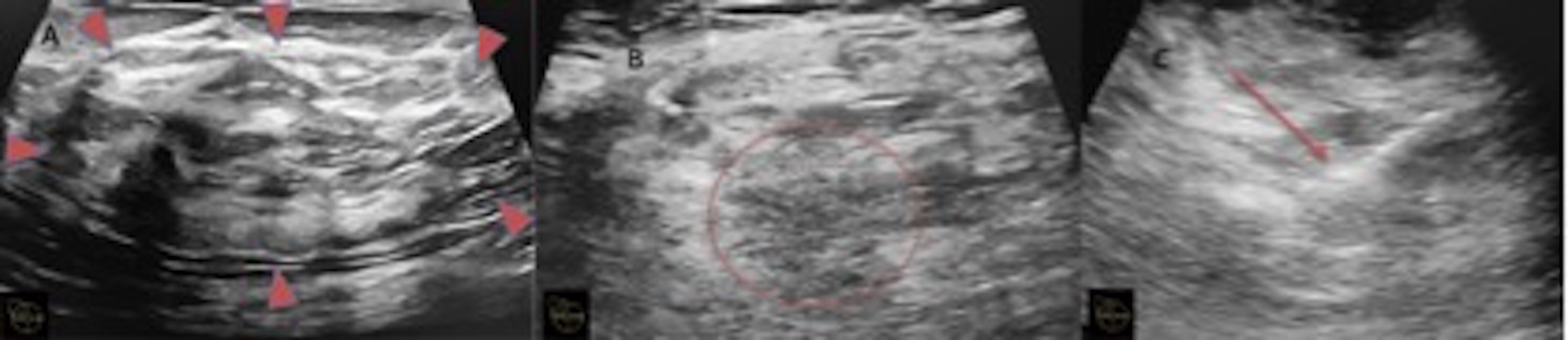
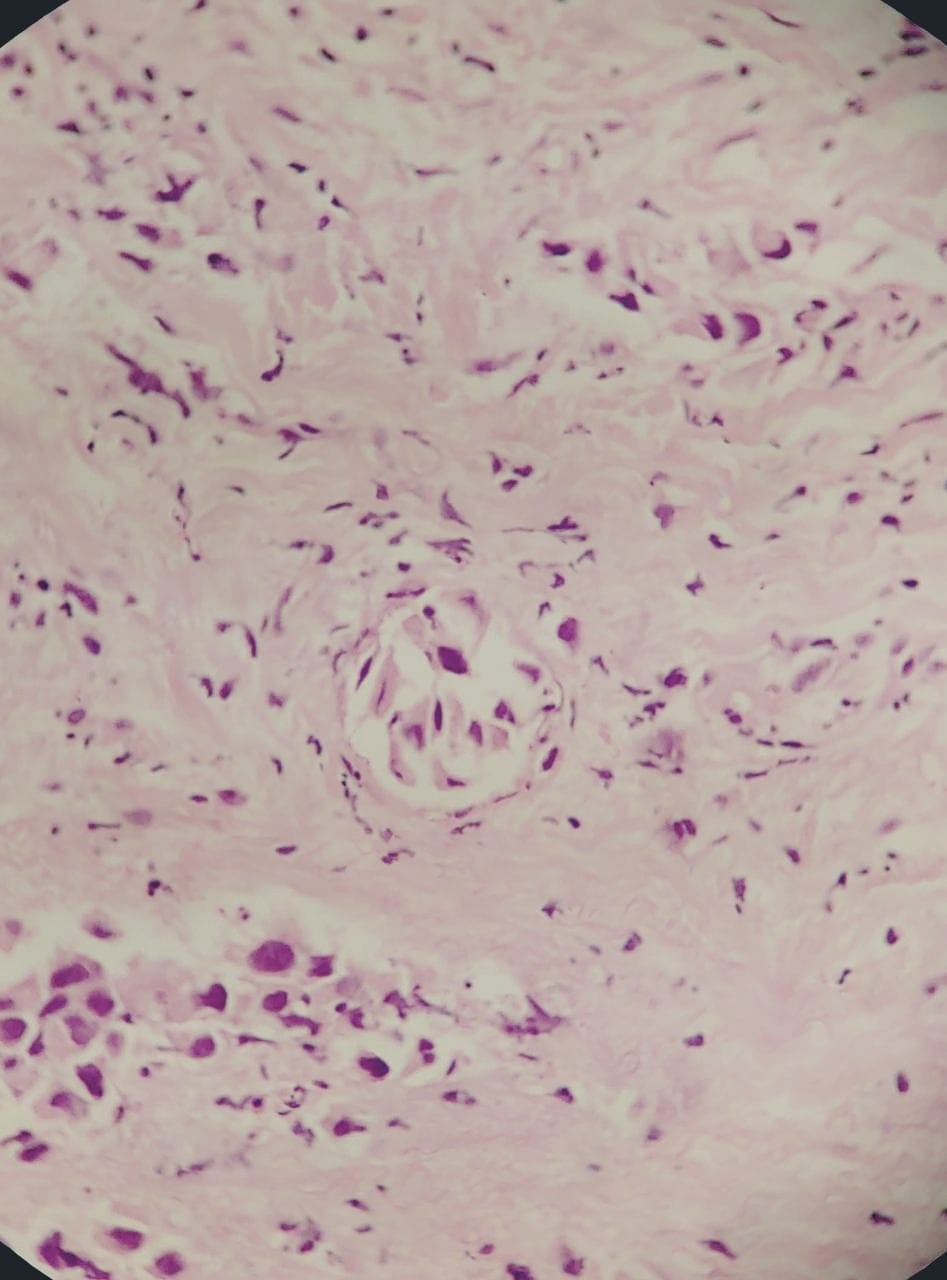
Based on clinical, mammography and ultrasonography findings, the patient was classified as Inflammatory Breast Carcinoma, Breast Imaging Reporting and Data System (BI-RADS) V. The patient underwent ultrasound guided tru-cut biopsy (Figure 3C) for grading characterization and immunohistochemical markers. On histopathology (Figure 4), the patient was confirmed with invasive ductal carcinoma of histological grade III and was negative for Estrogen, Progesterone and Human Epidermal Growth Factor Receptor 2 (HER2) immunohistochemical markers.

Figure 3: PET-CT scan - Axial Computed Tomography (CT) image: Image A confirmed a well-defined encapsulated mixed fibro glandular tissue with breast within breast appearance, suggestive of hamartoma. Within the hamartoma, an irregular shaped soft tissue density lesion and clumped soft calcification are seen separately. PET-CT image in B showed two heterogeneously enhancing hyper-metabolic soft tissue lesions within hamartoma showing intense FDG uptake with SUVmax of 12.8. Two hypermetabolic skin nodules were also confirmed.  Few heterogeneously enhancing hypermetabolic lymph nodes were present in right axilla at level I, II and IIIa.
Figure 4: Histopathology specimen confirms the Invasive Ductal Carcinoma.

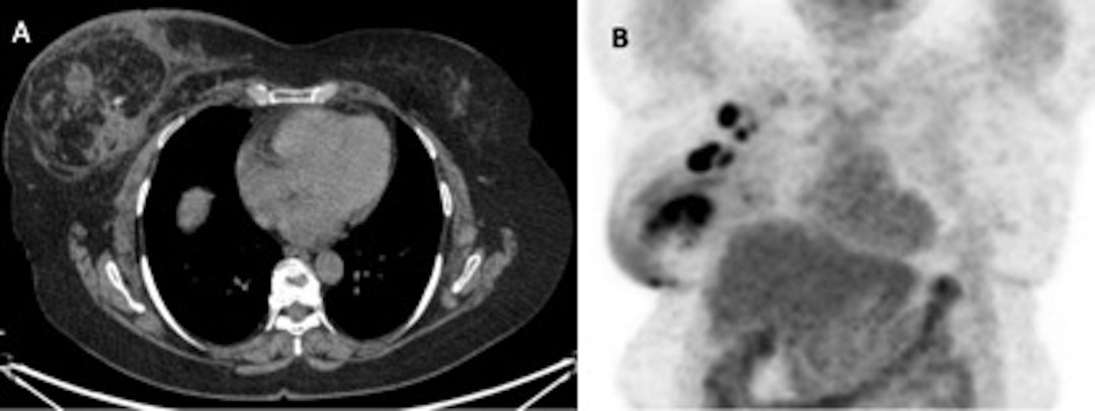
PET-CT (Figure 3) was done for staging, which shows mild increased uptake in entire right breast (as compared to left breast) with diffuse overlying skin thickening (more than 1/3rd of the skin of the breast) and nipple retraction. It also confirmed a well-defined encapsulated fibro glandular tissue same as breast with classical appearance of “Breast within Breast”, suggestive of breast hamartoma (Figure 3A). Within this lesion, two heterogeneously enhancing hyper-metabolic soft tissue lesions were noted, measuring 4.2 x 3.8 x 2.1 cm and 4.1 x 2.9 x 2.5 cm showing intense Fluorodeoxyglucose (FDG) uptake with the maximum standardized uptake value (SUVmax) of 12.8. Two hypermetabolic skin nodules were also confirmed. Few heterogeneously enhancing hypermetabolic lymph nodes were present in right axilla at level I, II and IIIa (Figure 3B). The clinical Tumor-Node-Metastasis (cTNM) scoring system classified as cT4 (m) N3M0 (m-modifier), that corresponds to stage IIIc according to 8th edition of American Joint Committee on Cancer (AJCC) staging system. The patient refused for genetic testing for Breast Cancer gene 1 and 2 (BRCA1, and BRCA2) for hereditary breast and ovarian cancer in spite of genetic counselling. The case was discussed in multidisciplinary meeting and advised to have neoadjuvant chemotherapy (Docetaxel, Anthracycline and Cyclophosphamide) for 6 cycles followed by surgery and radiation if required. 

Discussion

Arrigoni et al. reported mammary hamartoma first time in 1971 as a well circumscribed lesions comprising varying amount of benign fat and fibro glandular tissue [1]. He described a classical mammographic findings of a well circumscribed lesion comprising soft tissue density and fat surrounded by a pseudo capsule, which gives a “breast within a breast” or a “cut sausage” appearance [1,2]. Mammary hamartomas are well circumscribed, ‘wider than tall’ lesions with mixed echogenic internal echotexture without any microcalcifications or retro tumour acoustic shadowing on ultrasonography [3]. On Magnetic Resonance Imaging (MRI), a hamartoma is an encapsulated mass with mixed fat and fibro-glandular signal intensity with some contrast enhancement of fibro-glandular elements [4].

The incidence of malignancy within hamartomas is extremely rare. Very few case reports have described different types of malignancy including both invasive and non-invasive malignancies arising within hamartomas [5-9]. Therefore, the finding of breast hamartoma does not completely exclude the possibility of malignancy originating from benign lesion. Forte et al. in 2019 described invasive carcinoma arising from hamartoma or fibroadenolipoma [9]. Most of the reported cases were of invasive ductal carcinoma as in our case, however, few cases were also of lobular carcinoma and in situ carcinoma [7].

We are in agreement with Lee et al [10]. That any lesion that grows, or shows radiologic features atypical for a hamartoma, a histopathological diagnosis should be warranted. Atypical radiological features, which should raise suspicion of possible malignancy developing within hamartoma, includes suspicious additional mass that shows discriminating imaging features from the hamartoma or development of microcalcifications or increase in size of the mass during follow up.

In conclusion, although malignancy arising within hamartoma is an extremely rare entity, the radiologist should be aware and prudent of the atypical as well as suspicious features within hamartoma during interpretation of imaging modalities and should thoroughly investigate any architectural distortion or microcalcifications within an otherwise typical hamartoma.

Acknowledgement

We are very thankful of the medical oncology department and patient for their unconditional help while preparing this manuscript.

Funding and Conflict of Interest

This case report did not receive any specific grant from funding agencies in the public, commercial, or not-for-profit sectors.

The authors declare that they have no competing interests and all authors have approved the manuscript for submission.

The content of the manuscript has not been published or submitted for publication elsewhere.

The patient reported had signed a written informed consent form, in which the patient was informed that they may be subjects of scientific publication and is informed of the ethical codes of conducts.

All data is available based on a reasonable request. 

References

Clearly Auctoresonline and particularly Psychology and Mental Health Care Journal is dedicated to improving health care services for individuals and populations. The editorial boards' ability to efficiently recognize and share the global importance of health literacy with a variety of stakeholders. Auctoresonline publishing platform can be used to facilitate of optimal client-based services and should be added to health care professionals' repertoire of evidence-based health care resources.

img

Virginia E. Koenig

Journal of Clinical Cardiology and Cardiovascular Intervention The submission and review process was adequate. However I think that the publication total value should have been enlightened in early fases. Thank you for all.

img

Delcio G Silva Junior

Journal of Women Health Care and Issues By the present mail, I want to say thank to you and tour colleagues for facilitating my published article. Specially thank you for the peer review process, support from the editorial office. I appreciate positively the quality of your journal.

img

Ziemlé Clément Méda

Journal of Clinical Research and Reports I would be very delighted to submit my testimonial regarding the reviewer board and the editorial office. The reviewer board were accurate and helpful regarding any modifications for my manuscript. And the editorial office were very helpful and supportive in contacting and monitoring with any update and offering help. It was my pleasure to contribute with your promising Journal and I am looking forward for more collaboration.

img

Mina Sherif Soliman Georgy

We would like to thank the Journal of Thoracic Disease and Cardiothoracic Surgery because of the services they provided us for our articles. The peer-review process was done in a very excellent time manner, and the opinions of the reviewers helped us to improve our manuscript further. The editorial office had an outstanding correspondence with us and guided us in many ways. During a hard time of the pandemic that is affecting every one of us tremendously, the editorial office helped us make everything easier for publishing scientific work. Hope for a more scientific relationship with your Journal.

img

Layla Shojaie

The peer-review process which consisted high quality queries on the paper. I did answer six reviewers’ questions and comments before the paper was accepted. The support from the editorial office is excellent.

img

Sing-yung Wu

Journal of Neuroscience and Neurological Surgery. I had the experience of publishing a research article recently. The whole process was simple from submission to publication. The reviewers made specific and valuable recommendations and corrections that improved the quality of my publication. I strongly recommend this Journal.

img

Orlando Villarreal

Dr. Katarzyna Byczkowska My testimonial covering: "The peer review process is quick and effective. The support from the editorial office is very professional and friendly. Quality of the Clinical Cardiology and Cardiovascular Interventions is scientific and publishes ground-breaking research on cardiology that is useful for other professionals in the field.

img

Katarzyna Byczkowska

Thank you most sincerely, with regard to the support you have given in relation to the reviewing process and the processing of my article entitled "Large Cell Neuroendocrine Carcinoma of The Prostate Gland: A Review and Update" for publication in your esteemed Journal, Journal of Cancer Research and Cellular Therapeutics". The editorial team has been very supportive.

img

Anthony Kodzo-Grey Venyo

Testimony of Journal of Clinical Otorhinolaryngology: work with your Reviews has been a educational and constructive experience. The editorial office were very helpful and supportive. It was a pleasure to contribute to your Journal.

img

Pedro Marques Gomes

Dr. Bernard Terkimbi Utoo, I am happy to publish my scientific work in Journal of Women Health Care and Issues (JWHCI). The manuscript submission was seamless and peer review process was top notch. I was amazed that 4 reviewers worked on the manuscript which made it a highly technical, standard and excellent quality paper. I appreciate the format and consideration for the APC as well as the speed of publication. It is my pleasure to continue with this scientific relationship with the esteem JWHCI.

img

Bernard Terkimbi Utoo

This is an acknowledgment for peer reviewers, editorial board of Journal of Clinical Research and Reports. They show a lot of consideration for us as publishers for our research article “Evaluation of the different factors associated with side effects of COVID-19 vaccination on medical students, Mutah university, Al-Karak, Jordan”, in a very professional and easy way. This journal is one of outstanding medical journal.

img

Prof Sherif W Mansour

Dear Hao Jiang, to Journal of Nutrition and Food Processing We greatly appreciate the efficient, professional and rapid processing of our paper by your team. If there is anything else we should do, please do not hesitate to let us know. On behalf of my co-authors, we would like to express our great appreciation to editor and reviewers.

img

Hao Jiang

As an author who has recently published in the journal "Brain and Neurological Disorders". I am delighted to provide a testimonial on the peer review process, editorial office support, and the overall quality of the journal. The peer review process at Brain and Neurological Disorders is rigorous and meticulous, ensuring that only high-quality, evidence-based research is published. The reviewers are experts in their fields, and their comments and suggestions were constructive and helped improve the quality of my manuscript. The review process was timely and efficient, with clear communication from the editorial office at each stage. The support from the editorial office was exceptional throughout the entire process. The editorial staff was responsive, professional, and always willing to help. They provided valuable guidance on formatting, structure, and ethical considerations, making the submission process seamless. Moreover, they kept me informed about the status of my manuscript and provided timely updates, which made the process less stressful. The journal Brain and Neurological Disorders is of the highest quality, with a strong focus on publishing cutting-edge research in the field of neurology. The articles published in this journal are well-researched, rigorously peer-reviewed, and written by experts in the field. The journal maintains high standards, ensuring that readers are provided with the most up-to-date and reliable information on brain and neurological disorders. In conclusion, I had a wonderful experience publishing in Brain and Neurological Disorders. The peer review process was thorough, the editorial office provided exceptional support, and the journal's quality is second to none. I would highly recommend this journal to any researcher working in the field of neurology and brain disorders.

img

Dr Shiming Tang

Dear Agrippa Hilda, Journal of Neuroscience and Neurological Surgery, Editorial Coordinator, I trust this message finds you well. I want to extend my appreciation for considering my article for publication in your esteemed journal. I am pleased to provide a testimonial regarding the peer review process and the support received from your editorial office. The peer review process for my paper was carried out in a highly professional and thorough manner. The feedback and comments provided by the authors were constructive and very useful in improving the quality of the manuscript. This rigorous assessment process undoubtedly contributes to the high standards maintained by your journal.

img

Raed Mualem

International Journal of Clinical Case Reports and Reviews. I strongly recommend to consider submitting your work to this high-quality journal. The support and availability of the Editorial staff is outstanding and the review process was both efficient and rigorous.

img

Andreas Filippaios

Thank you very much for publishing my Research Article titled “Comparing Treatment Outcome Of Allergic Rhinitis Patients After Using Fluticasone Nasal Spray And Nasal Douching" in the Journal of Clinical Otorhinolaryngology. As Medical Professionals we are immensely benefited from study of various informative Articles and Papers published in this high quality Journal. I look forward to enriching my knowledge by regular study of the Journal and contribute my future work in the field of ENT through the Journal for use by the medical fraternity. The support from the Editorial office was excellent and very prompt. I also welcome the comments received from the readers of my Research Article.

img

Dr Suramya Dhamija

Dear Erica Kelsey, Editorial Coordinator of Cancer Research and Cellular Therapeutics Our team is very satisfied with the processing of our paper by your journal. That was fast, efficient, rigorous, but without unnecessary complications. We appreciated the very short time between the submission of the paper and its publication on line on your site.

img

Bruno Chauffert

I am very glad to say that the peer review process is very successful and fast and support from the Editorial Office. Therefore, I would like to continue our scientific relationship for a long time. And I especially thank you for your kindly attention towards my article. Have a good day!

img

Baheci Selen

"We recently published an article entitled “Influence of beta-Cyclodextrins upon the Degradation of Carbofuran Derivatives under Alkaline Conditions" in the Journal of “Pesticides and Biofertilizers” to show that the cyclodextrins protect the carbamates increasing their half-life time in the presence of basic conditions This will be very helpful to understand carbofuran behaviour in the analytical, agro-environmental and food areas. We greatly appreciated the interaction with the editor and the editorial team; we were particularly well accompanied during the course of the revision process, since all various steps towards publication were short and without delay".

img

Jesus Simal-Gandara

I would like to express my gratitude towards you process of article review and submission. I found this to be very fair and expedient. Your follow up has been excellent. I have many publications in national and international journal and your process has been one of the best so far. Keep up the great work.

img

Douglas Miyazaki

We are grateful for this opportunity to provide a glowing recommendation to the Journal of Psychiatry and Psychotherapy. We found that the editorial team were very supportive, helpful, kept us abreast of timelines and over all very professional in nature. The peer review process was rigorous, efficient and constructive that really enhanced our article submission. The experience with this journal remains one of our best ever and we look forward to providing future submissions in the near future.

img

Dr Griffith

I am very pleased to serve as EBM of the journal, I hope many years of my experience in stem cells can help the journal from one way or another. As we know, stem cells hold great potential for regenerative medicine, which are mostly used to promote the repair response of diseased, dysfunctional or injured tissue using stem cells or their derivatives. I think Stem Cell Research and Therapeutics International is a great platform to publish and share the understanding towards the biology and translational or clinical application of stem cells.

img

Dr Tong Ming Liu

I would like to give my testimony in the support I have got by the peer review process and to support the editorial office where they were of asset to support young author like me to be encouraged to publish their work in your respected journal and globalize and share knowledge across the globe. I really give my great gratitude to your journal and the peer review including the editorial office.

img

Husain Taha Radhi

I am delighted to publish our manuscript entitled "A Perspective on Cocaine Induced Stroke - Its Mechanisms and Management" in the Journal of Neuroscience and Neurological Surgery. The peer review process, support from the editorial office, and quality of the journal are excellent. The manuscripts published are of high quality and of excellent scientific value. I recommend this journal very much to colleagues.

img

S Munshi

Dr.Tania Muñoz, My experience as researcher and author of a review article in The Journal Clinical Cardiology and Interventions has been very enriching and stimulating. The editorial team is excellent, performs its work with absolute responsibility and delivery. They are proactive, dynamic and receptive to all proposals. Supporting at all times the vast universe of authors who choose them as an option for publication. The team of review specialists, members of the editorial board, are brilliant professionals, with remarkable performance in medical research and scientific methodology. Together they form a frontline team that consolidates the JCCI as a magnificent option for the publication and review of high-level medical articles and broad collective interest. I am honored to be able to share my review article and open to receive all your comments.

img

Tania Munoz

“The peer review process of JPMHC is quick and effective. Authors are benefited by good and professional reviewers with huge experience in the field of psychology and mental health. The support from the editorial office is very professional. People to contact to are friendly and happy to help and assist any query authors might have. Quality of the Journal is scientific and publishes ground-breaking research on mental health that is useful for other professionals in the field”.

img

George Varvatsoulias

Dear editorial department: On behalf of our team, I hereby certify the reliability and superiority of the International Journal of Clinical Case Reports and Reviews in the peer review process, editorial support, and journal quality. Firstly, the peer review process of the International Journal of Clinical Case Reports and Reviews is rigorous, fair, transparent, fast, and of high quality. The editorial department invites experts from relevant fields as anonymous reviewers to review all submitted manuscripts. These experts have rich academic backgrounds and experience, and can accurately evaluate the academic quality, originality, and suitability of manuscripts. The editorial department is committed to ensuring the rigor of the peer review process, while also making every effort to ensure a fast review cycle to meet the needs of authors and the academic community. Secondly, the editorial team of the International Journal of Clinical Case Reports and Reviews is composed of a group of senior scholars and professionals with rich experience and professional knowledge in related fields. The editorial department is committed to assisting authors in improving their manuscripts, ensuring their academic accuracy, clarity, and completeness. Editors actively collaborate with authors, providing useful suggestions and feedback to promote the improvement and development of the manuscript. We believe that the support of the editorial department is one of the key factors in ensuring the quality of the journal. Finally, the International Journal of Clinical Case Reports and Reviews is renowned for its high- quality articles and strict academic standards. The editorial department is committed to publishing innovative and academically valuable research results to promote the development and progress of related fields. The International Journal of Clinical Case Reports and Reviews is reasonably priced and ensures excellent service and quality ratio, allowing authors to obtain high-level academic publishing opportunities in an affordable manner. I hereby solemnly declare that the International Journal of Clinical Case Reports and Reviews has a high level of credibility and superiority in terms of peer review process, editorial support, reasonable fees, and journal quality. Sincerely, Rui Tao.

img

Rui Tao

Clinical Cardiology and Cardiovascular Interventions I testity the covering of the peer review process, support from the editorial office, and quality of the journal.

img

Khurram Arshad

Clinical Cardiology and Cardiovascular Interventions, we deeply appreciate the interest shown in our work and its publication. It has been a true pleasure to collaborate with you. The peer review process, as well as the support provided by the editorial office, have been exceptional, and the quality of the journal is very high, which was a determining factor in our decision to publish with you.

img

Gomez Barriga Maria Dolores

The peer reviewers process is quick and effective, the supports from editorial office is excellent, the quality of journal is high. I would like to collabroate with Internatioanl journal of Clinical Case Reports and Reviews journal clinically in the future time.

img

Lin Shaw Chin

Clinical Cardiology and Cardiovascular Interventions, I would like to express my sincerest gratitude for the trust placed in our team for the publication in your journal. It has been a true pleasure to collaborate with you on this project. I am pleased to inform you that both the peer review process and the attention from the editorial coordination have been excellent. Your team has worked with dedication and professionalism to ensure that your publication meets the highest standards of quality. We are confident that this collaboration will result in mutual success, and we are eager to see the fruits of this shared effort.

img

Maria Dolores Gomez Barriga