
Research Article | DOI: https://doi.org/10.31579/2578-8965/130
FSBEI VPO Novgorod University named after Yaroslav- the -Wise, GOBUZ "regional clinical oncological dispensary", Veliky Novgorod, Russia.
*Corresponding Author: Cherenkov V.G., FSBEI VPO Novgorod University named after Yaroslav- the -Wise, GOBUZ
Citation: Cherenkov V.G., Sannikova M.V., Alexandrova I.V. (2022) Immunotherapy and Control of Cervical Canal Crypts on HPV – Ways to Reduce Recurrence and Mortality from Cervical Cancer. J.Obstetrics Gynecology and Reproductive Sciences, 6(5); DOI: 10.31579/2578-8965/130
Copyright: © 2022, Cherenkov V.G., This is an open access article distributed under the Creative Commons Attribution License, which permits unrestricted use, distribution, and reproduction in any medium, provided the original work is properly cited.
Received: 11 August 2022 | Accepted: 29 August 2022 | Published: 26 October 2022
Keywords: HPV; associated pathology; PAP test; PCR treatment control
The results of HPV immunotherapy of associated cervical lesions (HSIL) after radio wave conization (534 cases) were analyzed in comparison with the control group (659 cases) in women with a verified process of CIN II-III and CIS, registered at the regional cancer center. In 9.55±1.3% of cases within 2-3 years after the radio wave conization with HSIL (CIN II-III, CIS), the progression of the disease (dysplasia, cancer in situ and in 2 cases cervical cancer of the 1st stage) was established in the control group. The latter was the need for the introduction of 2 cornerstone practices - the control of crypts of the upper cone of the cervix on HPV and immunotherapy. In the study group, HPV was detected in 23.7±1.8% of women after conization in the upper cone. All women in this group were prescribed inosine pranobex (isoprinosine). After the course of treatment, control for HPV and atypia was repeated after 6, 12 and 18 months. Relapses in the group of patients treated with isoprinosine amounted to 0.27 ± 0.5%. The difference is statistically significant - P less than 0.05.
At the same time, tracing the path from the moment of screening and detection of HPV-associated HSIL lesions, more than half of women do not receive treatment in a timely manner. For control and continuity between specialists, we have introduced signal cards with the informed consent of a woman for treatment when pathology is detected.
As is known, the main method of treatment of cervical intraepithelial neoplasia (CIN) is surgical, usually performed by the method of loop electroconization. In this case, the ectocervix epithelium is effectively removed, while a large number of deep "crypts" may not be removed and cause a recurrence of dysplasia. Detection of viral load in the cut edge was carried out by a number of studies [1,2,3].
Cervical cancer (СС) is one of the few nosologies that tends to rejuvenate and increase the frequency of diseases. СС develops gradually from previous lesions, and even earlier it is preceded by infection in almost all cases with viruses, mainly HPV types 16 and 18 [1]. Why not use this fact to warn the CС. What should it be like? To date, СС is a pathology whose development can be prevented. The greatest number of cervical lesions, including severe ones, occur in early reproductive age.
The mentality of adolescents who begin sexual activity at 14-16 years and earlier (M=15.6 years more than 55.7%, according to an anonymous survey of students) determines the frequent change of sexual partners, increases the risk of early HPV infection [2]. Therefore, the examination of the cervix should be started as early as possible: taking into account the age of the onset of sexual activity.
It was found that every third case of SS (30%) is found in women who have regularly undergone cytological screening [3,4]. The main way to increase the reliability of the cytological research method is to use the method of “liquid cytology". In addition, the material obtained by the “liquid cytology” method is stored for several months and can be used for HPV studies [5].
Meta-analysis of cohort studies with a high degree of reliability proved that HPV testing is a more sensitive method for detecting precancerous conditions of the cervix and cervical intraepithelial neoplasia (CIN II, III degree, CIS). Almost all cases of CIN II and higher are caused by persistent infection. Nevertheless, 43% of such patients have a positive result for HPV–BP, including those who have a normal cytogram. The HPV genotyping strategy was first presented by Thomas S. Wright from Columbia University based on a large-scale multicenter study (ATHENA). Screening for 16 and/or 18 HPV genotypes associated with high risk provides important additional information to the Pap test data for predicting the risk of precancerous conditions and cervical cancer in women. These studies served as the basis for WHO to include HPV-BP in the examination program for the detection of precancerous pathology of the cervix in 2014. From the standpoint of modern therapeutic and diagnostic tactics, we adhered to the International Cytological Classification of Bethesda (2001): ASCUS or AGUS - respectively, cells of the squamous or glandular epithelium.
LSIL - Low grade squamous intraepithelial lesion is a squamous intraepithelial lesion, including a low degree of dysplasia - CIN I and HPV infections. In most women, by the age of 28-30, the body independently gets rid of HPV – carrier. In the presence of other HPV infections, it persists and conservative therapy is required. In such cases, most Russian gynecologists recommend treatment with immunomodulators in combination with vitamin E intake and follow-up in a women's consultation. At the same time, the treatment of women with H SIL remains one of the topical and debatable problems [5,6].
Objective to conduct a comparative assessment of the results of HSIL treatment after radio wave conization of the cervix and ways to reduce the recurrence of HPV-associated pathology in comparison with the control group.
Solving organizational issues of improving the effectiveness of cervical screening and reducing mortality from breast cancer in the Novgorod region in accordance with the work of the staff of the N.N. Blokhin Federal State Budgetary Research Center [7,8], we analyzed the informativeness of smears taken from ecto- and endocervix in women with a verified process of CIN II-III and CIS. The material was taken from the cervical canal with a visually normal cervix. A brush with bristles under eye control was carefully directed to the central part of the cervical canal. A circular rotation of the brush was carried out – 3 in one direction and 2 in the other. The brush was removed from the vagina and dumped into a special BD Sure Path ™ bottle with preservative liquid for cytological examination and PCR.
Abnormal hybridomic cells were found in 167 (23.7%) of 534 women in the study group in smears from the cervix-brush endocervix (Table 1). In the rest, they were not detected, which indicates the need to take material with the capture of not only the transformation zone, but also the crypts of the cervical canal using the Cervex-Brush Combi.
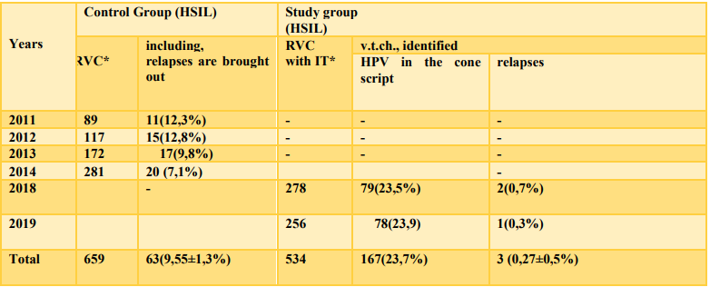
Table 1: Results of HPV immunotherapy for associated cervical pathology (HSIL) after radio wave conization in comparison with the control group
*RVC - radio wave conization; IT – immunotherapy (isoprinosine)
Since 2011, we have performed 1,193 conizations between the ages of 21 and 55. Until 2014, after conization, the cervical stump was treated with 10% potassium permanganate solution before scab formation and with swabs with syntomycin emulsion until it fell off (control group of 659 observations). A retrospective analysis of the treatment outcomes of this group showed that, on average, in 9.55±1.3%) of cases with slight fluctuations over the years, the progression of the disease (dysplasia, cancer in situ and in 2 cases cervical cancer of the 1st stage) was established for 2-3 years. In the latter cases, an increase in the volume of the operation was required before extirpation of the uterus or trachelectomy.
For electroconization of the cervix, we used the Surgitron radio wave apparatus and a special cone-shaped electrode, which allows to remove the cervix in the form of a cone in a single block by turning 360° degrees.
Taking into account the extremely high rate of relapses and persistent HPV in the cervical canal crypts (97.7±1.8%), starting in 2015 after conization with crypt removal (Fig. 1b) with HSIL (CIN II-III, CIS), control of the crypts of the upper cone of the cervix for atypia and HPV became mandatory manipulation. In addition, women were discharged with recommendations for treatment with inosine pranobex (isoprinosine), which, according to some authors, allows to
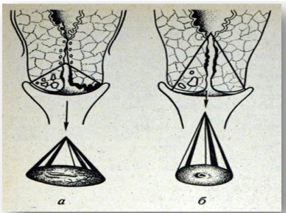
Figure 1: The scheme of conical removal of the cervix (a) is incorrect, б) is correct with the removal of crypts.
Achieve a cure of up to 96?ter conization [10]. The average duration of treatment was 28 days. While maintaining the HPV, the treatment was repeated, including the sexual partner. As a result, only 3 women (0.27±0.5%) showed progression of lesions during the study period, which may be due to non-compliance with the treatment regimen. Comparative statistical analysis of relapses of the two groups "Statistics - 6" showed that the difference in results is significant (P< 0.05). Interdisciplinary aspects of the fight against cancer are crucial factors in its effectiveness. Continuous monitoring, operational analysis and control are an integral part of anti-cancer measures. In order to ensure the continuity of the NOCOD with the cytological laboratories of the region, in addition to sending women in accordance with the "minimum", we have now introduced "signal cards" that must be filled in and sent to the Oncology Center within 10 days from the moment of the pathology detected by HSIL, which allows monitoring the timeliness of their treatment and reducing the category of women not covered treatment.
1. Screening for HPV-WRC (combination of 2 detection and quantification tests) associated with high risk provide important additional information to the Pap test data for predicting the risk of recurrence of cervical cancer in women.
2. In organizational terms, radio wave conization in HSIL pathology should be performed with the control of HPV crypts of the cervical canal, which are the source of 9.8 ± 1.3% of subsequent progression.
3. Relapses in the study group of patients amounted to 0.27 ± 0.5%. The difference is statistically significant – p less than0.05. Control on HPV-WRC should be carried out 6 and 12-18 months after treatment.
Dear Editorial Team, Clinical Medical Reviews and Reports. My experience with the journal was highly positive. The peer-review process was rigorous, constructive, and completed in a timely manner. The reviewers provided valuable comments that helped improve the quality and clarity of our manuscript. The editorial office was professional, responsive, and supportive throughout all stages of the publication process. Communication was clear and efficient, and any questions were addressed promptly. Overall, I found the journal to maintain high scientific standards and an excellent publication workflow. I would be pleased to consider submitting future work to this journal. Best wishes from, Elena Popa.

It was my pleasure to submit my testimonial concerning the Reviewer Board of our Scientific Journal “Brain and Neurological Disorders”. The Reviewers focused on some modifications and their contribution was helpful. The ladies of our Editorial Office were also supported my efforts. It was my honor to have such a co-operation and I am looking forward for more collaboration.

Dear Grace Pierce, Editorial Coordinator of Journal of Clinical Research and Reports, Thank you for the speedy and efficient peer review process. I appreciate the fact that your peer reviewers do not take months to respond like with some other journals. I would also like to thank the editorial office for responding quickly to my questions. It is an excellent journal. I plan to submit more manuscripts in the future. Best wishes from, Robert W. McGee

Dear Grace Pierce, Editorial Coordinator of Journal of Clinical Research and Reports, Working with you and your team on our recent publication in JCRR has been a truly wonderful and enjoyable experience. The responses were prompt, and the reviewers were patient, constructive, and highly professional. One reviewer in particular gave me the feeling that a professor was carefully reading and commenting on my coursework, which was deeply touching. The entire process was straightforward and hassle‑free, with no tedious online forms to complete. I highly recommend this journal. Best wishes from, DR Aibing Rao, Head of R&D

I Appreciate the Opportunity to Share my Experience with the Journal of Clinical Research and Reports. The peer review process was timely and constructive, and the feedback provided helped improve the quality of our manuscript. The editorial office was professional, responsive, and supportive throughout the process, ensuring smooth communication and efficient handling of the submission. Overall, it was a positive experience collaborating with your team.

Dear Mercy Grace, Editorial Coordinator of Obstetrics Gynecology and Reproductive Sciences, We would like to express our gratitude for your help at all stages of publishing and editing the article. The editors of the magazine answer all the necessary questions and help at every stage. We will definitely continue to cooperate and publish other works in the Obstetrics Gynecology and Reproductive Sciences! Best wishes from, Alla Konstantinovna Politova,
