
Case Report | DOI: https://doi.org/10.31579/2641-0419/419
1School of Medicine, University of Zagreb, Zagreb, Croatia.
2Department of Cardiology, Intensive Cardiac Care Unit, University Hospital Centre Sestre Milosrdnice, Zagreb, Croatia.
*Corresponding Author: Petra Knežević, School of Medicine, University of Zagreb, Zagreb, Croatia.
Citation: Petra Knežević, Zdravko Babić, (2024), Hypertrophic Cardiomyopathy and Cardiac Arrest Provoked by Low-Intensity Physical Activity – A Case Report, J Clinical Cardiology and Cardiovascular Interventions, 7(14); DOI: 10.31579/2641-0419/419
Copyright: © 2024, Petra Knežević. This is an open access article distributed under the Creative Commons Attribution License, which permits unrestricted use, distribution, and reproduction in any medium, provided the original work is properly cited.
Received: 26 September 2024 | Accepted: 22 November 2024 | Published: 20 December 2024
Keywords: activity; physical; cardiomyopathy; hypertrophic; death; sudden; cardiac
Hypertrophic cardiomyopathy (HCM) is the most prevalent genetic cardiac muscle disorder, predominantly inherited as an autosomal dominant trait due to mutations in sarcomere protein genes. Although HCM is genetically driven, its clinical presentation is marked by significant variability, with a wide range of phenotypic expressions. The condition often presents in childhood, with many patients facing a heightened risk of sudden cardiac death (SCD), particularly during high-intensity physical activity. Given the risks associated with HCM, particularly in the context of high-intensity competitive sports, it is generally recommended that affected individuals avoid such activities. However, even low-intensity physical activity has been associated with SCD, contributing to ongoing debates and unresolved questions within the field.
HCM – Hypertrophic Cardiomyopathy
SCD – Sudden Cardiac Death
LVOT – Left Ventricular Outflow Tract
LVEDd – Left Ventricular End-Diastolic Diameter
ETT - Exercise Tolerance Test
HF – Heart Failure
LGE – Late Gadolinium Enhancement
NSVT - Non-Sustained Ventricular Tachycardia
Hypertrophic cardiomyopathy (HCM) is the most common genetic disorder affecting the heart muscle, with prevalence of approximately 1:500 in the general adult population (1). The condition is typically inherited as an autosomal dominant trait caused by mutations in sarcomere protein genes. Despite its genetic basis, the clinical presentation of HCM is highly variable, with a broad spectrum of phenotypic expressions. This variability suggests that non-genetic or environmental factors may play a role in shaping the disease's phenotype, although these factors are not yet fully understood (2). HCM is characterized by unexplained left ventricular hypertrophy, which can lead to impaired diastolic function, left ventricular outflow tract obstruction, and cardiac arrhythmias. Most patients are diagnosed during childhood and face a significant risk of sudden cardiac death due to malignant ventricular arrhythmias, particularly during intense physical activity. In contrast, others may remain asymptomatic well into later life. In the past, upon diagnosis of HCM, it was crucial to advise against participation in competitive sports. However, this recommendation is not straightforward, as it raises ongoing debates and leaves several questions unanswered. Furthermore, differentiating between HCM and a benign athlete’s heart can be challenging, presenting both diagnostic and personal dilemmas (3). Systematic restriction from competitive sports in all affected individuals is probably unjustified and a more liberal approach to sports participation is reasonable in some individuals after careful evaluation. This is particularly important for most individuals with HCM who wish to participate in amateur sports or leisure-time exercise. Before recommending exercise intensity, physicians should carefully assess key factors such as symptoms, the ESC risk score, left ventricular outflow tract (LVOT) obstruction, blood pressure response to exercise, and arrhythmias. Although these guidelines promote a more flexible approach, it remains evident that the risk of sudden cardiac death cannot be eliminated, even in those without major risk factors (4). The 5-year SCD risk score, along with additional considerations like left ventricular dysfunction, apical aneurysm, extensive late gadolinium enhancement (LGE) on cardiac MRI, and genetic mutations, should guide tailored recommendations for exercise intensity and participation (5).
A 19-year-old, previously healthy female from France was admitted to the Sisters of Charity University Hospital Centre, Zagreb after an out-of-hospital cardiac arrest, that occurred on the street at the beginning of the planned hike. Cardiopulmonary resuscitation was started by laypersons and continued by the emergency medicine team. The first detected heart rhythm was ventricular fibrillation which was defibrillated once. Defibrillation was followed by the asystole which was treated with epinephrine intravenously (1 mg) resulting in the restoration of sinus rhythm. The patient was ventilated using iGEL. At the admission, the patient was unconscious (GCS 3), with spontaneous circulation and breaths. After placing the endotracheal tube in the Emergency Department, the patient underwent urgent CT angiography which ruled out aortic dissection and pulmonary embolism. CT scan revealed a „ground glass“ lung pattern with the zones of intrapulmonary hemorrhage (Figure 1). A brain CT scan was also performed and showed no pathological substrate. ECG showed sinus rhythm with a heart rate of 115 beats per minute, intermediate electrical axis, PQ interval of 0.16 s, QRS duration of 0.08 s, QT interval of 0.36 s, ST-segment elevation in aVR by 7 mm, diffuse ST segment depression up to 8 mm, and signs of left ventricular hypertrophy. Echocardiography revealed significant hypertrophy of the left ventricle walls (20 mm), with reduced cavity diameter (LVEDd 39mm) and mildly reduced ejection fraction (45-50%) with no visible left ventricular outflow tract obstruction or valvular pathology. Coronary angiography showed no signs of coronary artery stenosis or obstruction. Due to persistent neurologic impairment, "targeted-temperature management" with the goal body temperature less than 37.0°C during 48 hours was performed prompting partial neurological recovery, however, frequent epileptic seizures persisted. The patient opened her eyes spontaneously and moved her extremities on physical contact, but organized contact could not be made. Epileptic seizures were treated with diazepam intravenously and prevented with intravenous levetiracetam and lancosamide. Neuron Specific Enolase level after 72 hours was marginally elevated at 39 µg/L. A repeated brain CT scan revealed a new-onset ischaemic zone in the cerebellum (Figure 2). Brain Magnetic Resonance Imaging showed an acute ischaemic lesion in the right precentral cerebral gyrus and cerebellar hemisphere. Somatosensory evoked potential measurements showed severe conductance impairment of both median nerve sensory pathways, slightly more pronounced on the left side. During the hospitalization, the patient was treated with the thromboprophylactic dose of low-molecular-heparin, and additionally with acetylsalicylic acid after ischaemic lesions in the brain were visualized. 11 days after admission, the patient was discharged and transferred to the hospital in France. The patient's condition was complicated two days after the hospitalization by septic shock secondary to Enterobacter cloacae bacteriemia from an unknown source, treated with fluid resuscitation, norepinephrine, and cefepime antibiotic therapy for 7 days. A ventilator-associated pneumonia bacteriemia with Imipenem-resistant Acinetobacter baumannii was diagnosed and initially treated with colistin. Due to the persistent bacteriemia associated with Imipenem-resistant Acinetobacter baumannii on the central venous catheter, all vascular devices were replaced and antibiotic therapy was first switched to cefiderocol, and after ten days to ampicillin-sulbactam for seven days which led to conditions' improvement and negative hemocultures. Generalized tonic-clonic seizures were treated and resolved with a combination of levetiracetam and lancosamide. Upon cessation of sedation, an initially agitated awakening without contact was observed, managed with sedative treatment with neuroleptics, and eventually definitively stopped due to rapidly appearing extrapyramidal hypertonia under treatment, which resolved upon discontinuation. Treatment with benzodiazepines was initiated as a replacement, with gradually decreasing dosage. Several follow-up EEGs during the stay showed a reactive, reassuring trace, without underlying epileptic activity. An MRI of the brain did not show previously present ischaemic lesions in the cerebellum and precentral gyrus. A gradual calm awakening was observed, with progressively improving consciousness. Due to difficult ventilator weaning attributed to significant clinical malnutrition and probable associated diaphragmatic dysfunction, a surgical tracheotomy was performed and spontaneous ventilation through tracheostomy was initiated. Also, percutaneous endoscopic gastrostomy was performed due to the prolonged enteral feeding. Electrophoresis of plasma proteins and measurement of serum-free light chains for amyloidosis were normal. Measurement of alpha-galactosidase for Fabry's disease also returned normal (activity at 3.27 μmol/L/h). An implantable cardioverter defibrillator was placed under general anesthesia. At the time of the discharge, the patient was hemodynamically stable, with normal breathing and heart rate, moderate sensory-motor deficit with independent walking, and preserved muscle tone. The patient was referred for a cardiogenetic consultation and a follow-up cardiology examination in two months. It was also advised that first-degree relatives should be monitored every 2 to 5 years by ETT and ECG.
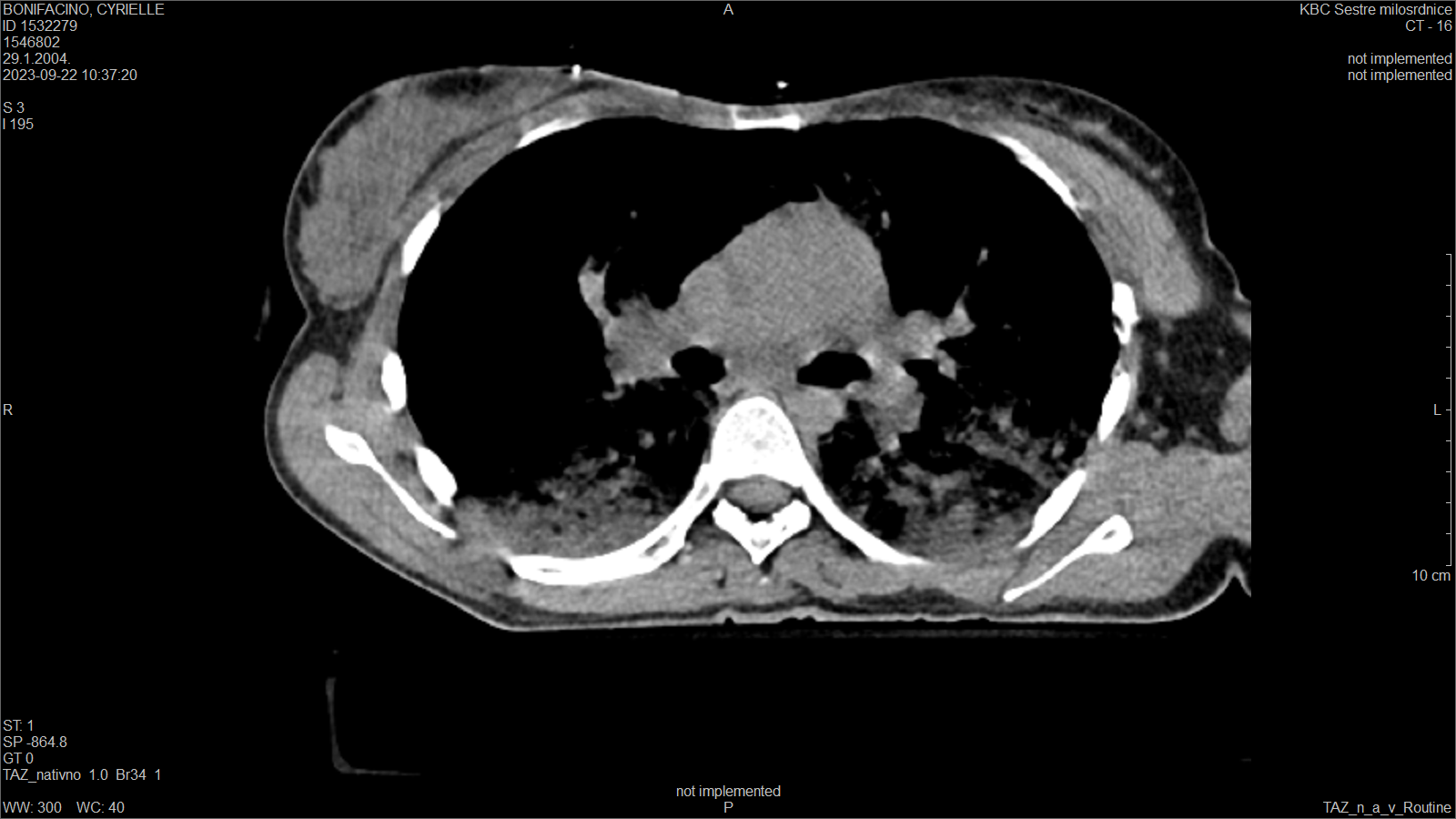
Figure 1: Urgent CT scan showing a „ground glass“ lung pattern with the zones of intrapulmonary hemorrhage.

Figure 2: A repeated brain CT scan revealing a new-onset ischaemic zone in the cerebellum.
The ESC score utilizes seven key variables, including age, family history of SCD, maximal LV wall thickness, left atrial diameter, and non-sustained ventricular tachycardia (NSVT), to assess SCD risk in HCM patients (5). In this case, the patient was classified as low-risk for SCD based on her ESC risk score of 2.5%, which falls below the 4% threshold for defining low-risk status. Despite this low-risk classification and minimal physical exertion, the patient unexpectedly suffered SCD, underscoring the limitations of existing risk stratification models and the unpredictable nature of HCM. Although current guidelines allow some low-risk HCM patients to engage in physical activity, this case highlights the dangers of sudden death even with minimal exertion. This outcome aligns with findings from Spirito et al., who reported that patients with HCM and no conventional risk factors still experienced an SCD rate of 0.6% per year, higher than expected for a low-risk population. These findings suggest that even patients with a favorable clinical presentation, characterized by mild or no symptoms, are not entirely free from the risk of sudden death (6). Furthermore, studies such as those conducted by Pelliccia et al. have analyzed the safety of physical activity in HCM patients, particularly athletes. Their cohort, which included athletes at low risk of SCD according to ESC and American Heart Association guidelines, showed no significant increase in cardiac events or symptoms for those who continued exercising. However, 0.3% of the athletes experienced cardiac arrest outside of athletic settings, and some reported symptoms like syncope and palpitations. Although the overall incidence of adverse events was low, the findings underscore that even in seemingly low-risk populations, SCD can still occur (7). Also, left atrial enlargement has been associated with higher SCD risk and poor outcomes in HCM, normal left atrial size is thought to indicate a more favorable prognosis (6). However, this patient had normal left atrial dimensions and still experienced SCD, further suggesting that factors beyond left atrial size and the conventional ESC criteria may contribute to sudden death in this population.
In conclusion, despite the trend toward permitting more athletes with HCM to engage in physical activity, including high-intensity competitive sports, according to ESC recommendations, this case illustrates that even patients classified as low-risk by the ESC risk score can experience SCD with minimal exertion. Therefore, careful consideration and individualized risk assessment are crucial when making decisions about sports participation for HCM patients.
Dear Editorial Team, Clinical Medical Reviews and Reports. My experience with the journal was highly positive. The peer-review process was rigorous, constructive, and completed in a timely manner. The reviewers provided valuable comments that helped improve the quality and clarity of our manuscript. The editorial office was professional, responsive, and supportive throughout all stages of the publication process. Communication was clear and efficient, and any questions were addressed promptly. Overall, I found the journal to maintain high scientific standards and an excellent publication workflow. I would be pleased to consider submitting future work to this journal. Best wishes from, Elena Popa.

It was my pleasure to submit my testimonial concerning the Reviewer Board of our Scientific Journal “Brain and Neurological Disorders”. The Reviewers focused on some modifications and their contribution was helpful. The ladies of our Editorial Office were also supported my efforts. It was my honor to have such a co-operation and I am looking forward for more collaboration.

Dear Grace Pierce, Editorial Coordinator of Journal of Clinical Research and Reports, Thank you for the speedy and efficient peer review process. I appreciate the fact that your peer reviewers do not take months to respond like with some other journals. I would also like to thank the editorial office for responding quickly to my questions. It is an excellent journal. I plan to submit more manuscripts in the future. Best wishes from, Robert W. McGee

Dear Grace Pierce, Editorial Coordinator of Journal of Clinical Research and Reports, Working with you and your team on our recent publication in JCRR has been a truly wonderful and enjoyable experience. The responses were prompt, and the reviewers were patient, constructive, and highly professional. One reviewer in particular gave me the feeling that a professor was carefully reading and commenting on my coursework, which was deeply touching. The entire process was straightforward and hassle‑free, with no tedious online forms to complete. I highly recommend this journal. Best wishes from, DR Aibing Rao, Head of R&D

I Appreciate the Opportunity to Share my Experience with the Journal of Clinical Research and Reports. The peer review process was timely and constructive, and the feedback provided helped improve the quality of our manuscript. The editorial office was professional, responsive, and supportive throughout the process, ensuring smooth communication and efficient handling of the submission. Overall, it was a positive experience collaborating with your team.

Dear Mercy Grace, Editorial Coordinator of Obstetrics Gynecology and Reproductive Sciences, We would like to express our gratitude for your help at all stages of publishing and editing the article. The editors of the magazine answer all the necessary questions and help at every stage. We will definitely continue to cooperate and publish other works in the Obstetrics Gynecology and Reproductive Sciences! Best wishes from, Alla Konstantinovna Politova,
