
Review Article | DOI: https://doi.org/10.31579/2693-7247/127
1 Department of Zoology, Gauhati University, Guwahati- 781014, Assam, India.
2 Postgraduate Department of Zoology, Darrang College (Affiliated to Gauhati University), Tezpur- 784001, Assam, India.
3 Darrang College, Tezpur- 784001, Assam, India.
*Corresponding Author: Chittaranjan Baruah, Postgraduate Department of Zoology, Darrang College (Affiliated to Gauhati University), Tezpur- 784001, Assam, India.
Citation: Pankaj Das, Ramashankar Bordoloi, Chittaranjan Baruah, Palash Moni Saikia, (2023), Human fungal disease and diagnosis in Northeast India: Zoonosis, antifungal resistance, and public health concern, J. Pharmaceutics and Pharmacology Research, 6(3); DOI:10.31579/2693-7247/127
Copyright: © 2023, Chittaranjan Baruah. This is an open access article distributed under the Creative Commons Attribution License, which permits unrestricted use, distribution, and reproduction in any medium, provided the original work is properly cited.
Received: 05 April 2023 | Accepted: 15 April 2023 | Published: 28 April 2023
Keywords: immunocompromised; invasive; antifungal; aspergillus; cryptococcus; candida; pneumocystis
Humans should avoid fungi, since they can infect both healthy and immunosuppressed hosts with a variety of diseases. Both yeast and filamentous/mold forms of fungi have the ability to infect human hosts. Fungi can be found in a wide range of habitats and occur in a wide range of shapes and sizes, from visible fungi such as mushrooms to microscopic yeasts and moulds. Although fungal infections cause the majority of deaths, public health officials neglect them. Invasive variants of these fungal infections can affect critically ill patients with major underlying immune system issues. Furthermore, people who have cancer, HIV/AIDS, organ transplants, or chronic respiratory disease are more susceptible to invasive fungal infections. The present paper addresses the different fungal diseases of northeast India. In the Northeast region of India, Aspergillus, Cryptococcus, Candida, or Pneumocystis species account for over 90% of fungal infection-related mortality, with fungi such as Coccidioides and Histoplasma posing an extra concern in endemic regions, infecting even immunocompetent hosts. Every year, the most frequent fungal species causing invasive infections in humans, Candida albicans, Aspergillus fumigatus, Cryptococcus neoformans, Pneumocystis jirovecii, endemic dimorphic fungi, and Mucormycetes, harm thousands of individuals worldwide. Other health problems caused by fungi include asthma and allergies. Antifungal drugs treat fungal infections in the body by killing or blocking the growth of harmful fungi. Fungi can develop antifungal medication resistance in the same manner as bacteria can acquire antibiotic resistance. A fungal infection can harm anyone, even those who are otherwise healthy. Higher doses of immune-suppressing medications increase the risk of fungal infection. Resistance to currently available antifungal drugs can develop because of drug-induced acquired mechanisms.
Fungi are eukaryotic organisms with one or more cells that can be found in many types of habitats. They come in a wide variety of shapes and sizes, from naked-eye fungi like mushrooms to microscopic yeasts and moulds. Fungi infections are common in a lot of the natural world. Humans get fungus infections when an invasive fungus colonizes a body component and develops into an aggressive form that the immune system can't control. Fungi infect more than a billion people and cause more than 1.5 million fatalities. Public health officials ignore fungal infections even though most deaths brought on by them may be avoided. Serious fungal infections might arise because of various medical illnesses such asthma, AIDS, cancer, organ transplantation, and corticosteroid therapy. (Felix et al.,2017) Even though most fungi are harmless to people, under certain conditions some of them can spread disease. Fungi reproduce by dispersing spores, which can be breathed or picked up through direct contact. The skin, nails, or lungs are the areas most affected by fungus infections. Infections caused by fungi can also harm organs, enter your skin and spread throughout entire body. Since invasive human fungal diseases, unlike bacterial and viral disorders, are seldom communicable, public health authorities have shown less interest in monitoring and as a result, individuals have limited understanding about the frequency and prevalence of mycoses. 2018 (Arturo, 2018)
Aspergillus, Cryptococcus, Candida or Pneumocystis species are responsible for 90% of fungal infection-related mortality in northeastern India; additional dangers come from fungi like Coccidioides and Histoplasma which can infect even immune-competent hosts in endemic regions. (Mathew et al., 2016). Fungi can be found everywhere, although there are millions of fungal species, only a few hundred are known to cause disease. Some fungal diseases, such as ringworm are zoonotic, which means they can spread from animals to humans. Other fungal diseases, such as histoplasmosis, cannot be transmitted between animals and humans, but they can infect both animals and humans who are exposed to fungi in the environment. Fungi exposure can also lead to other health issues such as asthma and allergies. In the aftermath of natural disasters such as flooding, fungi may develop rapidly. Environmental changes caused by urbanization and intensive farming practices can also have an impact on fungi and their spread. Zoonotic fungi, which can pose serious public health risks, can spread naturally between animals and people. Several mycoses connected to zoonotic transmission are among the most common fungal illnesses in the world. However, it is worth noting that several zoonotic-potential fungal illnesses have received insufficient attention from global public health programmes, resulting in a lack of focus on preventative measures. (Seyedmojtaba et al.,2015).
Antifungal medications treat fungal infections by killing or inhibiting the growth of harmful fungi in the body. Fungi can develop resistance to antifungal drugs in the same way that bacteria can develop antibiotic resistance. Resistance occurs when germs develop the ability to resist drugs that are designed to kill them. This means that the germs are not killed and can continue to grow. Because there are currently only a few antifungal drug types, resistance can severely limit treatment options. Some fungi, such as Candida auris, can develop resistance to all antifungal drugs used to treat these infections (Belinda et al., 2020). Patients with invasive fungal infections, which are serious diseases that affect the blood, heart, brain, eyes, or other organs, should be especially concerned about resistance
Fungal diseases are silent killers that are not prioritised in public health:
Even though they are hidden by simply identifying the primary underlying diseases, fungi are silent killers that kill over two million people each year (Felix et al., 2017). As a result, fungal diseases have surpassed tuberculosis and malaria as the leading causes of death worldwide (i.e., leukemia or COPD). Over the past few decades, advancements in cancer chemotherapy, solid organ and stem cell transplants, increased survival of patients with acute respiratory distress syndrome and other critically ill patients in intensive care, and the widespread use of indwelling medical devices have led to a significant increase in the overall number of patients and the percentage of those susceptible to serious invasive systemic fungal diseases. (Marcio et al., 2021). People who are vulnerable are disproportionately affected, and those who suffer from chronic fungal disease and its consequences live in misery due to illness, inability to work, and shame. According to (Ameet et al., 2011), People in the developing world and the global south are disproportionately affected by fungal keratitis, which is thought to cause nearly a million eyes to lose their vision as a result of agricultural activities. Healthy people who live and work in particular environmental niches are primarily affected by fungi that cause skin and subcutaneous diseases, endemic fungal infections, fungal rhinosinusitis, and other fungal disease. Millions of adults and children have fungus-induced asthma, which is aggravated by airborne fungi such as Aspergillus spp. Because of limited diagnosis and insufficient illness morbidity, the true scope of the problem is only partially known. (Marcio et al., 2018).
Infectious diseases are the world's second leading cause of death, after cardiovascular disease (World Health Organization 2018). Nonetheless, compared to bacterial and viral infections, fungal infections are relatively uncommon in healthy and immunocompetent humans and animals, even though fungal infections propagules are abundant in the environment, making contact with them inevitable (Köhler et al., 2015; Gnat et al., 2020). Furthermore, despite their low prevalence, a growing trend of recalcitrant and recurrent fungal infectious diseases caused by true and opportunistic pathogens has been observed in the northeastern region of India in both humans and animals over the last decades (Fisher et al., 2012; Bishnoi et al., 2018; Shenoy and Jayaraman, 2019; Gnat et al., 2020). Among the millions of common fungal species that pose no risk to humans, a small but growing number of species cause devastating diseases in immunocompromised people (Köhler et al., 2015). It's interesting to note that literature has shown that even more than 50% of known human-infecting diseases are zoonotic in origin (Jones et al. 2008). Additionally, once a zoonotic pathogen has infected humans and broken down the barrier between species, the spread of the disease within the human population depends on the ability of additional human-to-human transmission (Richard et al., 2017; Agowski et al., 2019; Gnat et al., 2019). Multiple channels, including aerosols and respiratory droplets, faecal-oral contact, direct or indirect contact, and vector-borne routes, can be used to transmit infectious pathogens between people and from animals to people (Seyedmousavi et al.,2015; Richard et al., 2017; Gnat et al.,2019). Additionally, reports about the potential for the transfer of fungal diseases from humans to animals have been published in the literature.
Different fungal disease in the Northeast region of India:
Blastomycosis:
The fungus Blastomyces causes the infection Blastomycosis. The fungus is found in the environment, specifically in moist soil and decomposing organic matter such as wood and leaves (Harbans et al.,2013). People can contract blastomycosis by inhaling microscopic fungal spores in the air, which occurs frequently after engaging in activities that disturb the soil. Although most people who breathe in the spores don't get sick, some people will develop symptoms like fever and cough. The infection can be severe in some people, such as those with weakened immune systems, especially if it spreads from the lungs to other organs. Fever is a common blastomycosis symptom. Approximately half of those infected with the fungus Blastomyces will experience symptoms. Blastomycosis symptoms are frequently similar to those of other lung infections, and can include fever, cough, night sweats, muscle aches or joint pain, weight loss, chest pain, and fatigue (extreme tiredness). Symptoms of blastomycosis usually appear 3 weeks to 3 months after inhaling the fungal spores. Blastomycosis cannot be transmitted between people or between humans and animals. (Michael et al., 2010)
Prevention of blastomycosis: There is no vaccine to prevent blastomycosis, and it may not be possible to completely avoid being exposed to the fungus that causes blastomycosis in areas where it is common in the environment. People who have weakened immune systems may want to consider avoiding activities that involve disrupting soil in these areas (Stanley et al., 2008).
Diagnosis of blastomycosis: Healthcare providers use patient medical and travel history, symptoms, physical examinations, and laboratory tests to diagnose blastomycosis. A doctor will likely test for blastomycosis by taking a blood sample or a urine sample and sending it to a laboratory.
Healthcare providers may do imaging tests such as chest x-rays or CT scans of patient lungs. They may also collect a sample of fluid from patient respiratory tract or perform a tissue biopsy, in which a small sample of affected tissue is taken from the body and examined under a microscope. Laboratories may also see if Blastomyces will grow from body fluids or tissues (Catharina et al., 2005, Kathleena et al.,2021)
Candidiasis
Candidiasis is a fungal infection caused by a yeast (a type of fungus) called Candida (Singh et al., 2020). Candida albicans is the most common Candida species that can infect humans. Candida normally lives on the skin and inside the body, including the mouth, throat, gut, and vagina without causing problems. Candida can cause infections if it grows out of control or enters the body deeply. It can, for example, cause infections in the bloodstream or internal organs such as the kidney, heart, and brain. White patches on the inner cheeks, tongue, roof of the mouth and throat are only a few of the signs of candidiasis in the mouth and throat. Inflammation or pain feeling of cotton in the mouth, loss of flavor eating or swallowing, as well as cracking and a reddening of the mouth's corners The most common signs of candidiasis in the esophagus are swallowing pain and difficulties. (Marcio and others, 2001)
Prevention of throat or mouth candidiasis: Maintaining good oral health is one way to help prevent candidiasis in the mouth and throat. After using inhaled corticosteroids, rinse your mouth or brush your teeth. (Peter et al., 2016)
Candidiasis Diagnosis and Testing: By simply looking inside, healthcare providers can usually diagnose candidiasis in the mouth or throat (Laura et al., 2013). A healthcare provider may occasionally take a small sample from the mouth or throat. The sample is sent to a laboratory for testing, usually to be examined under a microscope. An endoscopy is typically used to diagnose candidiasis in the esophagus.
An endoscopy is a procedure that uses a tube with a light and a camera to inspect the digestive tract. Without performing an endoscopy, a doctor may provide antifungal medication to a patient to observe whether their symptoms improve (Marta et al., 2018).
Mucormycosis
Mucormycosis (formerly known as zygomycosis) is a deadly but uncommon fungal infection caused by a group of moulds known as mucoromycetes (Upadhyay et al. 2019). These moulds can be found in almost any environment. Mucormycosis is more common in people who have health problems or who take medications that reduce the body's ability to fight infection and disease. When people inhale fungus spores from the air, it usually affects sinuses or lungs. It may also appear on the skin after a burn, cut, or other type of skin damage (Joshua et al., 2018)
Diagnosis of mucormycosis: When diagnosing mucormycosis, medical professionals take into account our medical history, symptoms, physical exams, and laboratory tests. A fluid sample from the patient's respiratory system may be taken and sent to a lab if the doctor suspects the patient has mucormycosis in the sinuses or lungs. Healthcare provider might conduct a tissue biopsy, in which a small sample of the diseased tissue is examined under a microscope or in a fungal culture in a lab to check for signs of mucormycosis. Depending on where the infection is thought to be, patient may also require imaging tests, such as a CT scan of your lungs, sinuses, or other areas of your body (Anna et al., 2020).
Cryptococcosis
A fungus called Cryptococcus neoformans is present in the environment cause cryptococcosis. It is possible for people to contract C. neoformans following inhalation of the tiny fungus, despite the majority of those who are exposed to it never get sick. Infection with a Cryptococcus fungal infection (either C. gattii) is referred to as cryptococcosis (Uejio et al. 2015). Typically, cryptococcosis affects the lungs or the brain and spinal cord, however it can also impact various body regions (Wilber et al.,2012). Cryptococcal meningitis is the name given to infections of the brain caused by the fungus Cryptococcus. C.neoformans infections are uncommon in normally healthy individuals. The majority of the people with compromised immune systems, especially those with diabetes and advanced AIDS/HIV are more susceptible to neoformans infection (Zhang et al. 2019)
Prevention of C. neoformans infection: It's challenging to hold breath in C. neoformans since it is believed to be widespread in the environment. many individuals who breathe in C. neoformans are never ill with it. C. neoformans is more potent in persons with compromised immune systems. neoformans can remain undetected in the body and eventually infect the body when the immune system is weak and unable to defend oneself from it. This creates a window of opportunity for early detection and treatment of the silent infection before symptoms appear. (John et al.,2010)
Diagnosis of C. neoformans infection: To diagnose a C. neoformans medical professionals rely on medical history, symptoms, physical exams, and laboratory tests. Doctor will collect a sample of body's tissue or bodily fluid (such blood, cerebrospinal fluid, or sputum) and send it to a lab for testing either microscopically inspected, subjected to an antigen test, or grown. Additionally, a doctor might perform tests like a chest x-ray or a CT scan of the patient's lungs, brain, or other organs (Eileen et al., 2016).
Pneumocystis Pneumonia:
Inflammation and fluid buildup are symptoms of the severe infection Pneumocystis pneumonia (PCP)within the lungs. It is caused by the Pneumocystis jirovecii fungus, which spreads throughout theair. PCP can occur to the people having week immune system, like from HIV or other severe health problems. PCP can have an impact on different areas of body, including your liver, bone marrow, and lymph nodes (Alison et al.,2004). Pneumocystis jiroveci is an unusual fungus that primarily causes PCP in HIV-infected people. PCP is still a major cause of morbidity and mortality in HIV/AIDS patients and is a global issue (Ravinder et al.,2015).
Prevention of PCP: No vaccination exists to protect against PCP. For those who are more likely to contract PCP, a doctor may recommend medication to avoid the condition. The Trimethoprim/sulfamethoxazole (TMP/SMX), also referred to as co-trimoxazole and by various other names, is a medication that is most frequently used to prevent PCP. Several brand names, such as Cotrim, Septra, and Bactrim. For those who cannot take TMP/SMX, there are other medications available. (Zeremy et al., 2020)
Diagnosis and Testing of PCP: A patient's lung sample is used to diagnose PCP. Typically, the sample is mucus that has been either coughed up by the patient (called sputum) or gathered by a professional. The bronchoalveolar lavage technique. PCP can occasionally be diagnosed with a small sample of lung tissue (a biopsy). A is sending the patient's sample to a often to be inspected under a microscope in a laboratory. Pneumocystis DNA can also be found using the Polymerase Chain Reaction (PCR) in a variety of samples. PCP can also be identified via a blood test to look for -D-glucan, a component of numerous forms of fungi's cell walls. (Sadatomo et al., 2015: Oliver and others et al., 2020). To diagnose pneumocystosis, DFA is an easily available method in resource-limited settings (Ravinder et al.,2016)
Coccidioidomycosis:
Valley fever, also known as coccidioidomycosis, is caused by the fungus Coccidioides. Valley fever can be contracted by inhaling minute fungus spores in the air, even if most people who do so do not become ill (Crum et al., 2004). Some patients who contract Valley fever will require antifungal medication, but most will recover on their own in a few weeks to months. Various demographic groups are greater chance of developing a serious illness. In locations where it is prevalent in the environment, it is challenging to prevent exposure to coccidioides, Although, Some patients who get Valley fever will require antifungal medication, but most will recover on their own in a few weeks to months. Various demographic groups are greater chance of developing a serious illness in locations where coccidioides are prevalent in the environment, if you're in these places, try to avoid inhaling a lot of dust to reduce your risk of developing severe Valley fever. Coccidioides, the fungus that causes Valley fever, spread between individuals or between people and animals via the lungs. However, a Coccidioides wound infection can spread in extremely uncommon circumstances. Valley fever can spread to another person or through organ transplantation using an infected organ (Kerry et al., 2012)
Prevention and diagnosis of Valley fever: In locations where the fungus Coccidioides is prevalent in the environment, it is quite difficult to avoid breathing it in. Residents of these places should attempt to stay away from spending as much time as possible in dusty areas. Those who are susceptible to developing severe Valley fever (such as those with compromised immune systems, pregnant women, people who have diabetes or people who black or filipino) may attempting to avoid breathing in fungal spores which will lower the chances of developing infection by the fungus(David et al., 2009).
To check for Valley fever pneumonia, doctors may perform imaging tests such as chest x-rays or lung CT scans. A tissue test could also be done. Biopsy: the removal of a small piece of tissue from the body for microscopic examination. Additionally, labs may check to see if Coccidioides can grow from a body tissues or fluids (Samantha et al.,2022).
Histoplasmosis
Histoplasmosis is an infection caused by the fungus histoplasma. The habitat is favourable to the fungus, especially in soil that contains a lot of bird or bat droppings. (Jatin et al.,2020). Histoplasmosis can be contracted by inhaling minute fungus spores from the air, frequently following soil-upsetting activities however most, those that inhale the spores may not become ill; nevertheless, those who do may experience fever, coughing, and exhaustion. Many sick patients will recover without the need for medical intervention or medication. In some people, such as those with weakened immune systems, the infection can become severe, especially if it spreads from the lungs to other organs. (Arghadip et al., 2019)
Prevention of histoplasmosis: In locations where Histoplasma is prevalent in the environment, it can be challenging to avoid breathing it in. People who have Histoplasma infection are at higher risk of Activities should be avoided by those with impaired immune systems (caused, for instance, by HIV/AIDS, organ transplantation, or medication, such as corticosteroids or TNF-inhibitors) activities have been linked to developing histoplasmosis such as: 10 Cleaning coops, exploring caves, and working with disturbing materials (like cutting wood or digging in soil where there are birds' or bats' droppings) as well as cleaning, restoring, or destroying historic structures (Tafireyi et al., 2014)
Diagnosis of histoplasmosis: The most common methods for diagnosing histoplasmosis are blood or urine tests. Medical and travel history, symptoms, physical examinations, and laboratory testing are used by healthcare professionals to make the diagnosis of histoplasmosis. Health care professionals frequently send a urine or blood sample to a lab in order to test for histoplasmosis. Medical professionals may perform imaging tests including chest x-rays or lung CT scans. Additionally, they could take a tissue sample or take a sample of fluid from respiratory system biopsy: the removal of a small piece of the diseased tissue from the body for microscopic examination. Additionally, laboratories may test the growth of Histoplasma tissues or bodily fluids (Marwan et al., 2020, Chadi et al., 2015)
Aspergillosis
Aspergillosis is caused by the filamentous fungi of the genus Aspergillus (Segal et al., 2009). These diseases encompass a wide range of infections in humans and animals, ranging from localized conditions caused by allergic reactions to fatal disseminated infections (Tell 2005; Seyedmousavi et al. 2015; Mohamed et al., 2020). Aspergillus co-infection has recently been reported in patients with severe coronavirus disease 2019 (COVID-19) pneumonia, resulting in acute respiratory distress syndrome (Song et al., 2020). However, those who have lung diseases or compromised immune systems increase the risk of developing Aspergillus-related health issues the various health issues lung infections, infections, and allergic responses are all brought on by Aspergillus in many organs (Michaelia et al.,2022).
Diagnosis of aspergillosis: Medical professionals look at a patient's medical history, risk factors, symptoms, physical examinations, and lab tests when diagnosing aspergillosis. Depending on where the infection occurred, a person may require imaging tests like a chest x-ray or a CT scan of their lungs or other parts of their body. A sample of fluid from the respiratory system may be taken by a medical professional and sent to a lab if they have reason to believe that a patient has an aspergillus infection in their lungs. Medical professionals may also perform a tissue biopsy, in which a small sample of the damaged tissue is examined for aspergillus or under the microscope of a fungus culture. An early diagnosis of invasive aspergillosis in those with severely compromised immune systems can be made with the aid of a blood test. (Thomas et al., 2016).
Fungal Eye Infections
Fungus-related eye infections can be quite dangerous, despite being extremely rare. Having an eye injury is the most common way for someone to develop a fungal eye infection, especially if the injury was caused by plant material like a stick or thorn. Endophthalmitis is the term used to describe inflammation or infection inside the eye, whereas keratitis is used to describe inflammation or infection on the cornea (the clear, front layer of the eye). Numerous types of fungi have the potential to infect the eyes.
Prevention fungal eye infection: Wear safety goggles if work on farms or with plants frequently to help prevent eye injuries. Individuals who use contact lenses must should take excellent care of their lenses (Barry et al.,2002)
Diagnosis and Testing: Eye doctor will checkeye to determine if people have a fungal eye infection and could perhaps extract a tiny sample of fluid or tissue from your eye. The instance will be delivered to a lab where it will either be cultured or inspected under a microscope. Additionally, PCR and confocal microscopy are being used. employed as more recent, quicker methods of diagnosis, but culture is still the gold standard a technique for making a final determination that an eye infection is caused by a fungus (Philip et al., 2013)
| Sl.No: | Fungal Disease | Pathogen | Symptoms | References |
| 1 | Histoplasmosis | Histoplasma capsulatum | Fever, cough, Fatigue, Chills, Headache, Chest pain, Body aches | Ram et al., 2012 Jatin et al., 2020 |
| 2 | Fungal eye infection a)Keratitis b) Endophthamitis | Fusarium Aspergillus Condida | Eye pain, Eye redness, Blurred vision sensitivity to light, Excessive tearing, Eye discharge | Gita et al., 2019 Ml Durand, 2013 |
| 3 | Cryptococcosis
| Cryptococcusgattii C.neofermans | In lungs: Cough shortness of breath chest pain fever
In CNS: Headache, fever confusion or change in behavior, Neck pain | Aroop et al., 2019 Kerri et al., 2012 Wilberg et al., 2012 |
| 4 | Pneumocystis pneumonia (PCP) | Pneumocystis jirovecii | Fever, Cough, Difficulty in breathing, chest pain, chills, Fatigue | Ravinder et al., 2016 Colleen et al., 2009 |
| 5 | Coccidioidomycosis | coccidiodes | Fatigue, cough, fever shortness of breath, Headache, Night sweats, Muscle aches, Rush on upper body or legs. | Vikram et al., 2014 |
| 6 | Mucormycosis
| Mucormycetes Rhizopus species Muccor species Rhizomucor Species Syncephalastum species | Rhinocerebral mucormycosis: One side facial swelling headache, Nasal or sinus congestion Pulmonary mucormycosis: Fever, cough, chest pain, shortness of breath Cutaneous mucormycosis: Blister or ulcers and the infected area may turn black Gastrointestinal mucormycosis: Abdominal pain, Nausea and vomiting, Gastrointestinal bleeding | Hariprasath et al., 2021 Joshua et al., 2018 Julie et al., 2000 |
| 7 | Blastomycosis | Blastomyces dermatitidis B. glichristii | Fever, cough, night sweat, muscle aches or joint pain, weight loss, chest pain, fatigue |
Harris et al.,2022 |
| 8 | Candidiasis | Candida albicans | White patch on the inner cheecks, tongue roof of the mouthg | Immaculata et al., 2007 |
| 9 | Aspergillosis | Aspergillus fumigatus
A. Flavus | Allergic Bronchopilmonary aspergillosis(ABPA): Wheezing, shortness of breath, cough, fever Allergic Aspergillus sinusitis: Runny nose, Stuffiness Headache reduced ability to smell Aspergilloma (Fungus ball): Shortness of breath Cough, coughing up blood, Chorionic pulmonary Aspergillosis: eight loss, cough, Coughing up blood, Fatigue, Shortness of breath. Invasive Aspergillosis: Fever, chest pain coughing up blood, shortness of breath
| Ritish et al., 2014
Chandramani et al., 2011
Priti et al., 2010
Ritesh et al., 2014
Shivprakash et al., 2019 |
Table 1. Human fungal disease in Northeast India
Who gets fungal infection:
A fungal infection can affect anyone, including people who are generally healthy. Every day, people breathe in or come into contact with fungal spores without becoming ill. These fungi, however, are more common in patients with compromised immune systems Infection (Fig. 1). (Olivier and colleagues et al., 2013).
Causes of weakened immune system:
Some people have a compromised immune system from birth. Others may be suffering from a condition that targets the immune system, such as HIV. Some medications, such as corticosteroids and cancer treatment, can impair the body's capacity to fight infections.
Many fungal infections are referred to as opportunistic infections because they typically affect people with weakened immune systems. Because HIV weakens the immune system, you are more likely to contract fungal infections such as cryptococcosis, coccidioidomycosis, histoplasmosis, and pneumocystis pneumonia (PCP). Markus et al., 2004).
Small bowel transplant recipients are most likely to develop fungal infections, followed by lung, liver, and heart transplant recipients (Peter et al.,2010). Some pathogenic fungi are more common in some parts of the world. If patient have had an organ transplant and live or visit these areas, they are more likely than the general population to contract these infections (Rachel et al.,2019).
People with blood cancers, such as leukemia or myeloma, may be more susceptible to fungal infections than those with other types of cancer. Some cancers, particularly blood cancers, may necessitate more aggressive chemotherapy treatment than others. This is referred to as aggressive chemotherapy. Chemotherapy can weaken your immune system and put you at risk for a fungal infection (V Krcmery et al.,1996)
People may require a central venous catheter during your hospital stay, which is a tube inserted into a vein to provide drugs or beverages. This makes it easier for fungi to enter body and increases chances of contracting a fungal infection. Candidaemia is a type of bloodstream infection. (M. A. Pfaller and D.J. Diekema et al.,2007). Fungi are naturally present on your skin and on the hands of healthcare workers. These fungi can enter body through cuts and wounds and cause infection while paitient are in the hospital, especially if wounds are serious. (George et al., 2011)
A stem cell transplant from a donor increases the risk of fungus more than a patient's own stem cells do (Dimitrios et al., 2010). If stem cells are transferred from a donor, they may attack the patient's body. This is known as graft-versus-host disease, and it can increase the patient's risk of fungal infection (Takahiro et al.,2003). Treatment for graft-versus-host disease with steroids increases the risk of developing a fungal infection (Thursky et al.,2004). Aspergillosis is the most common type of fungal infection in stem cell transplant patients, followed by Candida infection and mucormycosis, but other types of fungal infections are also possible. (Dimitrios, et al.,2010).
Higher doses of immune-suppressing drugs are more likely to raise risk of a fungal infection. R. K. Avery and M. G. Michaels et al., 2013)Some pathogenic fungi are more prevalent in particular places of the world, ifpeople live or frequently visit these places and are taking immune-suppressing drugs then people are more likely to contract these infections than the general population (Tamra and colleagues et al., 2009) Corticosteroids and TNF (tumor necrosis factor) inhibitors, in particular, are two types of drugs that can raise chances of having a fungal infection (Tauseef et al., 2013).
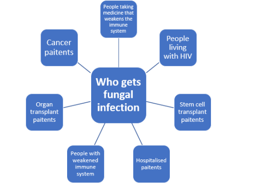
Figure 1: People with high risks of fungal infections
Antifungal resistance of fungal disease:
Antifungal resistance is becoming a major issue for doctors treating patients who are at high risk of invasive mycoses. Resistance to currently available antifungal medications can develop because of drug-induced acquired mechanisms. Aspergillus fumigatus azole resistance, Candida glabrata echinocandin resistance, and azole resistance in non-Candida albicans isolates are recent examples of acquired antifungal resistance (Snigdha et al., 2015, Paul et al., 2016, and Olga et al., 2016). Some fungal species, on the other hand, are intrinsically resistant to specific drugs (e.g., Candida krusei and fluconazole, or Candida lusitaniae and amphotericin B), whereas others show microbiologic resistance to all clinically available antifungals (e.g., Lomentospora (formerly Scedosporium) prolificans and Fusarium solani) (Michaela et al.,2014, T.J. Wash et al.,2004). New species are also emerging that may be resistant to many classes of available drugs (for example, C. auris) (Shawn et al.,2017). Despite the fact that antifungal resistance is not as prevalent as previously thought, treatment options for invasive fungal infections are limited for some microorganisms against different medicines, and patients at highest risk frequently have several comorbidities, including immunosuppression, which may impair therapy effectiveness even in the absence of drug resistance. To address this issue, new therapeutic techniques are required, in addition to overcoming the toxicities/adverse effects and medication interactions associated with currently available antifungals, which can limit therapy effectiveness. Several novel antifungals are currently being tested in preclinical and clinical trials to aid in the fight against antifungal resistance.
Fungal diseases used to be primarily inconveniences or, in rare cases, life-threatening illnesses. Fungi have become significant problems in both the hospital and the community, with increasing morbidity, mortality, and economic costs due to an increase in the number of immune-compromised hosts and an increase in antifungal resistance. These emerging pathogens will necessitate increased awareness as well as a collaborative effort on the part of clinicians, researchers, the pharmaceutical industry, and public health officials. A timely and accurate diagnosis is critical for a positive outcome; therefore, a high level of suspicion is required, based on knowledge of risk factors, disease manifestations, and local epidemiology.
Dear Editorial Team, Clinical Medical Reviews and Reports. My experience with the journal was highly positive. The peer-review process was rigorous, constructive, and completed in a timely manner. The reviewers provided valuable comments that helped improve the quality and clarity of our manuscript. The editorial office was professional, responsive, and supportive throughout all stages of the publication process. Communication was clear and efficient, and any questions were addressed promptly. Overall, I found the journal to maintain high scientific standards and an excellent publication workflow. I would be pleased to consider submitting future work to this journal. Best wishes from, Elena Popa.

It was my pleasure to submit my testimonial concerning the Reviewer Board of our Scientific Journal “Brain and Neurological Disorders”. The Reviewers focused on some modifications and their contribution was helpful. The ladies of our Editorial Office were also supported my efforts. It was my honor to have such a co-operation and I am looking forward for more collaboration.

Dear Grace Pierce, Editorial Coordinator of Journal of Clinical Research and Reports, Thank you for the speedy and efficient peer review process. I appreciate the fact that your peer reviewers do not take months to respond like with some other journals. I would also like to thank the editorial office for responding quickly to my questions. It is an excellent journal. I plan to submit more manuscripts in the future. Best wishes from, Robert W. McGee

Dear Grace Pierce, Editorial Coordinator of Journal of Clinical Research and Reports, Working with you and your team on our recent publication in JCRR has been a truly wonderful and enjoyable experience. The responses were prompt, and the reviewers were patient, constructive, and highly professional. One reviewer in particular gave me the feeling that a professor was carefully reading and commenting on my coursework, which was deeply touching. The entire process was straightforward and hassle‑free, with no tedious online forms to complete. I highly recommend this journal. Best wishes from, DR Aibing Rao, Head of R&D

I Appreciate the Opportunity to Share my Experience with the Journal of Clinical Research and Reports. The peer review process was timely and constructive, and the feedback provided helped improve the quality of our manuscript. The editorial office was professional, responsive, and supportive throughout the process, ensuring smooth communication and efficient handling of the submission. Overall, it was a positive experience collaborating with your team.

Dear Mercy Grace, Editorial Coordinator of Obstetrics Gynecology and Reproductive Sciences, We would like to express our gratitude for your help at all stages of publishing and editing the article. The editors of the magazine answer all the necessary questions and help at every stage. We will definitely continue to cooperate and publish other works in the Obstetrics Gynecology and Reproductive Sciences! Best wishes from, Alla Konstantinovna Politova,
