
Research Article | DOI: https://doi.org/10.31579/2693-2156/064
1 Lausanne University, Faculty of biology and medicine, Lausanne, Switzerland.
2 Centre Hospitalier Universitaire Vaudois, Department of cardiology, Lausanne, Switzerland.
3 Centre Hospitalier Universitaire Vaudois, Department of cardio-vascular surgery, Lausanne, Switzerland.
4 Lausanne University, Plateform of biostatistics, Lausanne, Switzerland.
5 Centre Hospitalier Universitaire Vaudois, Department of cardio-vascular surgery, Lausanne, Switzerland.
*Corresponding Author: Elsa Hoti, Lausanne University, Faculty of biology and medicine, Lausanne, Switzerland.
Citation: Elsa Hoti, Roger Hullin, Matthias Kirsch, Frédéric Schütz, Piergiorgio Tozzi, (2023), How Donor Profile Affects the Outcome of Heart Transplant: A New Score to Predict Primary Graft Failure, J Thoracic Disease and Cardiothoracic Surgery, 4(5); DOI:10.31579/2693-2156/064
Copyright: © 2023, Elsa Hoti, This is an open access article distributed under the Creative Commons Attribution License, which permits unrestricted use, distribution, and reproduction in any medium, provided the original work is properly cited.
Received: 04 September 2023 | Accepted: 12 September 2023 | Published: 21 November 2023
Keywords: heart transplantation; organ donor; primary graft failure; data base analysis
Aim: This study aimed to identify demographic and clinical parameters in donors that could predict the ischaemic time tolerated by the heart transplant in order to increase the number of heart transplantation performed in our centre by expanding the area of graft recruitment.
Methods: A single site retrospective study was conducted on 116 heart transplantations performed in our centre between 2013 and 2021. Multivariate logistic regression analyses were used to identify interactions between the different donor parameters.
Results: Sex, weight, height, total graft ischemic time were the donor parameters independently correlated with primary graft failure (p < 0.1). The logistic regression model included 11 variables, with an AUC of 0.81, this model appears to perform well in the prediction of primary graft failure in the recipient.
Conclusions: A predictive score of primary graft failure was created based on identified donors parameters that may help identify grafts with a higher tolerance to ischemia.
In Switzerland in 2021, 126 patients were placed on the waiting list for a heart transplant but only 33 were able to benefit from a transplantation based on the annual report of Swisstransplant. Recently, the number of patients on the waiting list has been constant and it is known that the longer the wait, the greater the risk that heart failure will negatively impact the functioning of other organs such as the kidneys [1]. Knowing that heart transplant is still the gold standart treatement for end stage heart failure, this mismatch between donors and receivers needs to be improved [2]. Moreover, we live in a time where cardiovascular diseases are the leading cause of death and the ageing of the population suggests that the needs for heart transplants are likely to increase [3]. Since the first heart transplantation performed by Barnard in 1967 [4], many advances have been made in organ preservation [5]. Thanks to all this discoveries the ischemic time that the myocardium can tolerate is now approximately four hours. During this time, the heart of the donor is flushed in-vivo with a cardioplegic solution and then stored at 4°C during the transport to the centre where it will be re-implanted into the receiver. Although this high potassium solution allows diastolic cardiac arrest by inducing membrane depolarisation, the induced calcium overload, myocardial ischemia and hypothermia result in time dependent injury to the heart. Therefore, the commonly accepted total ischemic time is 4 hours, thus limiting the distance at which heart grafts can be procured [4]. Although some studies have already looked at the impact of certain donor parameters (e.g. ABO group [6]) on receiver survival, there are still no guidelines for estimating the ischemic time that a graft can withstand. However, identifying biological and clinical parameters of the donor that would allow to predict the ischemic time that the graft can tolerate, could possibly reduce this mismatch between donors and receivers by increasing the area of graft recruitment. Grafts refused due to ischemia time exceeding 4 hours could be accepted if the donor meets the identified criteria. This could lead to an increase in the number of heart transplants per year and therefore reduce the number of patients who die while on the waiting list for a transplant by practising a more personalised medicine [5].
Study design
A single center retrospective study was conducted on heart transplants performed in our center between January 2013 and December 2021. After identifying the main relative knowledge of the subject by reviewin grelevant scientific publications. Clinical and biological parameters in the donors were selected and compared with the total ischemic time (cold + warm ischemic time corresponding to the implant time) that their graft underwent and with the outcomes in the recipients. This was to determine whether an association between the criteria identified in the donor and the ischemic time that the graft could tolerate existed. Given that the negative effects of longer graft ischemic time are mainly manifested in the early post-transplant period, the outcome was defined by whether or not Extra Corporeal Membrane Oxygenation (ECMO) was required following transplantation, allowing to determine indirectly the presence of a primary graft failure (PGF). Indeed, the marker used for the tolerance of the graft to ischaemia was primary graft failure. As primary graft failure occurs within 24 hours post-transplant and requires the use of ECMO for the cases of a severe presentation of the disease, the use of ECMO in the recipient was recorded only within 24 hours post-transplant. [7]
Data
After the acceptance of the project by the Ethics Commission of the Canton of Vaud (CER-VD 2022-00562), the data of the heart transplants performed in our center from January 2013 to December 2021 were collected. Including all adult donnors and receivers ( 18 years old) and excluding both donnors and receivers for whom a document attesting to a refusal existed (N = 116). The mean donnor age was 46 +/- 14.4 years, 64 (55%) were men. The data collected are presented in Table 1.
| Donor variable | Definition |
| Age | |
| Gender | |
| BMI | |
| Weight | |
| Height | |
| Clinical parameters | |
| Ejection Fraction (EF) | As reported by organ procurement center |
| Blood type | A, B , AB, O |
| Medical history | |
| Diabetes | Positive diabetes history includes type 1 and type 2 with or without treatement. |
| Hypertension | As reported by organ procurement center |
| Malignancy | Defined positive if a history of tumor exists with a 5 year remission phase before the transplantation |
| Active smoker | Includes donnors who were active smokers |
| Biological parameters | |
| Troponins | |
| Creatin Kinase (CK) | |
| Creatinin | |
| Graft ischemic time | Cold + warm meaning the time interval between the application of aortic clamping during cardiac harvesting in the donor and reperfusion of the graft in the recipient |
| Recipient variable | Definition |
| ECMO | Yes/No |
Table 1: Collected data
Analysis were conducted using RStudio version 2021.09.2. First, an independant correlation between each donnor parameter and the use of ECMO in the recipient was investigated in order to evaluate if the association found in our data set are alligned with literature. Therefore boxplots were drawn for continuous values (age, weight, height, BMI, EF, creatinin, troponins, CK and total graft ischemic time). The association was tabulated for categorical values (gender, diabetes, hypertension, malignancy, active smoker and ABO group). For ease of analysis, the values in the tables have been converted to percentages. The continuous variables were transformed using a logarithmic scale to get a better representation for parameters with highly skewed data (troponins, CK, creatinin). T-tests determined wether a statistically significant difference existed between the ECMO and non-ECMO group. The data all followed a normal distribution except for the “age” variable.
At a second phase, multivariate logistic regression tests were performed to create a score that could predict the use of ECMO in the recipient. The parameters included in the second phase were all the relevant parameters found in previous research as good predictors of the tolerance to ischemia of the graft and parameters that had a significant independant correlation with use of ECMO in our data set. Finally, a model was created with the logistic regression test, trained on a fraction equal to 70% of our sample and then tested on the ramaining 30 %. Taking into account our sample size, spliting randomly in half our data set could not provide enough data to the model to train. The model performance was evaluated using ROC analysis and a score was created based on the coefficients of each parameter of the model.
Between 2013 and 2021, 116 patients underwent heart transplatation in our center and met our inclusion criteria. The mean donnor age was 46 +/- 14.4 years, 64 (55%) were men. 25% of recipients were on ECMO within 24 hours post-transplant, i.e. 29 recipients.
After performing the t-tests, the results with p ≤ 0.1 were considered significant. The median of age in both groups was around 50 years old although 75% of the donors of recipients who needed ECMO are above 40 years old (figure 1a). However, there was not a significant interaction between the age of the donor and the use of ECMO in the recipient (p = 0.20). The weight of the ECMO group is slightly higher with an outlier exceeding 140 kg. 50% of the values in the ECMO group are above 80 kg (figure 1b). Likewise, in general, donors in the ECMO group were taller with values ranging from 155 cm to almost 200 cm compared to the non-ECMO group where values ranged from 140 cm to 190 cm. In addition, the median height of the ECMO group was 175 cm compared to 170 cm for the non-ECMO group (figure 1c).These two last parameters were significatly correlated with ECMO use in the recipient (p = 0.06 and p = 0.09, respectively). No significant interaction was observed between the BMI of the donors and a PGF in the recipient (p = 0,50) and not for the ejection fraction either (p = 0.73).The BMIs in both groups were similar with a median at 25 kg/m2 and a single outlier in the ECMO group at more than 50 kg/m2 (figure 1d). The median of the ejection fractions of both groups was around 60% with a single outlier at 80% for the non-ECMO group (figure 1e). The analysis show the total graft ischemic time to have a significant interaction with the use of ECMO in the recipient (p = 0.06). In fact, for half of the patients who were under ECMO, the graft had undergone more than 160 minutes of ischemia with a maximum of 275 minutes. The median stood at 152 minutes for the non-ECMO group and the maximum was 252 minutes (figure 1f).
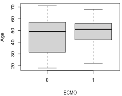
Figure 1a: Boxplot representing the independent association between the age of the donor and the use of ECMO in the recipient
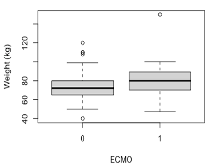
Figure 1b: Boxplot representing the independent association between the weight of the donor and the use of ECMO in the recipient
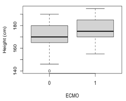
Figure 1c: Boxplot representing the independent association between the height of the donor and the use of ECMO in the recipient
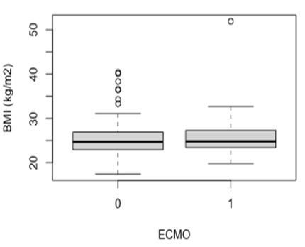
Figure 1d: Boxplot representing the independent association between the BMI of the donor and the use of ECMO in the recipient
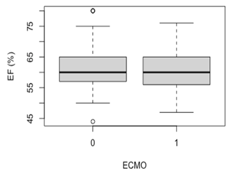
Figure 1e: Boxplot representing the independent association between the ejection fraction of the donor and the use of ECMO in the recipient
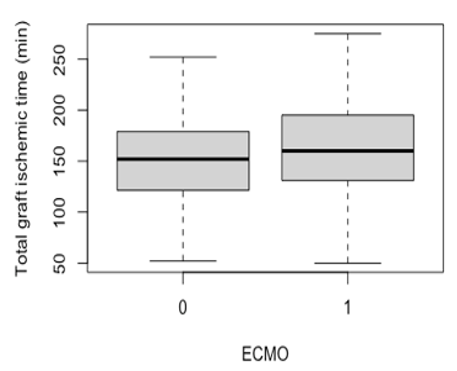
Figure 1f: Boxplot representing the independent association between the total graft ischemic time and the use of ECMO in the recipient
Box plots representing the independent association between creatinine, troponins, CK levels in the donor and ECMO use in the recipient are presented with numerical values (left box plots) and with logarithmic values (right box plots), the transformation of these values into logarithmic scale allows to better observe the distribution of the data. The creatinin levels in the non-ECMO group appears to be more spread with higher outliers. However, the median of both groups is close to 90 umol/l (figure 2a, 2b). Creatinin levels of the donors appears to not be correlated with an ECMO use in the recipient (p = 0,78) as well as troponins and CK levels (p > 0.5 for both parameters). Although the median troponins levels are similar in the 2 groups, maximum values are higher in the non-ECMO group (figure 2c, 2d). As for the CK level, the differences between the 2 groups are minimal except for an outlier which clearly stands out in the ECMO group at over 1000 microg/l (figure 2e, 2f).
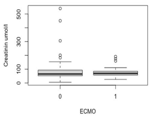
Figure 2a: Boxplot representing the independant correlation between the creatinin level of the donor and the use of ECMO in the recipient (numeric values)
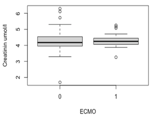
Figure 2b: Boxplot representing the independant correlation between the creatinin level of the donor and the use of ECMO in the recipient (logarithmic values)
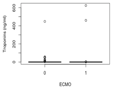
Figure 2c: Boxplot representing the independant correlation between the troponins level of the donor and the use of ECMO in the recipient (numeric values)
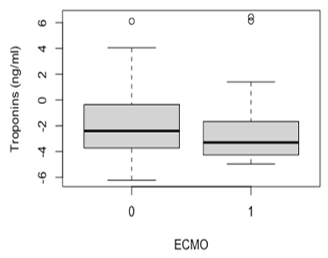
Figure 2d: Boxplot representing the independant correlation between the troponins level of the donor and the use of ECMO in the recipient (logarithmic values)
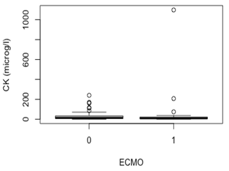
Figure 2e: Boxplot representing the independant correlation between the CK level of the donor and the use of ECMO in the recipient (numeric values)
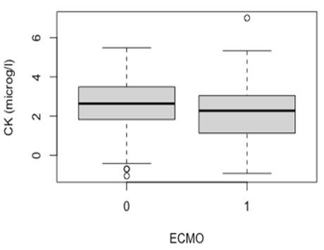
Figure 2f: Boxplot representing the independant correlation between the CK level of the donor and the use of ECMO in the recipient (logarithmic values)Categorical values
After tabulating the independent correlations between the categorical donor parameters and the occurrence of primary graft failure in the recipient, as well as for the continuous parameters, we assessed whether the difference between the 2 groups was significant (table 2). Male donors seem to be significantly more associated with an ECMO use in the recipient (p = 0.07). Although we observed higher proportions of PGF in recipients with a diabetic, hypertensive, active smoker and/or AB blood group donor, the differences between the 2 groups are not significant with p-values ranging from 0.22 to 0.52. The proportions being almost the same in both groups, shows that the history of malignancy in the donor does not seem to be correlated with the use of ECMO in the recipient.
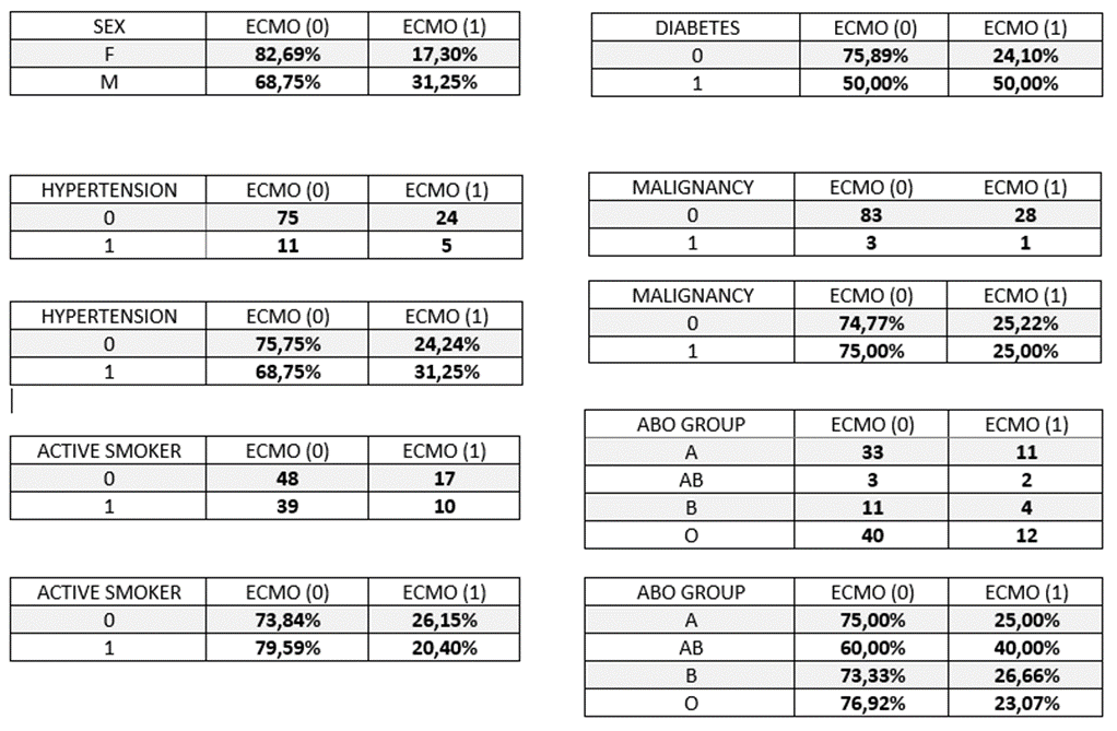
Table 2: The independant correlation between the categorical parameters of the donnor and the use of ECMO in the recipient
Multivariate analysis
In order to create a model based on which a score will be derived, multivariate logistic regression tests were performed. The first model included all donor parameters for which we a significant interaction was found with ECMO use in the recipient (weight, height, total graft ischemic time, gender) as well as donor age. The interaction between these parameters does not appear to play a role in the prediction of a primary graft failure in the recipient. The predictions of the model only slightly match the actual use of ECMO with an accuracy of 0.641 and this is confirmed by the ROC curve of the model, AUC = 0,68 (figure 3a and 3b).
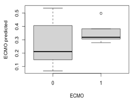
Figure 3a: Boxplot reprensenting the distribution of the predicted values of ECMO for each group with the first model
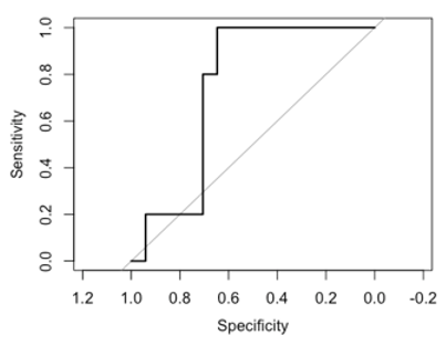
Figure 3b: ROC curve based on the predicted values of ECMO with the first model and the actual values of ECMO in our dataset
The second model included age, gender, total graft ischemic time, height, weight, BMI, EF, hypertension, malignancy, diabetes, troponins, creatinin and CK. With an accuracy of 0.719, this model seems to perform better in the prediction of ECMO use in the recipient. Indeed, as shown in the boxplot (figure 4a), not only the second model predicts more often ECMO in the recipient when it actually was used but is also better to predict when the recipient will not suffer from a primary graft failure as shown by the ROC curve, AUC = 0,81 (figure 4b). The specificity and the sensitivity of the model are respectly of 0,64 and 1.
Scoring system
Based on the ROC curve of the second model, an optimal threshold was calculated at 0.19. The coefficients of each parameter were extracted from the logistic regression model (table 3) which provides the weight of each parameter and allows to calculate a score that could predict the occurrence of PGF in the recipient.
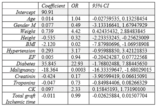
Table 3 : coefficients, OR and 95% CI of each variable of the logistic regression model
By multiplying each parameter of the donor by the corresponding coefficient and additionning all the products and the intercept, a value is obtained. This value is passed through a sigmoid function to obtain a probability between 0 and 1. If the result is under 0.19, the graft can be accepted even with a longer ischemic time with high reliability since the negative predictive value of the score is 100%. With a positive predictive value of 43.75%, if the score is greater than 0.19, the recipient has a 43.75% risk of suffering from PGF. For continuous paramters (age, weight, height, BMI, EF, creatinin, troponins, CK and total graft ischemic time), the value of the donor would be entered in the formula. For the binomial parameters, the coefficient would be multiplied by 1 or 0 wether the medical condition is present in the donor or not. With regard to the sex of the donor, the coefficient is multiplied by 1 if the donor is male and by 0 if the donor is female.
The formula for the score calculation :
x = 90.91 + (age * 0.014) + (sex * 0.077) + (weight * 0.739) + (height * -0.535) + (BMI * -2.120) + (hypertension * 0.293) + (EF * 0.005) + (diabetes * 35.845) + (malignancy * -18.364) + (creatinine * -0.424) + (troponins * -0.047) + (CK * 0.097) + (total graft ischemic time*-0.011) prob = 1 / (1 + exp(-x))
Graft allocation is a complex process faced by a heart transplant clinician. Many parameters have to be taken into account both in the donor (organ function, circumstances of death, medical history) and the recipient. It is therefore quite often difficult to predict the interaction of these different factors and the impact on post-transplant outcome. The current landscape of heart transplantation makes decision making even more complicated. Indeed, with the current demographic shift towards an older population and advances in heart failure management, the number of patients reaching an end stage disease is increasing [8]. However, this increase in patients on the waiting list is not accompanied by an increase in the number of donors. Faced with this discrepancy, different solutions have been proposed. One of them is to adopt extended criteria in organ allocation by accepting transplants from older donors, diabetics or with transmissible diseases [9]. Other teams suggested a scoring system that could predict the perceived risk of graft failure [10,11]. Although there are no standardised guidelines to help the clinician decide whether to accept or reject a graft and this is reflected in organ acceptance rates that vary from one institution to another [12], certain clinical and demographic parameters of the donor are known to affect the outcome in the recipient. One of the major parameters commonly accepted to affect post-transplant mortality in the recipient is the age of the donor. Older age (>40 or >50 years old depending on the study) is associated with a decrease in recipient survival [7, 10, 13, 14, 15]. In our study, the age of the donor does not seem to have a significant interaction with the use of ECMO in the recipient, taking into account our relatively small sample size, the results with a p-value ≤ 0.1 were considered significant. However, some of the studies previously mentioned look at the impact on medium to long term survival (3 months to 5 years) and not directly on primary graft failure which may explain the difference in results obtained, due to a different timing of occurrence [16]. In view of the scientific consensus on this parameter, the age was nevertheless included in our model. The impact of the sex of the donor on the outcome in the recipient is a source of disagreement in the literature. With fairly equal proportions of both sexes in our sample, the association of male sex with ECMO use in the recipient can be considered reliable. On the contrary, in the studies of Kuzemchak MD and al. and Sorabella RA and al., female sex seems to be associated with complications in the recipient in the short term [2,13]. An other study suggested that the mismatch of the sex between the donnor and the recipient depending on the age of the donnor had an impact on the survival of the recipient. Female sex donor was associated with worse long term survival in male recipients when they received a heart from an older donor (>45 years old) [17]. The study of Stehlik J and al. shows that history of hypertension and diabetes mellitus in the donor compromises the survival of the recipient when the heart came from a male donor [12]. The interactions between the different donor parameters mentioned above could explain our results. History of diabetes mellitus and hypertension in the donor is commonly associated with worse survival in the recipient as mentioned also in the report-2020 of the the International Society for Heart and Lung Transplantation [14]. No significant interaction was observed between the presence of these medical conditions in the donor and the developpement of a PGF in the recipient, however, as shown in table 2, the proportions of donors in our sample presenting a history of diabetes mellitus or hypertension are low.
As these diseases are cardio-vascular risk factors, this could lead to a more frequent refusal of hearts from donors with diabetes and/or hypertension, thus explaining the low prevalence in our sample. An other cardio-vascular risk factor is smoking and the smoking history of the donor is also considered in the decision making for the use of a graft [14, 18]. In our study, only the prevalence of active smoker in our sample was accessible, which doesn’t provide information about the history of tobacco use of the donor or the quantity smoked, however these factors are known to play an important role [19]. Therefore, this parameter was considered unreliable. As well as for hypertension and diabetes mellitus, the proportion of donors with malignancy history in our sample is very low (table 2). The severity of the disease could explain the low number of people who are able to fulfil the conditions for being a donor, including being in remission for at least a period of 5 years [20]. The results found for ABO group weren’t significant because of the low number of patients in the AB group. This is compatible with the distribution of blood groups in the general population in Switzerland. Our sample contained 38% of patients with A group, 13% of B, 4% of AB and 45% of O. The distribution in the general population is of 45% of group A, 9% of group B, 5% of AB and 41% of O [21]. This small difference in proportions in our sample compared to the general Swiss population can be explained by the fact that not all hearts come from Swiss centres. Moreover, hearts from O-donors need only ABO-compatible matching and not ABO-identical matching [22] which might explain the higher proportion of O-donors in our sample. Nevertheless, in the literature, O donors appear to be associated with poorer recipient outcomes [6,23]. Size mismatch, BMI, weight, height are well studied and as mentioned in the 2019 report of the International Society of Heart and Lung Transplanation, BMI is not an optimal metric to assess the size match between the donor and the recipient [24]. Although overweight and obesity are associated with cardiovascular risk, it is also known that a high BMI is not necessarily a manifestation of one of these medical conditions. This could explain why no significant difference in BMI was found between the 2 groups in our study. On the contrary, a higher weight and height in the donor seems to be associated with PGF in the recipient. The weight of male recipients shouldn’t be greater than 30 percentage above the weight of the donor and for female recipients the threshold is at 20 percentage according to the recommandations of the International Society of Heart and Lung Transplanation [25]. However, body weight is not well correlated with heart weight neither for women nor for men [26] which may explain our results. In the search for good predictors of outcome in the recipient, biological parameters were also investigated, the transformation of these values into logarithmic scale allows to better observe the distribution of the data. The higher level of troponins found in the non-ECMO group may be the result of the myocardiac suffering as well as any skeletal muscle suffering [27], therefore troponins levels in the donor were not considered to be good predictors of the occurrence of PGF in the recipient. The literature is also sceptical about this parameter with contradictory studies [28,29]. Knowing the relationship between kidney injury and heart failure [30], it could be assumed that the donor's creatinine level is an indirect reflection of their heart health. However, the creatinin levels are similar in both groups, this parameter appears to not be correlated with an ECMO use in the recipient. Indeed, some studies show that creatinin alone is not a good predictor of the outcome, they identified the ratio BUN (Blood Urea Nitrogen) / creatinin to be more representative of the cardiac performance [31]. The final biological parameter analysed was CK level and similarly to troponins level, a slightly higher level of CK was found in the non-ECMO group. This could be explained by the fact that, as with troponins, CKs are not specific to myocardial tissue and can therefore also be a manifestation of skeletal muscle injury or brain damage [32]. And finally, a significant interaction was found between longer total graft ischemic time and the occurrence of PGF in the recipient. As mentioned previously, a total graft ischemic time of more than 4 hours is associated with a worse survival in the recipient [7,13,14].
It is important to mention that genetic studies have also been carried out in the area of tolerance to graft ischaemia. The role of the anti-aging gene Sirtuin 1 for example is critical to heart function and Sirtuin downregulation is associated with endothelial dysfunction and cardiovascular disease. Sirtuin 1 activators versus inhibitors may determine the success of heart transplantation [33-38].
As other teams have done before us, we created a score based on our second model and applicable to the data in our center. The score developped by Eric S Weiss and al was predictive for 30-day mortality with an OR = 0.11 and includes 4 parameters : ischemic time, donor age, race mismatching, and blood urea nitrogen (BUN)/creatinine ratio [11]. Although this score is easier to use and can help in the allocation of donor hearts, we believe that our score brings accuracy in predicting the outcome in the recipient. Firstly, by entering the values of the donor variables directly into the formula instead of assigning points by value range. Secondly, by taking into account more parameters that reflect the donor's cardiac performance. Finally, to ensure the safety of accepting grafts with longer ischemic times, a threshold of 0.19 was fixed in the predictive score. This threshold allowed to reach a negative predictive value of 100% for the prediction of the occurrence of a PGF in the recipient. This is based on our data only, therefore, studies with larger sample sizes and data from other centres would be needed. However, it was ensured that most recipients who were predicted to not require ECMO, will not experience complications such as a PGF by setting the threshold that would minimise the occurrence of false negatives.
This study was conducted in order to consider a possible increase in the number of heart transplants performed in our centre by increasing the area of graft recruitment. Therefore, the score was developed with data from heart transplants performed in our center and may not be applicable in other centres or countries, especially as the sample size was limited to the experience of our centre. Missing data is another limitation inherent to databases such as SOAS, it cannot be excluded that these data would have influenced the results. Due to our sample size, 70% of our data set was used to allow robust training of the model. This implies that our model was tested on the remaining 30%. Therefore, it would be necessary to collect new data in order to not only test the model on a larger sample but also to provide statistically stronger results. And finally, the aim of the study was to identify donor parameters that could affect the time of ischaemia tolerated by the graft by overlooking recipient parameters that could influence post-transplant outcome. It would also be relevant to study factors in the recipient that might predict tolerance to a graft with prolonged ischaemic time.
In this study, demographic and clinical parameters were identified in donors that could predict the ischaemic time tolerated by the heart transplant. The aim was to create a score that would allow the acceptance of transplants from centres further away from ours. The clinicians in charge of heart transplants could easily input donor values in the score calculation formulated in a user-friendly tool such as Excel and obtain a result ranging from 0 to 1. The score threshold being predetermined at 0.19. Although our score is an aid to decision making, it is based on data from heart transplants performed at our centre, further studies are needed before its potential application on a larger scale.
Dear Editorial Team, Clinical Medical Reviews and Reports. My experience with the journal was highly positive. The peer-review process was rigorous, constructive, and completed in a timely manner. The reviewers provided valuable comments that helped improve the quality and clarity of our manuscript. The editorial office was professional, responsive, and supportive throughout all stages of the publication process. Communication was clear and efficient, and any questions were addressed promptly. Overall, I found the journal to maintain high scientific standards and an excellent publication workflow. I would be pleased to consider submitting future work to this journal. Best wishes from, Elena Popa.

It was my pleasure to submit my testimonial concerning the Reviewer Board of our Scientific Journal “Brain and Neurological Disorders”. The Reviewers focused on some modifications and their contribution was helpful. The ladies of our Editorial Office were also supported my efforts. It was my honor to have such a co-operation and I am looking forward for more collaboration.

Dear Grace Pierce, Editorial Coordinator of Journal of Clinical Research and Reports, Thank you for the speedy and efficient peer review process. I appreciate the fact that your peer reviewers do not take months to respond like with some other journals. I would also like to thank the editorial office for responding quickly to my questions. It is an excellent journal. I plan to submit more manuscripts in the future. Best wishes from, Robert W. McGee

Dear Grace Pierce, Editorial Coordinator of Journal of Clinical Research and Reports, Working with you and your team on our recent publication in JCRR has been a truly wonderful and enjoyable experience. The responses were prompt, and the reviewers were patient, constructive, and highly professional. One reviewer in particular gave me the feeling that a professor was carefully reading and commenting on my coursework, which was deeply touching. The entire process was straightforward and hassle‑free, with no tedious online forms to complete. I highly recommend this journal. Best wishes from, DR Aibing Rao, Head of R&D

I Appreciate the Opportunity to Share my Experience with the Journal of Clinical Research and Reports. The peer review process was timely and constructive, and the feedback provided helped improve the quality of our manuscript. The editorial office was professional, responsive, and supportive throughout the process, ensuring smooth communication and efficient handling of the submission. Overall, it was a positive experience collaborating with your team.

Dear Mercy Grace, Editorial Coordinator of Obstetrics Gynecology and Reproductive Sciences, We would like to express our gratitude for your help at all stages of publishing and editing the article. The editors of the magazine answer all the necessary questions and help at every stage. We will definitely continue to cooperate and publish other works in the Obstetrics Gynecology and Reproductive Sciences! Best wishes from, Alla Konstantinovna Politova,
