
Research Article | DOI: https://doi.org/10.31579/2690-4861/499
1 Department of Gynecology and Obstetrics, The Second Affiliated Hospital of Anhui Medical University, Hefei, Anhui, 230601, China.
2 The School of Life Science, Anhui Medical University, Hefei, Anhui, 230601, China.
*Corresponding Author: Wenyan Wang, Furong Road, Economic and Technological Development District, Hefei, Anhui, 230601, China and Enlin Wang, Tanghe Road, Yaohai District, Hefei, Anhui, 230032, Chian.
Citation: Xiaofeng Ma, Yun Lu, Bing Wei, Wenyan Wang, Enlin Wang, (2024), High Expression of KIF4A Predicts Poor Prognosis Hallmark and Is Correlated with Immune Infiltrates in Cervical Cancer, International Journal of Clinical Case Reports and Reviews, 19(4); DOI:10.31579/2690-4861/499
Copyright: © 2024, Wenyan Wang and Enlin Wang. This is an open-access article distributed under the terms of the Creative Commons Attribution License, which permits unrestricted use, distribution, and reproduction in any medium, provided the original author and source are credited.
Received: 01 July 2024 | Accepted: 19 July 2024 | Published: 14 November 2024
Keywords: kif4a; cervical cancer; bioinformatics; hallmark; immune infiltration
Cervical cancer (CC) has become the fourth most common cancer among women and cause a larger number of deaths in worldwide. Screening at the early stage of CC is an effective precaution. Discovery of the new hallmark of CC will provide a guidance for CC screening. Kinesin family member 4A (KIF4A) expressed in a variety of tissues and also contributed to development of several cancers, however its function in CC remains unclear. In this study, high-expression of KIF4A was observed in the CC patients according to the bioinformatics analysis and clinical test. Additionally, loss-function of KIF4A with shRNA abrogated cervical cell proliferation, migration and invasion. We also found that the difference expression genes were identified between KIF4A-high and KIF4A-low CC patients among with abundant mutation of several genes occurred in the CC progression. Finally, we also proved that KIF4A was involved in the immune cell infiltration in the CC patients by clinical information analysis. These demonstrated that the dys-expression of KIF4A may be used for the CC screening and clinical therapy.
Cervical cancer (CC) is the fourth most common cancer among women worldwide and causes over 300,000 deaths in worldwide [1]. Although the high-risk human papilloma virus (hrHPV) infection is a major pathogenic factor for CC development, but several other pathogenic factors also led to CC progression in the non-hrHPV infected patients [2]. Therefore, investigation of development mechanisms and urgent diagnostic markers in CC are an important work for the disease prevention and clinical therapy. The dysfunctional alterations of genome stability, transcriptional control, translation regulation and posttranslational modification in CC generation were occurred. This alteration was regulated by abundant regulatory factor, such as the novel proteins, miRNAs and lncRNAs. The diagnostic biomarkers may be applied to be a future target for the new therapy in the rational drug design [3-5]. In recent years, the bioinformation analysis and clinical classification study is widely used in the CC biomarker screening and attestation, which received high-considerable attention on new-emerging molecular marker discovery and was widely applied in clinical practice [6, 7]. Kinesin family member 4A (KIF4A) belongs to the chromosome-associated kinesin superfamily and contains 1232 amino acids encoded by KIF4A, located on chromosome Xq13.1[8]. Structurally, KIF4A mainly contained the motor domain and microtubule-binding motif, which regulated multiple cellular progresses, including chromosomes rearrangement, sister chromatids separation and associated protein movement. Accumulating evidences reported that KIF4A expressed in a variety of tissues and the overexpression of KIF4A may contribute several diseases and cancer development. In hepatocellular carcinoma, it has been proved that KIF4A promoted hepatoma carcinoma cell (HCC) over-proliferation via activating PI3K/AKT-pathway activity [9]. Besides, KIF4A also acted as the target molecular in HCC progression. Hu et al demonstrated FOXM1 enhanced KIF4A expression and provoked HCC progression by binding to the promotor region of KIF4A, directly[10]. In addition, Hepatitis B virus (HBV) infection, one major causes of HCC, upregulated KIF4A expression and promoted HCC [11, 12]. Taken together, KIF4A plays a critical role in HCC, but the function of KIF4A in CC is still less-known. In this study, we first screened the upregulated genes in the CC patients and found KIF4A was overexpressed in CC according with TCGA, GTEx and GEOs datasets. Besides, we demonstrated KIF4A was indeed upregulated in the malignant CC patient samples with biochemical test. Loss-function of KIF4A inhibited cell proliferation, migration and invasion. Finally, we identified the multiple signaling pathways and cellular progresses between KIF4A-high and KIF4A-low group, which may indicate KIF4A modulated these pathways to regulated CC development.
2.1 Cell culture and transfection
Human embryonic kidney cell HEK293T and cervical cancer cell Hela cell lines were purchased from the American Type Culture Collection (ATCC) and both cell lines were cultured in the DMEM medium supplemented with 10?tal bovine serum (FBS; Gibco) and 1% penicillin/streptomycin (Solarbio) at 37 ℃ under 5% CO2. For establishment of KIF4A RNAi cell lines with short hairpin RNA (shRNA), the Hela cells were transfected with Lipofectamine 2000 (Invitrogen, USA) according to the manufacturer’s handbook and screened with puromycin. Two shRNA-KIF4A sequences are: shKIF4A-1# (5’-3’)- CCTCAGGAATGAGGTTGTGAT,shKIF4A-2# (5’-3’)- CTTACTGAAGTGCGTGGTCAA.
2.2 Immunoblotting
Cultured cells and cervical tissue samples were lysed with lysis buffer RIPA contained proteinase inhibitor PMSF on ice for 30 min. After centrifugation at 12,000g for 15 min, the supernatant was boiled with loading buffer at 100 ℃ for 5 min. The lysates were subjected into sodium dodecyl sulphate-polyacrylamide gel electrophoresis (SDS-PAGE) and transferred polyvinylidene fluoride (PVDF) membranes according to a standard protocol. The membranes were blocked with 5% slim milk for 1 h at room temperature and incubated with primary antibodies overnight at 4°C. On the next day, the membranes were washed with TBST and incubated with the HRP-conjugated secondary antibodies for 1 h at room temperature. After being washed with TBST, the membrane was drenched in ECL HRP substrate (Solarbio) for signal generation.
2.3 Immunohistochemistry
All tissue samples were formalin-fixed and tissue sections were deparaffinized in xylene, boiled in 10 mM citrate buffer (pH 6.8) at 100 ℃ for 5 min. Tissue sections were blocked with 3% BSA for 30 min and probed with KIF4A (ABclonal, 1:100 for IHC, 1:2000 for WB) antibody at 4 ℃ overnight. Next day, all sections were washed with TBST and immunoreacted with the biotinylated secondary antibody at room temperature for 15 min. Finally, signals were raised by DAB substrate kit and pictures were visualized under microscopy (Leica, LeicaDFC420C).
2.4 Cell colony formation and cell proliferation assay
The scramble, shKIF4A-1# and shKIF4A-2# Hela cells (1 × 10³) respectively were seeded in the 6-well plates and replaced with the fresh medium every 2 days until the clones were noted (about 2 weeks). After being washed with PBS, the clones were fixed with 4% paraformaldehyde (PFA) for 15 min and stained with 1% crystal violet for 2 h at room temperature. After the staining, the plates were rinsed with distilled water and dried. The images of the plates were collected and clones were counted directly. The scramble, shKIF4A-1# and shKIF4A-2# Hela cells (1 × 10⁴) were cultured in the 96-well plates and counted at the indicated time points.
2.5 Cell Migration and Invasion Analysis
For the migration assay, Transwell inserts (pore size: 8 μm, Corning, USA) in 24-well plates were applied. The Hela cells (2 × 10⁴) were seeded into upper chamber and cultured in DMEM medium with 20?S in the lower chamber. After 24 h, the migrated cells on the lower side were fixed with 4% paraformaldehyde, stained with 0.1% crystal violet for 10 min and eluted in 250 μL of 10?etic acid for 10 min. The absorbance was measured at 570 nm. The cell invasion assay was carried out similarly, except that pre-coating with 100 μL of 1:8 DMEM-diluted Matrigel (BD Biosciences, Franklin Lakes, NJ, USA) was added to each well at 37℃ for 6 hours before cells were seeded onto the membrane, followed by incubation for 48 hours. An equal number of seeded cells was also stained and measured for absorbance, and the reading was used to divide that of migrated cells.
2.6 Data collection and statistical analyses
The RNA-seq data and clinical data of cervical cancer patients from TCGA and GTEx datasets were downloaded in the online website Sangerbox (http://sangerbox.com). The data of mutations were downloaded and visualized using the maftools package in R software. Genes with higher mutational frequency detected of cervical cancer patient in histogram was showed. The RNA-seq data of Gene Expression Omnibus (GEO) were downloaded from GEO Dataset (https://www.ncbi.nlm.nih.gov/gds) and analyzed with online analyses tool GEO2R.
2.7 Statistical analysis
GraphPad Prism 5.0 was used to performed statistical analysis. Student t-test was applied to analyze the data. The data were presented as mean ± SD of two independently experiments in triplicate. The p<0>
3.1 High-expression of KIF4A in the cervical cancer patients from TCGA, GTEx and GEOs datasets
We screened difference expression genes (DEGs) in CC patients from TCGA and GTEx databases. The genes mRNA profile that contained CC samples (n=306) and normal samples (n=22) was used for classification. The adjusting criteria was set as adj. p‐value <0>p<0>p<0>
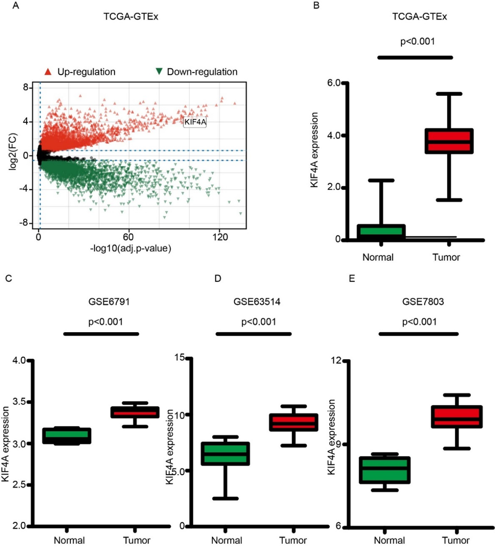
Figure1. High-expression of KIF4A in the cervical cancer patients from TCGA, GTEx and GEOs datasets. (A). The volcano plot of DEGs in the cervical cancer patients compared with the normal. (B). The high-expression of KIF4A was detected in CC patients from TCGA and GTEx. (C). The high-expression of KIF4A was calculated in CC patients according to GSE6791 database. (D). The high-expression of KIF4A was calculated in CC patients according to GSE63514. (E). The high-expression of KIF4A was calculated in CC patients according to GSE7803 database.
3.2 The clinical expression pattern of KIF4A in cervical cancer
In order to determine the KIF4A expression pattern in the cervical cancer patients, we collected the cervical cancer and the normal cervical tissues from the Second Hospital of Anhui Medical University. Immunohistochemistry staining (IHC) shown that the higher expression of KIF4A was revealed along with morphology alteration of cancer cell nucleus and performed statistical analysis according to histochemical staining criteria (Figure 2A-B). In addition, we detected the KIF4A protein level in cervical cancer tissues and the adjacent normal tissues. Consistent with IHC result, the KIF4A protein level was also up-expressed in cervical cancer samples (Figure 2C-D). Overall, the KIF4A was indeed upregulated in the cervical cancer.
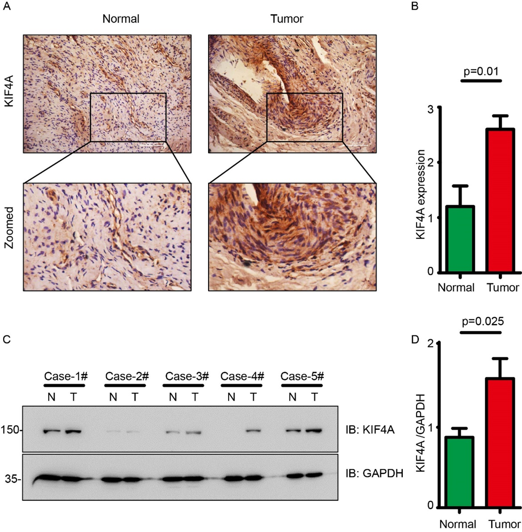
Figure 2. The clinical expression pattern of KIF4A in cervical cancer. (A). Immunohistochemistry staining of KIF4A in cervical cancer tissues and paired normal tissues. (B) Quantitive analysis of immunohistochemistry staining in panel A. (C) Western blot of KIF4A in the cervical cancer tissues and paired normal tissues. N: normal, T: tumor. (D) Quantitive analysis of western blot in panel C. (normalized with GAPDH).
3.3 Inhibition of cervical cancer cell proliferation and migration with loss-function of KIF4A
To investigate the function of KIF4A in the cervical cancer, shRNAs against KIF4A were performed to knockdown KIF4A in Hela cell (Figure 3A). After transfection, clone formation assay, Transwell assays were used to assess cell proliferation, migration and invasion. As shown in Figure 3B knock-down of KIF4A reduced the cell proliferation significantly compared with the control group. Consistently, the colony formation assays also showed that the number of cell clone was decreased in the KIF4A-RNAi cell lines (Figure 3C), which suggesting deletion of KIF4A inhibited cell proliferation. In the transwell assay, KIF4A silencing reduced the cells migration and invasion (Figure 3D-3E). Collectively, these findings indicate that attenuation of KIF4A in cervical cancer cells prevents from cells proliferation, migration and invasiveness.
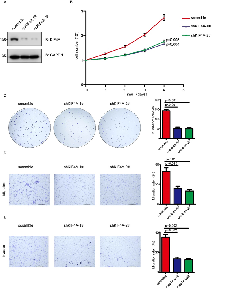
Figure 3. Inhibition of cervical cancer cell proliferation and migration in loss-of function of KIF4A in Hela cells. (A) The expression of KIF4A was detected by western blot in Hela cells transfected with the scramble, shKIF4A-1# and shKIF4A-2#. (B) The cell number of scramble, shKIF4A-1# and shKIF4A-2# was counted at indicated time points. (C) The clone formation assay detected the cell proliferation after knowdown of KIF4A. (D-E) Transwell assay detected the migration and invasion of Hela cell after knowdown of KIF4A.
3.4 Identification of DEGs between KIF4A-High and KIF4A-Low cervical cancer patients
In order to investigate how KIF4A mediates cell proliferation and migration, we explored the difference pathway and functional biological mechanism occurred between the KIF4A-High expression and KIF4A-Low expression patients. There are 483 differentially expressed genes (DEGs) between the KIF4A-High and KIF4A-Low groups, including 380 up-regulated genes and 84 down-regulated genes (|Log2-FC| > 1.5, adjusted p value <0>NCAPH, BUB1, CENPI, RACGAP1 and DEPDC1, the top 5 down-regulated genes included PDZK1IP1, SLPI, LCN2, TCN1 and ARL14. GO enrichment and KEGG signal-pathway analysis were used to elaborate potential pathway and functional biology involving the entire 483 DEGs (Figure 4B). Generally, in the KEGG pathway enrichment, the DEGs mainly participate in cell cycle control, human immunity response and DNA replication. Besides, the GO enrichment analysis shown the DEGs regulated cell cycle and cell division (Figure 4C). Hypothetically, the feature of KIF4A in promoting cervical cancer development may participate in the cell cycle anddivision control and tumor immunity response.
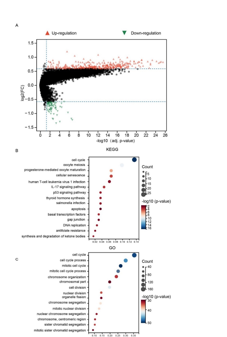
Figure 4. DEGs were identified between KIF4A-High and KIF4A-Low cervical cancer patients. (A) Volcano plots of DEGs in the KIF4A-High expression and KIF4A-Low expression patients. (B) KEGG pathway analysis of DEGs. (C) GO enrichment analysis of DEGs.
3.5 The alterations in mutations between KIF4A-High and KIF4A-Low patients
Analysis of the gene mutation data from TCGA showed that a total of 248 in 286 samples existed abundant mutations. Several genes in cervical cancer (TTN, PIK3CA, KMT2D, and FBXW7) contained the most common missense mutation. In the KIF4A-high group, 48 patients carried TTN mutations, 47 patients carried PIK3CA mutations, 23 patients had KMT2D mutations, and 19 patients harbored FBXW7 mutations. In the KIF4A-low group, 40 patients had TTN mutations, 47 patients harbored PIK3CA mutations, 20 patients carried KMT2D mutations, and 17 patients harbored FBXW7 mutations (Figure 5A). Further analysis focused on the somatic mutations in the cervical cancer patients according to the massively parallel sequence and missense mutations. SNV occurrence, with nucleotide mutation were obtained. All mutation information were listed in (Figure 5B-5G).
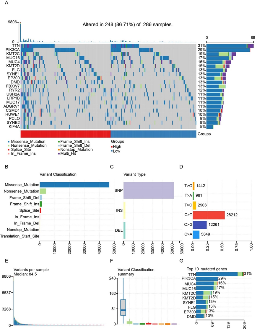
Figure 5. The alterations in mutations between KIF4A-High and KIF4A-Low patients. (A) Onco-plot of somatic landscape in the KIF4A-high and KIF4A-low groups. (B-G) Cohort summary plot of variant classification, type and SNV class, mutation load of each sample, variant classification and the top ten mutated genes.
3.6 The correction of immune infiltration with KIF4A expression
As found in the KEGG pathway analysis, we hypothesis the KIF4A may affect tumor immune activity and regulate cervical cancer progression. Therefore, we used the TIMMER tool to test the correlation between KIF4A expression and infiltration levels of six immune cell subtypes. As
shown in the (Figure 6A), the KIF4A expression was positively associated with the infiltration of CD4+T cells (Cor=0.188 p=1.63e-03) and dendritic cells (Cor=0.134 p=2.57e-02). Moreover, copy number variation (CNV) of KIF4A always significantly related to B cell, CD8+ T cells, CD4+ T cells and dendritic cells infiltration (Figure 6B), suggesting the expression of KIF4A modulate immune cell infiltration and may be implicated in the tumor immunology.
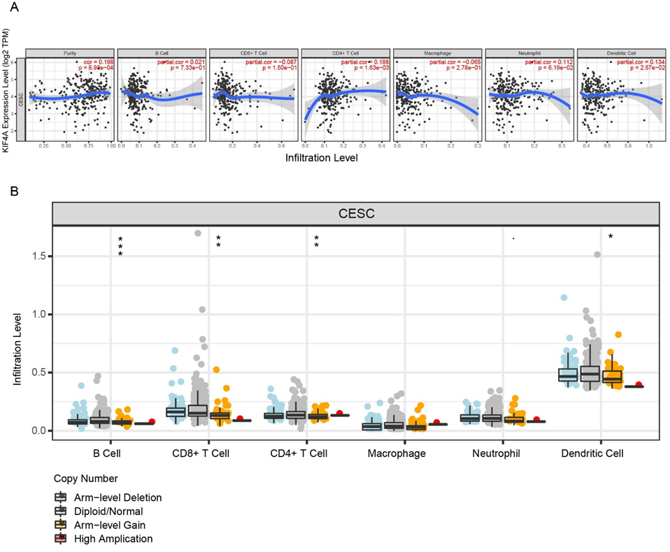
Figure 6. The correction of immune infiltration with KIF4A expression. (A) The correction of B cell, CD8+ T cells, CD4+ T cells, macrophage, neutrophil and dendritic cells infiltration with KIF4A expression. (B) The copy number variation of KIF4A affected B cell, CD8+ T cells, CD4+ T cells, macrophage, neutrophil and dendritic cells infiltration.
Cervical cancer is a malignant epithelial tumor that forms in the uterine cervix. The HPV vaccination has greatly prevented its occurrence [13], However, it remains the fourth-most common cancer in women globally and a major cause of mortality among women in developing countries due to inadequate screening protocols [14]. Advanced cervical cancer usually requires total hysterectomy with chemotherapy or radiotherapy[15, 16]. Despite the available standard treatment of choice, outcome is suboptimal among patients with CC and there are no satisfactory treatments for patients with late-stage disease [17]. Therefore, early diagnosis (or precancer screening) and assessment of disease progression trends are effective to improve the prognosis of CC patients and important for the discovery of novel molecular targets for treatment. Recently, with the development of molecular bioinformatics, the application of the expression of related genes in the diagnosis and prognosis evaluation of malignant tumors has gradually increased, because these methods provide a certain reference for the prevention and treatment of diseases. KIF4A is the members of the Kinesin (KIF) superfamily proteins which are microtubule-dependent molecular motors [18]. As an oncogene, KIF4A is abnormally expressed in a variety of cancers. Interestingly, KIF4A is often up-regulated but can also be down-regulated in some cancers [19].. In our study, the expression of KIF4A was up-regulated in CC tissues. This suggests distinctive regulatory mechanisms for different cancers. KIF4A participated in a variety of cellular processes, such as DNA repair, mitosis [20], genetic stability [21], virus infection [22] and tumor progression [23]. For instance, intracellular KIF4A can lead to mitotic defects and DNA damage repair, and chromosome instability and the formation of aneuploidy, cause abnormal cell proliferation, differentiation and cause tumor formation [24, 25]. In accordance with our current results showing the robust elevation of KIF4A, enhance proliferation in in both Hela cells and SiHa cells. Additionally, analysis of the gene mutation data from TCGA showed that TTN, PIK3CA, KMT2D, and FBXW7 are the most abundant mutation genes both in KIF4A-high group and KIF4A-low group. In the KEGG pathway enrichment, the DEGs mainly participate in cell cycle control, human immunity response and DNA replication. Besides, the GO enrichment analysis also shown the DEGs regulated cell cycle and cell division. Tumor immune escape is the key step of initiation of tumor metastasis, which is response for the failure of some cancer therapy and poor prognosis [26]. Currently, outcomes for women with node-positive and metastatic cervical cancer remain poor [27]. KIF4A is closely associated with the activation of immunocytes. For instance, the high expression of KIF4A is always correlated with the fewer CD8+ tumor-infiltrating lymphocytes (TILs) and a much worse prognosis in patients of bladder cancer [28]. Similarly, the immune-related cells such as B cell, CD4+ T cell, CD8+ T cell, Neutrophil and Myeloid dendritic cells levels also were decreased as KIF4A high-expressed, which promoted occurrence of breast cancer metastasis [29]. The result from TIMER tool showed in the tested six immune cell subtypes, KIF4A expression was positively associated with the infiltration of CD4+T cells and dendritic cells. Moreover, copy number variation (CNV) of KIF4A always significantly related to B cell, CD8+ T cells, CD4+ T cells and dendritic cells infiltration, suggesting the expression of KIF4A modulate immune cell infiltration and may be implicated in the tumor immunology. Clearly, the results of this study are contradictory to previous results in breast and bladder cancer [28, 29]. This suggests distinctive regulatory mechanisms for in CC. This study may have several limitations. We were limited by the sample size of TCGA and clinical specimens, which may has resulted in a slight bias. Further research will help to further validate our findings and better understand the mechanisms of KIF4A in CC.
In current study, we identified 8844 differentially expressed genes (DEGs) by analyzing CC mRNA expression profiling datasets in the TCGA plus GTEx datasets. Based on the above analysis, KIF4A was selected as the key CC gene in our study. Western blot and IHC staining were used to detect the expression of KIF4A in collected clinical CC and adjacent tissues., It was found that the expression level of KIF4A in CC tissues was significantly higher than that in corresponding adjacent tissues. IHC staining revealed that KIF4A was mainly positively expressed in the cytoplasm and the expression level of KIF4A in patients with advanced CC was significantly higher. In a series of cell function experiments, Knockdown KIF4A was found to decrease the migratory and proliferation ability of CC cells, suggesting that KIF4A may be an oncogene of CC. Extensive investigation revealed KIF4A may exert its capacity to control cell cycle and division, DNA stability and tumor immune, highlighting the potential of KIF4A as a new biomarkers and therapeutic targets for the monitoring and treating CC.
Funding:
This study was supported by Science Foundation of Anhui Medical University (grant number. 2023xkj016).
Xiaofeng Ma, Yun Lu, Bing Wei and Wenyan Wang and Enlin Wang were contribute to the conception of the work, acquisition, analysis and interpretation of the data, and drafted the work. All authors approved the version to be published and are accountable for all aspects of the work in ensuring that questions related to the accuracy or integrity of any part of the work are appropriately investigated and resolved.
all author declares that there is no conflict of interest in this article.
Ethical approval to report this case was obtained from the Institutional Review Board of Anhui Medical University (No: 20180023).
Data will be made available on reasonable request.
Dear Editorial Team, Clinical Medical Reviews and Reports. My experience with the journal was highly positive. The peer-review process was rigorous, constructive, and completed in a timely manner. The reviewers provided valuable comments that helped improve the quality and clarity of our manuscript. The editorial office was professional, responsive, and supportive throughout all stages of the publication process. Communication was clear and efficient, and any questions were addressed promptly. Overall, I found the journal to maintain high scientific standards and an excellent publication workflow. I would be pleased to consider submitting future work to this journal. Best wishes from, Elena Popa.

It was my pleasure to submit my testimonial concerning the Reviewer Board of our Scientific Journal “Brain and Neurological Disorders”. The Reviewers focused on some modifications and their contribution was helpful. The ladies of our Editorial Office were also supported my efforts. It was my honor to have such a co-operation and I am looking forward for more collaboration.

Dear Grace Pierce, Editorial Coordinator of Journal of Clinical Research and Reports, Thank you for the speedy and efficient peer review process. I appreciate the fact that your peer reviewers do not take months to respond like with some other journals. I would also like to thank the editorial office for responding quickly to my questions. It is an excellent journal. I plan to submit more manuscripts in the future. Best wishes from, Robert W. McGee

Dear Grace Pierce, Editorial Coordinator of Journal of Clinical Research and Reports, Working with you and your team on our recent publication in JCRR has been a truly wonderful and enjoyable experience. The responses were prompt, and the reviewers were patient, constructive, and highly professional. One reviewer in particular gave me the feeling that a professor was carefully reading and commenting on my coursework, which was deeply touching. The entire process was straightforward and hassle‑free, with no tedious online forms to complete. I highly recommend this journal. Best wishes from, DR Aibing Rao, Head of R&D

I Appreciate the Opportunity to Share my Experience with the Journal of Clinical Research and Reports. The peer review process was timely and constructive, and the feedback provided helped improve the quality of our manuscript. The editorial office was professional, responsive, and supportive throughout the process, ensuring smooth communication and efficient handling of the submission. Overall, it was a positive experience collaborating with your team.

Dear Mercy Grace, Editorial Coordinator of Obstetrics Gynecology and Reproductive Sciences, We would like to express our gratitude for your help at all stages of publishing and editing the article. The editors of the magazine answer all the necessary questions and help at every stage. We will definitely continue to cooperate and publish other works in the Obstetrics Gynecology and Reproductive Sciences! Best wishes from, Alla Konstantinovna Politova,
