
Case Report | DOI: https://doi.org/10.31579/2690-8808/187
1 Senior Resident MD obstetrics and Gynaecology Department of Obstetrics and Gynaecology Lady Hardinge Medical College, New Delhi, India.
2 Director Professor and Head of Department MD obstetrics and Gynaecology Department of Obstetrics and Gynaecology Lady Hardinge Medical College, New Delhi, India.
3 Professor MD, obstetrics and Gynaecology Department of Obstetrics and Gynaecology Lady Hardinge Medical College, New Delhi, India.
4 Junior Resident Obstetrics and Gynaecology Department of Obstetrics and Gynaecology Lady Hardinge Medical College, New Delhi, India.
*Corresponding Author: Anuradha Singh. Professor MD, obstetrics and Gynaecology Department of Obstetrics and Gynaecology Lady Hardinge Medical College, New Delhi, India.
Citation: Noopur Chawla, Kiran Aggarwal, Anuradha Singh, Prabha Lal and Kavita Badhal, (2024), Herniation of Gravid Uterus: Case Report. J.Clinical Case Reports and Studies, 5(1); DOI:10.31579/2690-8808/187
Copyright: © 2024, Anuradha Singh. This is an open access article distributed under the Creative Commons Attribution License, which permits unrestricted use, distribution, and reproduction in any medium, provided the original work is properly cited.
Received: 01 March 2024 | Accepted: 11 March 2024 | Published: 18 March 2024
Keywords: pregnancy; incisional hernia; abdominal surgery; herniation of uterus; skin ulcers
Incisional hernia is the most common long-term complication of abdominal surgery. After midline laparotomy, more than 10% of patients develop an incisional hernia. Anterior abdominal wall hernias are uncommon in pregnancy. Hernias complicating pregnancy can be an obstetric as well as a surgical challenge to manage especially if the hernia is complicated or the patient comes into labor at term gestation. We reported 2 cases of a young woman who presented to us with term gestation in labor with a large anterior abdominal wall hernia and the challenges we faced in managing the cases.
Herniation of Gravid Uterus: Report of 2 Cases
Incisional hernia is the most common long-term complication of abdominal surgery. After midline laparotomy, more than 10% of patients develop an incisional hernia. Anterior abdominal wall hernias are uncommon in pregnancy. Hernias complicating pregnancy can be an obstetric as well as a surgical challenge to manage especially if the hernia is complicated or the patient comes into labor at term gestation. We reported 2 cases of a young woman who presented to us with term gestation in labor with a large anterior abdominal wall hernia and the challenges we faced in managing the cases
Case 1:
A 21-year-old lady, G2P1L0 at 37 weeks gestation presented to us in an emergency in early labor with a large incisional hernia. She gave a history of having undergone an exploratory laparotomy for suspected tubercular pyoperitoneum 10 days after her first normal vaginal delivery. She had stitch line sepsis and had a prolonged hospital stay of 3 months after the laparotomy due to poor healing of the stitch line for which a skin graft was placed. She was consequently started on antitubercular treatment. After 7-8 months of surgery, she started developing a gradually increasing swelling in the abdomen arising from the surgical scar which was then diagnosed to be an incisional hernia for which she did not take any treatment. She conceived again 1 month after developing the hernia. The patient also gave a history of occasional pain abdomen which was resolved with analgesics.
She was admitted at 24 weeks of gestation to our hospital with pain abdomen. Ultrasonography showed the gravid uterus to be the content of the incisional hernia sac. She was managed conservatively with bed rest and analgesics and was discharged after 2 days. The patient was lost to follow-up after 24 weeks of period of gestation.
At 37 weeks the patient presented in early labor. Her vitals were stable and per abdominal examination revealed a large 20 x 20 cm incisional hernia arising due to a defect from the midline incision. It was globular in shape and an old skin graft was present on its surface. The skin was dry and scaly with a large superficial ulcer measuring 7 x 2 cm on the surface of the hernia with unhealthy margins (Fig 1) On palpation, the uterus was present in the hernial sac but it was reducible without any complication, abdomen had a doughy consistency and was non-tender. It was difficult to palpate the uterine contour and contractions which made monitoring of labor difficult and the fetal heart could only be heard by on CTG. Her investigations showed mild anemia Hb 10gm%, and liver and kidney functions were normal. Since the patient had no hernia-related or obstetric complications the patient was allowed for normal vaginal delivery. She delivered a baby girl weighing 1.8 kg uneventfully. The post-delivery patient was planned for the repair of a hernia.
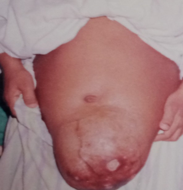
Figure 1
A 24-years old G2P1L1 with previous LSCS done for Cephalopelvic disproportion with obstructed labor, was admitted to our hospital at 36 weeks POG in early labor. On examination her vitals were stable. Abdominal examination revealed gravid uterus eventrating out through previous LSCS midline scar area (Fig 2). Her investigations showed mild anemia. The patient underwent emergency LSCS for CPD in labor along with repair of incisional hernia by the surgical team using the double breasting technique of rectus sheath. The postoperative course was uneventful and the patient was discharged on the 10th postop day. Few similar case reports have been described in the literature, that highlight the risk factors, complications during pregnancy, timing, and method of repair. Multiple risk factors for incisional hernia have been identified, including obesity, infection, diabetes, smoking, immunosuppression medication, ascites, advanced age, and poor nutritional status [1,2]. Pregnancy itself is also associated with an increased risk of abdominal hernia recurrence [3]. Midline incisions are associated with a higher incidence of incisional hernia than transverse incisions which was present in our patient as well [4]. In the cases reported in the literature, the Cesarean section accounts for approximately 60% of the surgeries that lead to incisional hernia whose incidence is 3%. [5-9] However in our case preceding surgery was laparotomy with pyoperitoneum The risk factors following cesarean sections which are associated with hernia formation include the need for additional operative procedures, postoperative abdominal distension, intra-abdominal sepsis, residual intra-abdominal abscess, Surgical site infection and wound dehiscence [10] Our patient had a history of midline incision laparotomy followed by wound infection and intraabdominal sepsis, a poor nutritional status which was the probable reasons for the incisional hernia. As the uterus is very much enlarged to fit inside the hernial sac by the time it reaches the level of the hernial aperture, herniation of the gravid uterus is probably uncommon. However, in both instances, the uterus was a component of the hernial sac. Although initially reducible, late in the course of pregnancy, incarceration and subsequent strangulation within the hernial sac may complicate the herniation of the pregnant uterus.
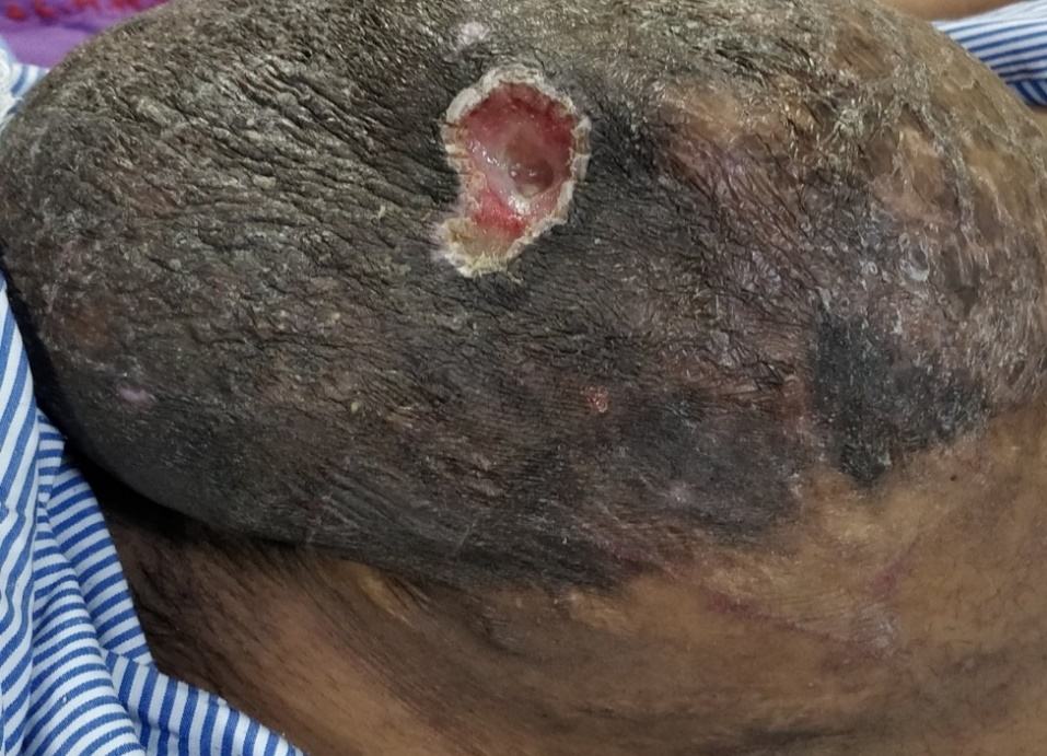
Figure 2
The usual complications encountered include burst abdomen, incarceration, strangulation or evisceration of the uterus in the hernial sac complicating the obstetric management [12-16] The skin overlying the distended hernia can ulcerate and act as a source of infection which was present in our patients [17]. The other problem encountered is physical discomfort caused by a gravid uterus hanging down in the abdomen. Hernias have previously been estimated to account for under 5 % of the cases of bowel obstruction during pregnancy [18]. There may be potential obstetrics complications like spontaneous abortion, preterm labor, accidental hemorrhage, intrauterine fetal death, and rupture of the lower uterine segment during labor [19] Hence, such patients require multidisciplinary teamwork.
The management includes reduction of gravid uterus in abdominal cavity if uncomplicated, Obstetrical management and treatment of ulcers.It depends on the symptoms and the stage of pregnancy at the time of diagnosis. It may be preferable to postpone surgery for pregnant patients with minor, asymptomatic hernias until after birth or after the last pregnancy. It may be also advisable to delay the repair until the second trimester or after delivery if complications do not arise if the hernia is symptomatic and appears to be affecting the patient's quality of life. If the hernia is significant and causing symptoms in individuals who are not pregnant, it may be preferable to perform an elective repair and then postpone the next pregnancy for a year or two. The recurrence of Hernia is significantly increased by pregnancy. When possible, laparoscopic mesh repair should be offered; however, in complex cases, the open method may be preferable. In cases of severe contamination as well as small hernias, the suture repair may be appropriate [20].
In strangulation occurring in early pregnancy immediate repair is required and the pregnancy may be carried to term. Pregnancy-related incisional hernia does not necessarily require a caesarean section. Regardless of the extent of the hernia, mesh surgery is more effective than suture repair at preventing recurrence. After surgery, an abdominal binder may be worn. While complications are possible, careful management, as in our situation, can result in a successful pregnancy.
The patient in our first case had a fetal growth restriction, was in spontaneous labor, had an incisional hernia, and the uterus had eventrated into a sac with trophic ulcers. Manual repositioning of the uterus during labor enabled for a successful vaginal delivery for the patient. Post-delivery patient was [planned for hernia repair by the Surgical team. In the second case-patient had to undergo emergency LSCS so it was combined with mesh repair of the Incisional hernia done by the surgical team Despite the limited long-term follow-up, future pregnancy following mesh surgery of an abdominal wall hernia appears safe and without a significantly increased risk of hernia. No consensus can be drawn regarding pregnancy following suture repair for recurrence, based on the literature found, however, the overall risk of recurrence following suture repair in non-pregnant individuals indicates that this practice should be avoided [21].
Primary prevention of incisional hernia entails thorough preoperative preparation of the patient before primary surgery, correcting anemia and hypoproteinemia and treatment of comorbidities, careful closure of the abdomen using evidence-based techniques, use of monofilament sutures, minimal tissue handling, meticulous hemostasis, prevention of hematoma formation and surgical site infection along with strict asepsis.
Depending on the nature of the complications and the gestational age at presentation, the treatment of pregnant patients with uterus lying in incisional hernia necessitates special attention. The history, detailed clinical, and ultrasound examination are the three key elements of diagnosis. It is advised to adopt conservative management until term, and herniorrhaphy should wait until after birth because the gravid uterus prevents optimal repair throughout the prenatal period. However, if the uterus is strangled at or close to term, an emergency laparotomy cesarean delivery followed by hernia surgery is the preferred treatment
Authors Noopur Chawla, Kiran Agarwal, Anuradha Singh, Prabha Lal declare that they have no conflict of interest.
Funding:
Nil.
Dear Editorial Team, Clinical Medical Reviews and Reports. My experience with the journal was highly positive. The peer-review process was rigorous, constructive, and completed in a timely manner. The reviewers provided valuable comments that helped improve the quality and clarity of our manuscript. The editorial office was professional, responsive, and supportive throughout all stages of the publication process. Communication was clear and efficient, and any questions were addressed promptly. Overall, I found the journal to maintain high scientific standards and an excellent publication workflow. I would be pleased to consider submitting future work to this journal. Best wishes from, Elena Popa.

It was my pleasure to submit my testimonial concerning the Reviewer Board of our Scientific Journal “Brain and Neurological Disorders”. The Reviewers focused on some modifications and their contribution was helpful. The ladies of our Editorial Office were also supported my efforts. It was my honor to have such a co-operation and I am looking forward for more collaboration.

Dear Grace Pierce, Editorial Coordinator of Journal of Clinical Research and Reports, Thank you for the speedy and efficient peer review process. I appreciate the fact that your peer reviewers do not take months to respond like with some other journals. I would also like to thank the editorial office for responding quickly to my questions. It is an excellent journal. I plan to submit more manuscripts in the future. Best wishes from, Robert W. McGee

Dear Grace Pierce, Editorial Coordinator of Journal of Clinical Research and Reports, Working with you and your team on our recent publication in JCRR has been a truly wonderful and enjoyable experience. The responses were prompt, and the reviewers were patient, constructive, and highly professional. One reviewer in particular gave me the feeling that a professor was carefully reading and commenting on my coursework, which was deeply touching. The entire process was straightforward and hassle‑free, with no tedious online forms to complete. I highly recommend this journal. Best wishes from, DR Aibing Rao, Head of R&D

I Appreciate the Opportunity to Share my Experience with the Journal of Clinical Research and Reports. The peer review process was timely and constructive, and the feedback provided helped improve the quality of our manuscript. The editorial office was professional, responsive, and supportive throughout the process, ensuring smooth communication and efficient handling of the submission. Overall, it was a positive experience collaborating with your team.

Dear Mercy Grace, Editorial Coordinator of Obstetrics Gynecology and Reproductive Sciences, We would like to express our gratitude for your help at all stages of publishing and editing the article. The editors of the magazine answer all the necessary questions and help at every stage. We will definitely continue to cooperate and publish other works in the Obstetrics Gynecology and Reproductive Sciences! Best wishes from, Alla Konstantinovna Politova,
