
Case Report | DOI: https://doi.org/10.31579/2639-4162/225
1 Assistant Professor, Critical Care Quality Improvement Research Center, Shahid Modarres Hospital, Shahid Beheshti University of Medical Sciences, Tehran, Iran.
2 Resident of shahid beheshti university of medical sciences, Critical Care Quality Improvement Research Center, Shahid Modarres Hospital, Tehran, Iran.
3 Assistant Professor, Critical Care Quality Improvement Research Center, Shahid Modarres Hospital, Shahid Beheshti University of Medical Sciences, Tehran, Iran.
4 Associate professor, imam Khomeini hospital complex, Tehran university of medical sciences, Tehran, Iran
*Corresponding Author: Saberi Kianoush, imam Khomeini hospital complex, Tehran university of medical sciences, Tehran, Iran.
Citation: Lak. Mehran, Vosough. Farnaz, Sharifi. Shahnaz , Saberi. Kianoush, (2024), Haemorrhagic Multidermatomal Disparate Herpes Zoster in Immunocompotent Patient, J. General Medicine and Clinical Practice, 7(17); DOI:10.31579/2639-4162/225
Copyright: © 2024, Saberi Kianoush. This is an open-access article distributed under the terms of the Creative Commons Attribution License, which permits unrestricted use, distribution, and reproduction in any medium, provided the original author and source are credited.
Received: 04 September 2024 | Accepted: 24 October 2024 | Published: 08 October 2024
Keywords: herpes zoster;haemorrhagic;disparate;multidermatomes
Herpes zoster or shingles, is characterized by a unilateral vesicular eruption with a dermatomal distribution. . The onset of disease is heralded by pain within the dermatomes that precedes the lesions by 48 to 72 hours. more than one contiguous unilateral dermatome, called multidermatomal HZ. Multidermatomal haemorrhagic involvement is rare in immunocompetent patients. we report a 74 iranian immunocompotent male;a rare case of herpes zoster involving right C5-C7,T6,T7,L1,L2 and left T11.Herpes zoster infection was confirmed by polymerase chain reaction analysis. We prescribed ACYCLOVIR 500 MG IV TDS for 7 days. On outpatient follow up two weeks later the patient's clinical status improved and rashes resolved.
The Varicella-zoster virus (VZV) or human herpes virus 3 is a neurotropic human alpha herpes virus responsible for chickenpox/varicella and shingles/Herpes zoster (HZ). In adults, advanced age, distress, other infections (such as AIDS or COVID-19), and immunosuppression are the most common risk factors.[1] Herpes zoster (HZ) is a neurocutaneous disorder due to endogenous reactivation of the varicella-zoster virus (VZV). The typical clinical manifestation is an acute segmental eruption of herpetiform umbilicated vesicles associated with malaise, pain, dysaesthesia, allodynia and probably fever.[2] Herpes zoster (HZ) is primarily a disease of nerve tissue. Complications may be dermatological (e.g. secondary bacterial infection), neurological (e.g. long-term pain, segmental paresis, stroke), ophthalmological (e.g. keratitis, iridocyclitis, secondary glaucoma) or visceral (e.g. pneumonia, hepatitis.[3] Proportions of cutaneous, neurologic, and other complications of zoster were 6.40% , 0.77% , and 1.01%.The case-fatality rate was 0.04%.[4] The estimated average overall incidence of HZ is about 3.4–4.82 per 1000 person years which increases to more than 11 per 1000 person years in those aged at least 80 years.[3] The cornerstone of treatment is early intervention with acyclovir or brivudine. Second-line treatments are available. Pain management is essential.[5] We present a case of Haemorrhagic multidermatomal herpes zoster in an immunocompetent unvaccinated patient whose lesions were in arm, forearm, palm, back, abdomen and thorax.
A 74 years old immunocompotent man, known case of Hypertension presented to our hospital with papulovescular rashes on his right arm,forearm,limb, chest, abdomen and back. He had itching for a few days before admission. He had pain and fever before the rashes appear but after the appearance of rashes he had no pain. He had no nausea and vomiting.
on arrival he was afebrile with normal vital singes. On physical examination, papulovesicular rashes with blister had been seen in right C5-C6-C7, T6, T7, L1, L2 and left T11. (figure 1) (figure 2 middle of the treatment)
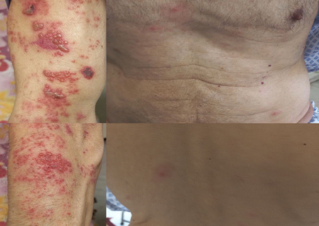
Figure 1
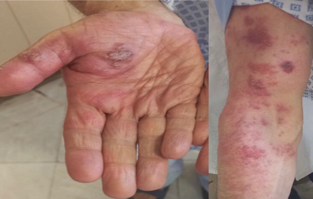
Figure 2
Lab Tests Results
WBC x10 3 /μl): 4.5, Hb (g/dl):14.4, plt:144, CRP (mg/l):5
VZV Ab IgM:1/8 (LESS THAN 9 NEGATIVE), VZV Ab IgG:15/1 (LESS THAN 9 NEGATIVE), PCR CMV: WAS NOT DETECTED, CMV Ab IgM: 0.4(<0>10(<0>
We prescribed ACYCLOVIR 500 MG IV TDS for 7 days. Also we prescribed Cefazolin 1 gr TDS, and and gabapentin 300 mg po daily. Fluid of palmar bulla was sent for VZV PCR. We thought about other differential diagnosis. for instance CMV disease AND HIV which were negative. Also recommended cold shower and cold compress to reduce the itching. On outpatient follow up two weeks later, he brought the biopsies results which revealed VZV PCR FLUID positive. The patient's clinical status improved and rash resolved.(figure 3)
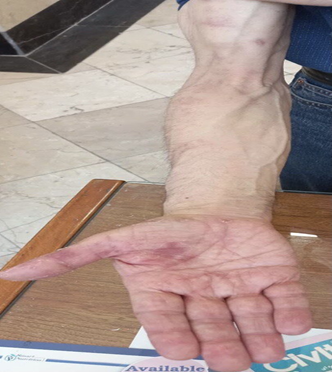
Figure 3
Herpes zoster is the result of reactivation of this residual latent virus. The first manifestation of zoster is usually pain, which may be severe and
accompanied by fever, headache, malaise and tenderness localized to one or more nerve roots. The lymph nodes draining the affected area are enlarged and tender.[7]
VZV is transmitted via the airborne route. The incidence of HZ, which causes significant morbidity, increases with age and reaches approximately 10 cases per 1,000 patient-years by age 80. Cell-mediated immunity (CMI) is known to decline with age as part of immunosenescence, and decreased CMI is associated with reactivation of VZV.[7]
The rash associated with HZ has a brief erythematous and macular phase, which is often missed, after which papules rapidly appear. These papules develop into vesicles within 1–2 days, and vesicles continue to appear for 3–4 days. The lesions tend to be grouped, and clusters are often seen where there are branches of the cutaneous sensory nerve. Pustulation of vesicles begins within 1 week of the onset of rash, if not sooner, and is followed 3–5 days later by lesion ulceration and crusting. Crusts usually are gone by the end of 3 or 4 weeks, but scarring and hypo- or hyperpigmentation may persist long after the HZ resolves. Fewer than 20% of patients have significant systemic symptoms, such as fever, headache, malaise, or fatigue.[8]
The phenomenon of zoster occurring in 2 noncontiguous dermatomes has been referred to as zoster duplex unilateralis or bilateralis, depending whether one half or both halves of the body are involved.[9]
Complications of herpes zoster are more common among elderly individuals and immunosuppressed patients. Herpes zoster and its complications can impact the patient's quality of life. In most patients, sleep and social activities are affected. Post-herpetic neuralgia is the most common complication of herpes zoster. The other complications noted following post-herpetic neuralgia, include secondary bacterial infections, ophthalmic complications, cranial and peripheral nerve palsies, and segmental zoster paresis.[10]
Typically, a single dermatome is involved, although two or three adjacent dermatomes may be affected. The lesions usually do not cross the midline. Herpes zoster can also present with unique or atypical clinical manifestations, such as glioma, zoster sine herpete and bilateral herpes zoster.[11]
A 30-year-old male presented to the emergency department with low-grade fever, sore throat and generalised malaise that started 3 days prior to presentation. he developed an erythematous pruritic warm raised rash on his left anterolateral chest that extended to the medial aspect of the axilla and mid-thoracic area on his back left to the mid-line.at first he was treated with the impression of an allergic reaction to ibuprofen. By worsening of itching and rash skin biopsy was done and reported positive for herpes zoster virus. He was then prescribed oral valacyclovir for 7 days with the diagnosis of multidermatomal herpes zoster (T1-T4).[12]
A 50-year-old man presented to clinic with a ten-day history of a painful, swollen red plaque on his right lateral eyebrow with overlying small coalescing erosions, as well as extensive soft tissue swelling and erythema of the right preauricular cheek with right submandibular lymphadenopathy. Findings included the maxillary and mandibular (V2/V3) dermatomes but excluded ocular involvement (V1). The rash had worsened despite a 5-day course of oral doxycycline 100 mg twice daily for presumed cellulitis. PCR studies returned positive for VZV. A lumbar puncture showed pleocytosis, and cerebrospinal fluid PCR was VZV positive. The patient was treated for VZV meningitis with intravenous acyclovir 10 mg/kg every 8 hours for 14 days.[13]
A 47 male patient presented to private practice with a rash, itching, and pain in the left lower extremity for 3 days. He had no underlyingdiseases and Chickenpox infection in the past. He was diagnosed with herpes zoster and symptoms were relieved by the administration of acyclovir five times daily for one week.[14]
A 26-year-old male presented to the Dermatology Outpatient Department with chief complaints of eruptive lesions (multiple papular and vesicular lesions) over the anterior chest, medial aspect of the arm, forearm palms of the right hand, and right upper back for four days which followed skin rashes with a burning sensation over the involved areas. The patient reported a heightened psychological stress state. VZV-specific IgM antibody came out to be positive.The patient was prescribed tablet acyclovir 800 mg five times a day for 5 days, topical antibiotic ointment mupirocin twice daily for 1 week to prevent secondary infection, analgesic like tablet pregabalin 75 mg as per the need and was advised to use vaseline over the affected area. Notably, a comprehensive stress management strategy.[15]
A 15-year-old Chinese boy presented with multiple vesicles/bullae on an erythematous base, distributed bilaterally and symmetrically in a band-like distribution along T7, T8, and T9 dermatomes. The patient was treated with acyclovir 800 mg five times a day for 7 days. The blistering and discomfort resolved in 14 days, and the secondary dyspigmentation took 3 months to completely fade.[16] Early diagnosis and treatment with antiviral agents plus intervention treatments is believed to shorten the duration and severity of acute HZ and reduce the risk of PHN. Prophylactic vaccination against VZV can be the best option to prevent or reduce the incidence of HZ and PHN.[17] Routine vaccination for individuals over 60 years has shown considerable effect in terms of reducing the incidence of herpes zoster and post-herpetic neuralgia.[10] The efficacy of antiviral therapy in patients with HZ has been demonstrated by multiple randomized controlled clinical trials. Acyclovir (800 mg 5 times daily for 7–10 days), famciclovir (500 mg 3 times daily for 7 days, the approved dosage in United States; 250 mg 3 times daily is approved in some other countries), and valacyclovir (1000 mg 3 times daily for 7 days) have been approved by the US Food and Drug Administration for the treatment of HZ. These antiviral agents are phosphorylated by viral thymidine kinase and cellular kinases to a triphosphate form that inhibits viral replication.[8]
Treatment of Zoster Duplex Bilateralis is the same as for the more common form of herpes zoster, which included an antiviral agent, pain management, and care of the skin lesions.[18]
Although diagnosis of herpes zoster is easy and can be diagnosed by general practitioners, sometimes delay in diagnosis can occur. The most important reason is atypical manifestation of zoster. Multidermal presentation is uncommon but disparate multidermatomal involvement is rare.Clinicians should be aware of rarer variants of herpes zoster in immunocompotent patients.
Informed consent was obtained from the patient for the publication of this case report and accompanying images.
The authors declare no conflicts of interest.
Dear Editorial Team, Clinical Medical Reviews and Reports. My experience with the journal was highly positive. The peer-review process was rigorous, constructive, and completed in a timely manner. The reviewers provided valuable comments that helped improve the quality and clarity of our manuscript. The editorial office was professional, responsive, and supportive throughout all stages of the publication process. Communication was clear and efficient, and any questions were addressed promptly. Overall, I found the journal to maintain high scientific standards and an excellent publication workflow. I would be pleased to consider submitting future work to this journal. Best wishes from, Elena Popa.

It was my pleasure to submit my testimonial concerning the Reviewer Board of our Scientific Journal “Brain and Neurological Disorders”. The Reviewers focused on some modifications and their contribution was helpful. The ladies of our Editorial Office were also supported my efforts. It was my honor to have such a co-operation and I am looking forward for more collaboration.

Dear Grace Pierce, Editorial Coordinator of Journal of Clinical Research and Reports, Thank you for the speedy and efficient peer review process. I appreciate the fact that your peer reviewers do not take months to respond like with some other journals. I would also like to thank the editorial office for responding quickly to my questions. It is an excellent journal. I plan to submit more manuscripts in the future. Best wishes from, Robert W. McGee

Dear Grace Pierce, Editorial Coordinator of Journal of Clinical Research and Reports, Working with you and your team on our recent publication in JCRR has been a truly wonderful and enjoyable experience. The responses were prompt, and the reviewers were patient, constructive, and highly professional. One reviewer in particular gave me the feeling that a professor was carefully reading and commenting on my coursework, which was deeply touching. The entire process was straightforward and hassle‑free, with no tedious online forms to complete. I highly recommend this journal. Best wishes from, DR Aibing Rao, Head of R&D

I Appreciate the Opportunity to Share my Experience with the Journal of Clinical Research and Reports. The peer review process was timely and constructive, and the feedback provided helped improve the quality of our manuscript. The editorial office was professional, responsive, and supportive throughout the process, ensuring smooth communication and efficient handling of the submission. Overall, it was a positive experience collaborating with your team.

Dear Mercy Grace, Editorial Coordinator of Obstetrics Gynecology and Reproductive Sciences, We would like to express our gratitude for your help at all stages of publishing and editing the article. The editors of the magazine answer all the necessary questions and help at every stage. We will definitely continue to cooperate and publish other works in the Obstetrics Gynecology and Reproductive Sciences! Best wishes from, Alla Konstantinovna Politova,
