
Case Report | DOI: https://doi.org/10.31579/2642-9756/123
1 Department of Obstetrics and Gynecology, University Clinical Hospital of Valladolid, Regional Health Management of Castilla y León (SACYL), Spain.
2 Department of Pediatrics and Immunology, Obstetrics and Gynecology, Nutrition and Bromatology, Psychiatry and History of Science, Faculty of Medicine, University of Valladolid, Spain.
*Corresponding Author: Sonia De-Miguel-Manso. Department of Pediatrics and Immunology, Obstetrics and Gynecology, Nutrition and Bromatology, Psychiatry and History of Science, Faculty of Medicine, University of Valladolid, Spain. International Hospital.
Citation: Sonia De-Miguel-Manso, Blanca Heras-Pérez, Victoria Pascual-Escudero, Dakota Viruega-Cuaresma, Álvaro Sanz-Díaz-Heredero.(2022). Gonococcal Pelvic Inflammatory Disease with Sepsis Criteria: Review of 2 Cases, J. Women Health Care and Issues, 5(5) DOI: 10.31579/2642-9756/123.
Copyright: © 2022 Sonia De-Miguel-Manso. This is an open-access article distributed under the terms of The Creative Commons Attribution License, which permits unrestricted use, distribution, and reproduction in any medium, provided the original author and source are credited.
Received: 01 June 2022 | Accepted: 12 June 2022 | Published: 15 July 2022
Keywords: neisseria gonorrhoeae; pelvic inflammatory disease; sepsis
Background: Infection caused by Neisseria Gonorrhoeae increases the risk of pelvic inflammatory disease (PID). Gonococcal PID tends to be clinically more severe than non-gonococcal ones. The main is to present two cases of gonococcal PID, with rapid clinical and analytic progression, leading to severe sepsis, but without imaging manifestations.
Clinical presentation:
1. 41-year-old patient with replacement of intrauterine releasing levonorgestrel device (IUD), presented abdominal pain and green vaginal discharge. Abdominal examination revealed signs of peritoneal irritation and blood test showed leukocytosis, increased C Reactive Protein and procalcitonin, as well as coagulation abnormalities. Imaging tests (vaginal ultrasound/tomography) revealed no structural pathology, without collections. Given the criteria of severe sepsis, broadspectrum intravenous (iv) antibiotic therapy was started and laparoscopy and IUD removal were performed. Cervical and IUD cultures were positive for Neisseria gonorrhoeae.
2. 20-year-old woman, with an IUD, consulted for abdominal pain, low-grade fever and green vaginal discharge. Abdominal examination suggested peritoneal sensitivity and laboratory tests leukocytosis, increased C Reactive Protein and procalcitonin with coagulation abnormalities. Imaging tests (vaginal ultrasound/tomography) showed no structural pathology, without collections. Despite analgesia and broad-spectrum iv antibiotics, the patient worsened, proceeding to remove the IUD. Given the criteria compatible with severe sepsis, laparoscopy was decided. Endocervical and IUD cultures revealed Neisseria gonorrhoeae.
Conclusions: Facing the situation of an acute PID with severe and fast clinical worsening even without findings in imaging tests, we should consider gonococcal ethiology as a possible cause. Surgical approach shouldn’t be delayed in order to control the infection and rule out other possible diagnosis.
Gonorrhea, an infection caused by gram negative cocobacillus Neisseria Gonorrhoeae, is a major cause of morbidity among sexually active men and women worldwide. It represents one of the main causes of urethritis in men and cervicitis in women. It increases the risk of pelvic inflammatory disease (PID), infertility, ectopic pregnancy and chronic pelvic pain, as well as complications during pregnancy [1].
PID is an acute infection of internal genital tract, involving uterus, fallopian tubes and ovaries. Frequently, it spreads to adjacent pelvic organs. A sexually transmitted pathogen triggers the infection, which secondarily rises to the superior genital system [2].
PID occurs in approximately 15% of women with cervical gonorrhea, and N. Gonorrhoeae is estimated to be the causative microorganism in 40 percent of PID cases [3]. Gonococcal PID tends to be clinically more severe than non-gonococcal ones [4], but the extent of tubal inflammation and scarring appears to be similar.
The main objective of this work is to present two cases of gonococcal PID, with rapid clinical and analytic progression, leading to severe sepsis, but without findings in imaging tests.
Case 1 :
41-year-old women, smoker, with a history of 3 vaginal deliveries and carrier of an intrauterine releasing levonorgestrel device (IUD) for 5 years ago. She didn’t use any barrier contraceptive method since she has a stable sexual partner. She consulted for lower abdominal pain, that has appeared sharply in the last 24 hours. It was associated with green vaginal discharge, loss of appetite and bilious vomiting within the last few hours. Two days ago, her IUD was replaced by her gynecologist.
During abdominal examination, she complained of intense hypogastric and mesogastric pain, as well as generalized defense. Also, positive Blumberg sign was present, suggesting peritoneal irritation. In vaginal examination, grayish vaginal discharge was noticed as well as correctly placed IUD threads. We took vaginal and endocervical microbiologic samples. Cervical motion was painful and no adnexal masses were palpable.
Vital signs were taken and blood and urine samples collected. Results are shown in table 1 and 2 respectively.


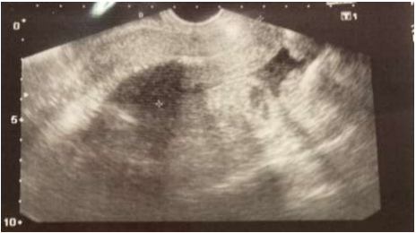
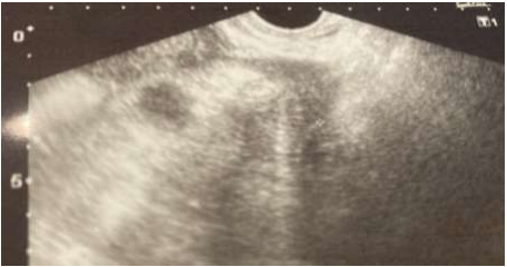
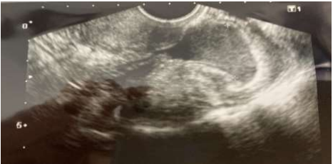
Vaginal ultrasound revealed regular uterus with normally positioned IUD (figure 1) and both ovaries without any ecographic findings (figures 2,3). A slight amount (23 x 18 mm) of free anechoic fluid with internal echos was noted in Douglas pouch (figure 4).


An abdominal CT was performed, with no findings in superior abdominal cavity as well as uterus and both ovaries, and correctly placed IUD. Small bowel dilatation was described from jejunum, with ileum loops in pelvis, showing 40 x 6 mm of mural thickening. This was attributed to a patched ileitis, with normal large bowel and small amount of free pelvic fluid.
We then suspected a pelvic inflammatory disease (PID), stage two (acute salpingitis associated with peritonitis) (5), without being able to rule out ileitis.
The patient was also evaluated by a general surgeon, who dismissed surgical approach at this moment.
Considering the presence of severe sepsis criteria (both clinical and analytic), pathologic abdominal and pelvic examination and the background of IUD replacement 48 hours ago, we started intravenous (i.e.) antibiotic treatment with Piperacillin-Tazobactam 4 g every 6 hours.
Diagnostic laparoscopic exploration was also performed, showing purulent pelvic fluid, omental inflammatory reaction and right fallopian tube edematous and hyperemic. We confirmed the presence of small bowel dilated loops in the proximity of right tube. Both uterus and left fallopian tube were macroscopically normal, such as the rest of the abdominal cavity. A bilateral salpingectomy was completed after profuse abdominal washing and aspiration and an abdominal drain was placed. We removed IUD after first antibiotic i.e. dose and it was sent for microbiological further studies.
Postoperative evolution was favourable. Endocervical and IUD cultures were positive for Neisseria Gonorrhoeae. After 48 hours of intravenous Piperacillin/Tazobactam, we switched antibiotic treatment to 500 mg ceftriaxone + 2 g azithromycin single oral dose. We alerted our patient that her sexual partner needed to be treated as well. He had a previous course of antibiotic 10 days ago, prescribed by his general doctor due to a possible urethritis. We recommended to avoid sexual intercourse during recovery.
At 30 days post operative check-up, patient was completely asymptomatic and new endocervical sample was negative for sexually transmitted germs.
20-year-old smoker and nulliparous woman. She was using since 4 years ago IUD as her contraceptive method and had a stable sexual partner at the moment of consultation.
She presented in the emergency room (ER) complaining of abdominal tenderness associated with greenish vaginal discharge for the last 4 days, as well as nausea and vomiting.
During abdominal examination, the patient complained of intense pain in left iliac fossa, along with signs of peritoneal irritation. Bilateral costovertebral angle percussion was negative.
In vaginal exam, IUD threads and pathological discharge were noticed and vaginal and endocervical samples we taken for microbiological testing. Palpation of left adnexal area showed tenderness as well as cervical painful motion.
Vital signs, blood and urine test results are shown in table 1 and 2, respectively.
Vaginal ultrasound showed regular uterus with normally positioned IUD and both ovaries without any sonographic findings (figures 5 and 6). A slight amount of free anechoic fluid with internal echo's was noted in Douglas pouch (figure 7).


Superior abdominal cavity as well as uterus, IUD and both ovaries were described as normal in CT abdominal scan. Thickened and enhancing pelvic peritoneum was observed without neither suspicious collection nor signs of bowel perforation. In addition, non-dilated but enhanced bowel loops were noted, with normal cecum and minimum amount of free pelvic fluid.
Stage II PID was suspected and patient was then admitted for observation and antibiotic treatment (ampicillin 2 g every 6 hours, clindamycin 900 mg every 8 hours and gentamicin 240 mg daily). A significant clinical and analytical worsening occurred during the following hours, leading to a severe sepsis. We then decided to remove IUD (sent to microbiological testing) and laparoscopic abdominal exploration. Patient was informed and consented the procedure.
Findings of laparoscopic examination consisted in hyperemic uterus, slightly swollen fallopian tubes but no pelvic abscesses. In addition, moderate amount of purulent free fluid was noted and important dilatation of bowel loops. At the end of the procedure, we placed an abdominal drain.
Postoperative evolution was without further complication. Both endocervical and IUD microbiological cultures were positive for Neisseria Gonorrhoeae and Chlamydia Trachomatis. We suspended antibiotic intravenous treatment. At the time of discharge, we prescribed both our patient and her sexual partner treatment with single oral dose of azithromycin 2 g and doxycycline 100 mg every 12 hours for 14 days. We advised them not to have sexual intercourse till the end of antibiotic course, and to switch to barrier method as contraception.
At 30 days postoperative check-up, patient was completely asymptomatic and endocervical sample resulted negative.
Ethical approval: for the preparation of this manuscript, both patients were informed of the scientific interest of their processes and their verbal consent was obtained, both for the publication of their data and images (Figures 1-7).
Genital infections, particularly cervicitis, are the most commonly associated with N. Gonorrhoeae and might spread to upper genital tract, causing PID.
Uterine cervix is the most frequent site of mucosal gonococcal infection in women. Up to 70 percent of these infections are asymptomatic [6,7]. Thus, the incubation period of gonorrhea is less well characterized in women than men. When present, genital symptoms tend to develop within ten days after exposure [8]. Symptomatic infection typically manifests as vaginal pruritus and/or a mucopurulent discharge. In rare cases, intermenstrual bleeding or menorrhagia can appear. The presence of abdomino-pelvic pain or dyspareunia should raise suspicion for upper genital tract involvement [9,10].
Given the high incidence of asymptomatic gonococcal infections, PID can be the first sign and these patients can end up being severely ill [4].
It is frequent that symptoms start with the beginning of menstruation [1]. This was first described back in 1979, when female rats were inoculated with N. Gonorrhoeae. A comparison between the pattern of infection was conducted, regarding if germs were suspended or not in mucine and hemoglobin. Those hanging in mucine and hemoglobine lead more frequently to gonococcal bacteremia due to phagocytosis interference and intracellular death of N. Gonorrhoeae. This model mimics the progression in women with PID from local peritonitis to blood spread with factors (mucine and hemoglobine) increasing infection [11].
More recent references also link menstruation to a higher risk of gonococcal dissemination, due to:
❖ Phenotypical switching of N. Gonorrhoeae from opaque strains to transparent ones (modified expression of transmembrane proteins) [12]. These modifications can reduce sensibility to trip sine lysis, neutrophil adhesion as well as increased adherence to fallopian tubes [13].
❖ Alkalinization of genital discharge, stimulating gonococcal growth [14].
❖ Increased mucosal concentration of transferrin and hemoglobin. This can be used as an iron source by N. Gonorrhoeae [16].
Normally, superior genital tract is protected by endocervix from germs of vaginal ecosystem. Endocervical infection by N. Gonorrhoeae can break this natural barrier, allowing the compromise of superior genital organs and adjacent ones. The resultant infection can be subclinical or in the way of pelvic inflammatory disease. Immune system response genetic variations, estrogen levels compromising viscosity of cervical mucus and bacterial charge, can explain this variable expression [9,17].
N. Gonorrhoeae and C. Trachomatis are frequently identified pathogens in PID among premenopausal sexually active women.
Nevertheless, in the majority of diagnosed cases, we are not able to clarify microbiological etiology. Despite the germ that triggers infection, we usually consider PID as a polimicrobian infection [2].
N. Gonorrhoeae is also a frequent cause of urethritis in men. Up to 60% of these patients can be asymptomatic or experience slight symptoms [18,19]. Among symptomatic ones, incubation period takes between 2 to 5 days [20].
Sexual intercourse is the main PID risk factor, especially in case of multiple partners. Younger patients, previous Chlamydia infection or PID episode and a sexual partner with a history of sexually transmitted disease (STI) are other significant risk factors. Contraception is also important in the prevalence of PID. The use of barrier method constitutes a protective factor [2].
There is a clear relation between PID and other contraceptive methods, as described below:
❖ Oral contraceptives (OCs) and PID have a complex interaction. Several publications have stated that OC use nearly doubles the prevalence of both chlamydia and gonococcal cervical infection [21,22]. Nevertheless, OC have traditionally been associated with a significant reduction of PID. Apparently, women using OCs tend to develop PID as frequently as other women, but the severity of the infection is substantially inferior [23,24].
❖ Modern intrauterine devices (IUD) don’t increase PID risk and this one is limited to the first 3 weeks after its insertion. There is evidence supporting leaving IUD inside the uterine cavity during the first 48 to 72 hours of acute PID antibiotic treatment, with close follow-up. If there is no response to treatment or in case of high-risk patients, IUD should be removed and microbiological studies can be performed to guide antibiotic regimen selection [25-27]. Although a case of infection of the IUD by actinomyces has been reported, leading to bilateral tubo-ovarian abscess in a postmenopausal woman with an IUD of 12 years of evolution [28].
❖ Tubal ligation may protect the distal oviducts from involvement, but the clinical syndrome of PID is otherwise unaffected.
Taking into account risk factors in our cases, we noted that patient 1 had a recent IUD replacement. We suggest that it is possible that cervical infection was already present at the day of cervical manipulation. This statement is based in her sexual partner previous symptoms (suspected urethritis) for which he was treated with oral antibiotics 7 to 10 days before patient admission.
Regarding patient 2, she was a young women aged 20, with several sexual partners in the past and IUD user, without barrier contraception associated.
In case 1, considering childbearing was completed, even though no tubal abscesses were present, we agreed with our patient to perform bilateral salpingectomy. This way we assured posterior contraception and reduced severity in case of PID relapse.
In both of our cases, we removed IUD after first dose of intravenous antibiotics (case 1 removal performed during surgical procedure, case 2 after no evidence of improvement with antibiotics), because of clinical severity. They both required hospital admission and parenteral antibiotics, being most frequent indications for this:
• Clinical severity (high fever > 38.5º, nausea, vomiting, intense abdominal pain…)
• PID complicated with the presence of pelvic abscess. • Surgical approach considered because of suspicion of ruptured pelvic abscess or confirming alternative diagnosis (appendicitis, ovarian torsion…).
• Inability to take oral medication because of intense nausea or vomiting. • Lack of response to oral antibiotics.
• Concern for non-adherence to therapy.
Antibiotic therapy is the cornerstone of PID treatment. For hospitalized patients, initial treatment consists in a parenteral regimen that provides antimicrobial coverage against a wide range of bacteria, including C. trachomatis and N. gonorrhoeae (most frequent STIs associated with acute PID), streptococci, gram-negative enteric bacilli (Escherichia coli, Klebsiella spp, and Proteus spp), and anaerobic organisms (bacterial vaginosis-associated flora) [29].
United States Centers for Disease Control and Prevention (CDC) recommends these following regimens [30]:
• Cefoxitin (2 g i.e., every six hours) or Cefotetan (2 g i.v. every 12 hours) plus doxycycline (100 mg every 12 hours oral or i.e.).
• Clindamicine (900 mg every 8 hours) plus gentamicin (3-5 mg/kg daily i.v. or 2 mg/kg i.v. followed by 1.5 mg/kg every 8 hours).
Regimens based on second generation cephalosporin plus doxycycline are preferred because of their tolerability and security. They also show excellent in vitro activity against N. gonorrhoeae and C. trachomatis. Regimen with gentamicin combined with clindamicine has only moderate in vitro activity against these bacteria [30,31]. However, both regimens have shown in several trials short-term clinical cure rates of more than 80 to 90 percent of cases [32,33].
Alternative regimens have more limited clinical data to support their use for hospitalized patients and less gonococcal activity
• Ampicillin-sulbactam (3 g i.v. every six hours) doxycycline (100 mg twice daily). This combination results in a similar clinical cure rate as cefoxitin plus doxycycline [33,34].
• Azithromycin (500 mg i.v. daily for one to two days followed by 250 mg orally daily to complete a five to seven-day course) with or without metronidazole (in case of pelvic abscess), lead to similarly high clinical and microbiologic cure rates (>95 percent) for mild to moderate PID compared with a combination beta-lactam and doxycycline regimen. Azithromycin was not evaluated in severe PID cases [35].
In patient 1, we combined penicillin with a beta lactamase inhibitor because diagnosis remained unclear, considering either grade II PID or ileitis. This regimen was not optimal in case of PID diagnosis. Doxycycline association would have been ideal in order to cover most frequent pathogens in PID, including STIs.
In contrast, parenteral treatment in patient 2 was adequate, combining ampicillin, clindamicine and gentamicin.
Both patients experienced a similar and adverse clinical evolution, suggesting severe sepsis because of the presence of the following criteria (table 3) [36]:
• White cell count over 12.000/mm3
• C- reactive protein (CRP) and procalcitonin (PCT) increase
• Coagulation disfunction (INR > 1.5)
• Systolic blood pressure < 90>

Even though neither structural nor organic pathology was observed in imaging tests, we decided that surgical approach was necessary because of adverse clinical evolution within the first hours of hospital admission and severe sepsis criteria. After at least 24 hours of clinical improvement, patients can usually be switched from parenteral therapy to oral antibiotics, consisting in administration of 100 mg doxycycline twice daily in order to complete a 14-day course. In case of not tolerating doxycycline, azithromycin (500 mg for 1 or 2 days followed by 250 mg once daily in a 7-day course) is an adequate option [37]. In our patients, after a consistent 48 hours clinical improvement and considering endocervical and IUD microbiological results, we stopped parenteral treatment. Patient 1 received intravenous extra 500 mg ceftriaxone and 2 g azithromycin single oral dose. In patient 2, we combined this same azithromycin single dose with 100 mg doxycycline twice daily for 14 days.
Patients diagnosed and treated for acute PID should be carefully advised on the following topics:
• Long antibiotic course of treatment: compliance with this kind of treatment can be problematic [37]. Patients should be educated about the importance of medication adherence and clinical outcomes.
• Sexual activity: patients with PID diagnosis should avoid sexual intercourse until they have completed therapy, their symptoms have disappeared and partners have been screened and treated for STIs. We should explain STIs way of transmission and counsel safe sex practices.
• STIs screening and prevention: patients should also be tested for HIV, syphilis… [37].
• Evaluation and treatment of sex partners: they should be treated in case of sexual intercourse with the patient during 60 days prior to the onset of symptoms, regardless of the woman's STI test results. Treatment regimens should include antibiotics with activity against N. gonorrhoeae and C. trachomatis [37]. If the patient denies sexual activity during previous 60 days, most recent sexual partner should be tested and treated [38]. Sexual partners, frequently showing no symptoms, are capable of reinfecting the index patient or spreading the infection to other sexual contacts. Expedited partner therapy (EPT) is a strategy to secure treatment of sexual partners of STI positive patients. Without any medical evaluation, we provide prescriptions or medication to the patient, who will deliver it to the partner(s) [38,39]. It is always preferable to evaluate before treatment, but EPT can be helpful in cases in whom clinical evaluation is unlikely. The benefits of EPT implementation are reduced rates of reinfection and increased number of patients receiving treatment. In randomized trials, EPT was more effective than traditional partner notification (the patient communicates his/her partner the need for clinical evaluation and treatment) in reducing persistent or recurrent urogenital gonococcal infection [40, 41]. Nevertheless, we can lose the chance for other STIs/HIV screening and it is possible that adverse effects caused by antibiotic treatment won’t reach a health-care professional [41,42].
First-line treatment for non-complicated gonococcal infections consists in third generation cephalosporins, due to increasing resistance rates to other antibiotics (penicillin, fluoroquinolones, tetracycline, sulfonamides) [43]. Among them, ceftriaxone is the most frequently used in a single high dose regimen. CDC recommendations are:
• 500mg intramuscular ceftriaxone, single dose if weight < 150>
• 1000 mg intramuscular ceftriaxone, single dose if weight > 150 kg.
These ceftriaxone regimens include higher doses in order to face rising gonococcal minimum inhibitory concentrations (MICs). Ceftriaxone + azithromycin was a frequent combination not long ago [43], but consistent data have demonstrated decreased susceptibility of N. Gonorrhoeae to azithromycin [43,44].
If concurrent C. trachomatis infection has not been ruled out by tests, we should prescribe treatment for Chlamydia at the same time as gonococcal regimen. Following CDC recommendations, we suggest doxycycline 100 mg orally twice a day during 7 days. Single-dose azithromycin is also a valid option. Nevertheless, considering the already mentioned decreased susceptibility of N. Gonorrhoeae to azithromycin, doxycycline regimen is preferred [44].
• Notification of N. Gonorrhoeae and C. Trachomatis infection as mandatory reported infectious diseases: these two infections are reported to public health system many countries. In Spain, cases are notified to “Sistema de Information Microbiological”. Spanish incidence rate per 100.000 inhabitants during 2008 was 13.78 for gonococcal infection and 12.81 for chlamydia [45].
• Reinsertion of IUD: in case previous IUD was removed during acute infection, the optimal moment for re-insertion remains unclear. We suggest to wait until antibiotic treatment is completed, all symptoms have disappeared and cervical screening is negative. Because of high rates of reinfection, retesting for N. gonorrhoeae and C. trachomatis is counseled within 3 months of the original infection [46].
• Test of cure: Asymptomatic patients following treatment for uncomplicated urogenital or anorectal gonococcal infections do not need to return for a microbiological culture [43]. This test is the preferred one and it should be performed at least 7 days after treatment [47,48]. Nucleic acid amplification tests (NAATs) are also a valid test of cure, but should be conducted at the minimum 14 days following therapy. If NAAT is performed too soon after treatment, false-positive results (from detection of nonviable organisms that may not represent persistent infection) can appear [49,50].
In our cases, we provided our patients with a recommendation of the antibiotic regimen their partners should follow (via EPT), considering microbiological tests results. Patient 1 sexual partner was treated with ceftriaxone (with activity proved against N. Gonorrhoeae) and azithromycin (useful for other germs such as C. trachomatis). In case 2, sexual partner was treated with azithromycin and doxycycline (first-line option for gonococcal infection would have been ceftriaxone, due to reduced susceptibility to azithromycin, but optimal coverage for C. trachomatis was assured by doxycycline).
Both our patients presented with gonococcal infection + acute pelvic inflammatory disease (PID), so we decided to perform endocervical culture as test of cure. This test was carried out one month after treatment and resulted negative. In patient 2, sexual counseling was particularly important ir order to avoid STIs reinfections (age, multiple sexual partners…). We didn’t perform other STIs (HIV, syphilis) testing.
• Gonococcal infection is an STI that can be asymptomatic in up to 70% of patients. This infection can lead to an acute PID that tends to be clinically more severe than non-gonococcal ones.
• We have resumed two cases of gonococcal PID with a fast clinical worsening, even meeting severe sepsis criteria.
• Therefore, facing the situation of an acute PID with severe and fast clinical worsening even without findings in imaging tests, we should consider gonococcal ethiology as a possible cause. Surgical approach shouldn’t be delayed in order to control the infection and rule out other possible diagnosis.
The authors have no relevant financial or non-financial interests to disclose
Dear Editorial Team, Clinical Medical Reviews and Reports. My experience with the journal was highly positive. The peer-review process was rigorous, constructive, and completed in a timely manner. The reviewers provided valuable comments that helped improve the quality and clarity of our manuscript. The editorial office was professional, responsive, and supportive throughout all stages of the publication process. Communication was clear and efficient, and any questions were addressed promptly. Overall, I found the journal to maintain high scientific standards and an excellent publication workflow. I would be pleased to consider submitting future work to this journal. Best wishes from, Elena Popa.

It was my pleasure to submit my testimonial concerning the Reviewer Board of our Scientific Journal “Brain and Neurological Disorders”. The Reviewers focused on some modifications and their contribution was helpful. The ladies of our Editorial Office were also supported my efforts. It was my honor to have such a co-operation and I am looking forward for more collaboration.

Dear Grace Pierce, Editorial Coordinator of Journal of Clinical Research and Reports, Thank you for the speedy and efficient peer review process. I appreciate the fact that your peer reviewers do not take months to respond like with some other journals. I would also like to thank the editorial office for responding quickly to my questions. It is an excellent journal. I plan to submit more manuscripts in the future. Best wishes from, Robert W. McGee

Dear Grace Pierce, Editorial Coordinator of Journal of Clinical Research and Reports, Working with you and your team on our recent publication in JCRR has been a truly wonderful and enjoyable experience. The responses were prompt, and the reviewers were patient, constructive, and highly professional. One reviewer in particular gave me the feeling that a professor was carefully reading and commenting on my coursework, which was deeply touching. The entire process was straightforward and hassle‑free, with no tedious online forms to complete. I highly recommend this journal. Best wishes from, DR Aibing Rao, Head of R&D

I Appreciate the Opportunity to Share my Experience with the Journal of Clinical Research and Reports. The peer review process was timely and constructive, and the feedback provided helped improve the quality of our manuscript. The editorial office was professional, responsive, and supportive throughout the process, ensuring smooth communication and efficient handling of the submission. Overall, it was a positive experience collaborating with your team.

Dear Mercy Grace, Editorial Coordinator of Obstetrics Gynecology and Reproductive Sciences, We would like to express our gratitude for your help at all stages of publishing and editing the article. The editors of the magazine answer all the necessary questions and help at every stage. We will definitely continue to cooperate and publish other works in the Obstetrics Gynecology and Reproductive Sciences! Best wishes from, Alla Konstantinovna Politova,
